Chinese Journal of Tissue Engineering Research ›› 2026, Vol. 30 ›› Issue (8): 1929-1939.doi: 10.12307/2026.017
Previous Articles Next Articles
Metal organic framework/carboxymethyl chitosan-oxidized sodium alginate/platelet-rich plasma hydrogel promotes healing of diabetic infected wounds
Liu Hongjie1, Mu Qiuju2, Shen Yuxue1, Liang Fei1, Zhu Lili1, 2
- 1Clinical Laboratory Science Teaching and Research Department, School of Clinical Laboratory Science, Guizhou Medical University, Guiyang 550004, Guizhou Province, China; 2Department of Blood Transfusion, Affiliated Hospital of Guizhou Medical University, Guiyang 550000, Guizhou Province, China
-
Received:2024-11-13Accepted:2025-01-06Online:2026-03-18Published:2025-07-15 -
Contact:Zhu Lili, Senior technologist, Clinical Laboratory Science Teaching and Research Department, School of Clinical Laboratory Science, Guizhou Medical University, Guiyang 550004, Guizhou Province, China; Department of Blood Transfusion, Affiliated Hospital of Guizhou Medical University, Guiyang 550000, Guizhou Province, China -
About author:Liu Hongjie, Master candidate, Junior technologist, Clinical Laboratory Science Teaching and Research Department, School of Clinical Laboratory Science, Guizhou Medical University, Guiyang 550004, Guizhou Province, China -
Supported by:Guizhou Provincial Science and Technology Plan Project, No. ZK[2024]184 (to ZLL)
CLC Number:
Cite this article
Liu Hongjie, Mu Qiuju, Shen Yuxue, Liang Fei, Zhu Lili. Metal organic framework/carboxymethyl chitosan-oxidized sodium alginate/platelet-rich plasma hydrogel promotes healing of diabetic infected wounds[J]. Chinese Journal of Tissue Engineering Research, 2026, 30(8): 1929-1939.
share this article
Add to citation manager EndNote|Reference Manager|ProCite|BibTeX|RefWorks
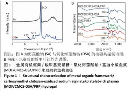
2.1 水凝胶结构及形貌表征 采用核磁共振氢谱观察氧化海藻酸钠的氧化过程(图1A),在5.15×10⁻⁶和5.75×10⁻⁶之间观察到羟基和醛基形成的半缩醛峰,表明海藻酸钠的α-羟基被成功氧化成二醛。傅里叶变换红外光谱分析以检查水凝胶形成(图1B),发现氧化海藻酸钠和羧甲基壳聚糖混合后它们特征性的峰值消失,在1 623 cm-1处出现了一个新的亚胺结构峰值,证实了席夫碱反应的发生;在氧化海藻酸钠、金属有机框架材料与富血小板血浆的凝胶反应中,于1 623 cm-1处也能够观察到席夫碱键的形成,从而证实了几种物质的成功交联。扫描电镜下可见羧甲基壳聚糖-氧化海藻酸钠、金属有机框架/羧甲基壳聚糖-氧化海藻酸钠和金属有机框架/羧甲基壳聚糖-氧化海藻酸钠/富血小板血浆水凝胶都具有相互交联且均匀的多孔结构,富血小板血浆的引入进一步提高了水凝胶的交联密度,使水凝胶内部结构更加紧密(图2)。"
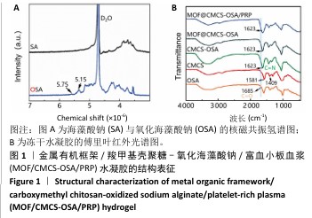
"

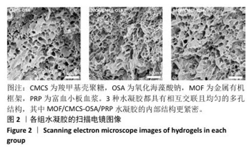
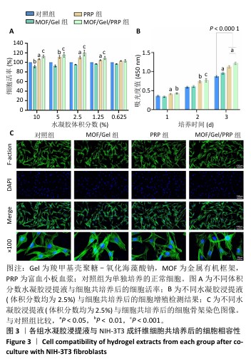
2.2 水凝胶的细胞相容性 CCK-8检测结果显示,不同体积分数水凝胶浸提液与NIH-3T3成纤维细胞共培养24 h后的细胞活率均在90%以上,其中以体积分数2.5% MOF/Gel/PRP水凝胶浸提液组细胞活率最高(图3A),所以后续实验中各水凝胶浸提液的体积分数均选择2.5%。CCK-8检测结果显示,富血小板血浆凝胶浸提液、MOF/Gel水凝胶浸提液与MOF/Gel/PRP水凝胶浸提液均可促进NIH-3T3成纤维细胞的增殖(图3B)。细胞骨架染色结果显示,4组细胞均呈现明显突起的纺锤形扁平状结构,并且细胞核呈规则的卵圆形(图3C)。"

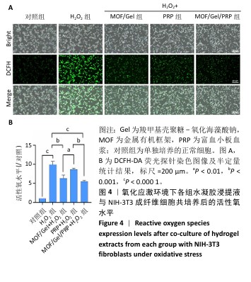
2.3 水凝胶的活性氧清除能力 DCFH-DA荧光探针检测结果显示,暴露于H2O2后,NIH-3T3成纤维细胞中活性氧水平增加(P < 0.000 1),达到对照组的9.9倍;与H2O2组相比,MOF/Gel+H2O2组和MOF/Gel/PRP+H2O2组细胞内活性氧水平显著下降(P < 0.001,P < 0.000 1),分别下降了1.5倍和1.8倍;与富血小板血浆+H2O2组相比,MOF/Gel+H2O2组和MOF/Gel/PRP+H2O2组细胞内活性氧水平下降(P < 0.01,P < 0.001),见图4,提示MOF/Gel水凝胶与MOF/Gel/PRP水凝胶具有强大的抗氧化能力。"

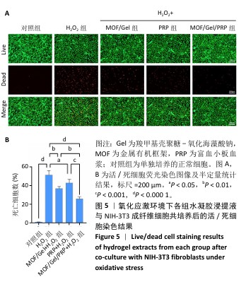
2.4 水凝胶对高浓度H2O2刺激下细胞死亡的影响 活/死细胞染色结果显示,与对照组相比,高浓度H2O2刺激后大量细胞死亡(P < 0.000 1),这是由于过量H2O2引发的细胞凋亡;与H2O2组相比,3个实验组均显示出有效的细胞保护作用,并且MOF/Gel/PRP+H2O2组死亡细胞数目明显少于MOF/Gel+H2O2组和富血小板血浆+H2O2组(P < 0.01,P < 0.001),见图5,表明MOF/Gel/PRP水凝胶对氧化刺激下细胞的保护能力更强。"
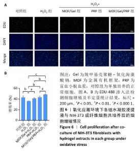
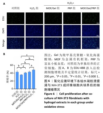
2.5 水凝胶对氧化应激条件下细胞增殖的影响 各组细胞增殖检测结果见图6。对照组、H2O2组、MOF/Gel+H2O2组、富血小板血浆+H2O2组和MOF/Gel/PRP+H2O2组细胞增殖率分别为(42.9±2.9)%,(35.8±0.5)%,(40.8±1.2)%,(43.3±2.0)%,(48.3±1.3)%,表明H2O2的加入抑制了细胞增殖率(P < 0.01),随着水凝胶的引入,细胞增殖率能力有所恢复,特别是MOF/Gel/PRP+H2O2水凝胶组细胞增殖率较H2O2组提高了约12.5%(P < 0.000 1)。"
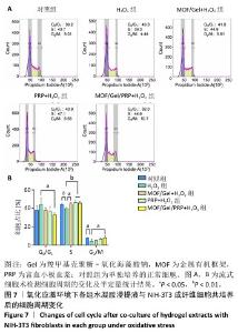
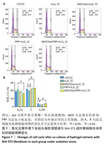
2.6 水凝胶对氧化应激条件下细胞周期的影响 流式细胞仪检测结果显示,与对照组相比,H2O2组G0/G1期细胞轻微增多(P > 0.05),S期与G2/M期细胞减少(P < 0.05),说明细胞周期被阻滞在G0/G1期;与H2O2组相比,各水凝胶浸提液能够改善氧化应激所造成的细胞周期停滞,特别是在MOF/Gel/PRP+H2O2组S期和G2/M期细胞比例相较于H2O2组显著提升(P < 0.01,P < 0.05),并且伴随着G0/G1期细胞比例减少(P < 0.05),见图7,表明MOF/Gel/PRP水凝胶能够有效改善氧化应激对细胞周期的影响,并促进细胞增殖。"
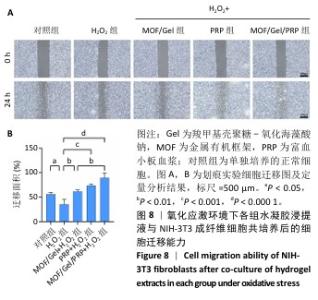
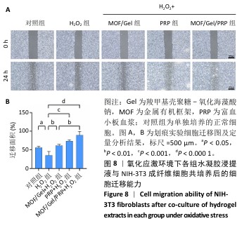
2.7 复合水凝胶对氧化应激条件下细胞迁移的影响 细胞迁移在组织修复和再生过程中发挥着关键作用,此次研究通过细胞划痕实验探究不同处理下NIH-3T3成纤维细胞的迁移能力。H2O2处理显著抑制了细胞迁移,仅有30.01%的细胞迁移面积,而对照组细胞迁移面积为55.89%;与H2O2组相比,MOF/Gel+H2O2组、富血小板血浆+H2O2组细胞迁移能力有所增强,细胞迁移面积分别达到了61.21%,73.66%,而MOF/Gel/PRP+H2O2组细胞迁移面积最高,达到了89.53%,表明MOF/Gel水凝胶与富血小板血浆具有协同作用,显著刺激了细胞在氧化应激状态下的细胞迁移能力,见图8。以上结果强调了MOF/Gel水凝胶协同富血小板血浆作为有效治疗剂的潜力,有助于提升细胞迁移能力,克服氧化应激对创面愈合的负面影响。"
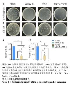
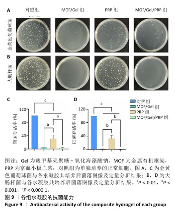
2.8 水凝胶的体外抗菌能力 各组水凝胶与金黄色葡萄球菌或大肠杆菌共培养后的菌落图像及定量分析结果,见图9。MOF/Gel组和MOF/Gel/PRP组对金黄色葡萄球菌的抗菌率分别为96.1%和97.0%,对大肠杆菌抗菌率为99.0%和99.6%,均明显高于对照组和富血小板血浆组(P < 0.05),MOF/Gel组和MOF/Gel/PRP组之间抗菌率比较差异没有显著性(P > 0.05)。"
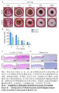
2.9 水凝胶对糖尿病大鼠感染创面的修复作用 两组大鼠治疗不同时间的创面大体观,见图10A,治疗第4天,对照组创面存在出明显的细菌感染,实验组未观察到细菌生物膜的形成。对照组创面愈合速率较慢,治疗第16天仍有16%的创面剩余面积,实验组治疗第16天的创面剩余面积仅为3.4%,实验组治疗第4,8,12,16天的创面剩余面积均小于对照组(P < 0.05或P < 0.01),见图10B。治疗第14天,苏木精-伊红染色结果显示,对照组上皮化不完全,实验组可见完整的上皮结构且与皮下组织连接紧密,见图10C;Masson染色结果显示,对照组胶原纤维数量较少且分布不均匀,实验组可见较多的胶原纤维且排列整齐、分布均匀,见图10D。这些结果提示MOF/Gel/PRP水凝胶展现出优异的促创面愈合效果。"

| [1] XIONG Y, CHU X, YU T, et al. Reactive Oxygen Species-Scavenging Nanosystems in the Treatment of Diabetic Wounds. Adv Healthc Mater. 2023;12(25):2300779. [2] DUNNILL C, PATTON T, BRENNAN J, et al. Reactive oxygen species (ROS) and wound healing: the functional role of ROS and emerging ROS-modulating technologies for augmentation of the healing process. Int Wound J. 2017; 14(1):89-96. [3] LIANG Y, HE J, GUO B. Functional hydrogels as wound dressing to enhance wound healing. ACS Nano. 2021;15(8):12687-12722. [4] SILVA JC, PITTA MG, PITTA IR, et al. New peroxisome proliferator-activated receptor agonist (GQ-11) improves wound healing in diabetic mice. Adv Wound Care. 2019;8(9):417-428. [5] LIU K, ZHANG H, WANG Z, et al. Carrier-Free, Injectable Hydrogel Formed by Self-Assembled Nanofibers of Antimicrobial and Anti-inflammatory Peptide for Wound Healing. ACS App Nano Mater. 2024;7(14):16585-16598. [6] CHEN J, LIU Y, CHENG G, et al. Tailored hydrogel delivering niobium carbide boosts ROS‐scavenging and Antimicrobial activities for diabetic wound healing. Small. 2022;18(27):2201300. [7] ZHENG SY, WAN XX, KAMBEY PA, et al. Therapeutic role of growth factors in treating diabetic wound. World J Diabetes. 2023;14(4):364. [8] XU D, WU L, YAO H, et al. Catalase‐like nanozymes: Classification, catalytic mechanisms, and their applications. Small. 2022;18(37):2203400. [9] WU A, LI M, CHEN Y, et al. Multienzyme Active Manganese Oxide Alleviates Acute Liver Injury by Mimicking Redox Regulatory System and Inhibiting Ferroptosis. Adv Healthc Mater. 2024;13(11):2302556. [10] HU S, WANG L, LI J, et al. Catechol-modified and MnO2-nanozyme-reinforced hydrogel with improved antioxidant and antibacterial capacity for periodontitis treatment. ACS Biomater Sci Eng. 2023;9(9):5332-5346. [11] SUN H, YU X, MA X, et al. MnOx-CeO2 catalyst derived from metal-organic frameworks for toluene oxidation. Catal Today. 2020;355:580-586. [12] PAN W, WANG W, WANG P, et al. Sustained delivery of extracellular vesicles using UiO-66-NH2 crosslinked hydrogel for accelerating chronic diabetic wound-healing. Mater Design. 2024;238:112688. [13] ZHU J, QIU W, YAO C, et al. Water-stable zirconium-based metal-organic frameworks armed polyvinyl alcohol nanofibrous membrane with enhanced antibacterial therapy for wound healing. J Colloid Interf Sci. 2021;603:243-251. [14] FAN YL, LIU HJ, WANG ZL, et al. A One-Nano MOF-Two-Functions Strategy Toward Self-healing, Anti-inflammatory, and Antibacterial Hydrogels for Infected Wound Repair. Chem Eng J. 2024;497:155037. [15] BI X, BAI Q, LIANG M, et al. Silver peroxide nanoparticles for combined antibacterial sonodynamic and photothermal therapy. Small. 2022;18(2): 2104160. [16] ZHANG Y, KANG J, CHEN X, et al. Ag nanocomposite hydrogels with immune and regenerative microenvironment regulation promote scarless healing of infected wounds. J Nanobiotechnol. 2023;21(1):435. [17] EVERTS P, ONISHI K, JAYARAM P, et al. Platelet-rich plasma: new performance understandings and therapeutic considerations in 2020. Int J Mol Sci. 2020; 21(20):7794. [18] FANG J, WANG X, JIANG W, et al. Platelet-rich plasma therapy in the treatment of diseases associated with orthopedic injuries. Tissue Eng Part B Rev. 2020;26(6):571-585. [19] CL K, JEYARAMAN M, JEYARAMAN N, et al. Antimicrobial Effects of Platelet-Rich Plasma and Platelet-Rich Fibrin: A Scoping Review. Cureus. 2023;15(12):e51360. [20] ZHANG Y, WANG ZL, DENG ZP, et al. An extracellular matrix-inspired self-healing composite hydrogel for enhanced platelet-rich plasma-mediated chronic diabetic wound treatment. Carbohyd Polym. 2023;315:120973. [21] SHANG S, ZHUANG K, CHEN J, et al. A bioactive composite hydrogel dressing that promotes healing of both acute and chronic diabetic skin wounds. Bioact Mater. 2024;34:298-310. [22] KŁOSIŃSKI KK, WACH RA, KRUCZKOWSKA W, et al. Carboxymethyl Chitosan Hydrogels for Effective Wound Healing—An Animal Study. J Funct Biomater. 2023;14(9):473. [23] JIANG Y, GUO S, JIAO J, et al. A biphasic hydrogel with self-healing properties and a continuous layer structure for potential application in osteochondral defect repair. Polymers (Basel). 2023;15(12):2744. [24] KIRK T, AHMED A, ROGNONI E. Fibroblast memory in development, homeostasis and disease. Cells. 2021;10(11):2840. [25] WOODLEY JP, LAMBERT DW, ASENCIO IO. Reduced Fibroblast Activation on Electrospun Polycaprolactone Scaffolds. Bioengineering (Basel). 2023; 10(3):348. [26] WOODLEY JP, LAMBERT DW, ASENCIO I O. Understanding fibroblast behavior in 3D biomaterials. Tissue Eng Part B Rev. 2022;28(3):569-578. [27] 梅和平,石孟琼,陈茂华,等.地连二心颗粒对H2O2 致小鼠成纤维细胞损伤的保护作用研究[J].中药药理与临床,2017,33(1):139-144. [28] WU M, TU J, HUANG J, et al. Exosomal IRF1-loaded rat adipose-derived stem cell sheet contributes to wound healing in the diabetic foot ulcers. Mol Med. 2023;29(1):60. [29] 胡晶晶 . 具有 ROS 清除作用的水凝胶包裹小檗碱 @ NMOF 促进糖尿病足创面愈合 [D]. 广州 : 南方医科大学 ,2022. [30] DENG L, DU C, SONG P, et al. The role of oxidative stress and antioxidants in diabetic wound healing. Oxid Med Cell Longev. 2021;2021(1):8852759. [31] LONG M, ROJO DE LA VEGA M, WEN Q, et al. An essential role of NRF2 in diabetic wound healing. Diabetes. 2016;65(3):780-793. [32] YAN R, ZHANG X, XU W, et al. ROS-Induced Endothelial Dysfunction in the Pathogenesis of Atherosclerosis. Aging Dis. 2024;16(1):250-268. [33] WANG Y, XIE R, LI Q, et al. A self-adapting hydrogel based on chitosan/oxidized konjac glucomannan/AgNPs for repairing irregular wounds. Biomater Sci. 2020;8(7):1910-1922. [34] ZHENG Z, LI M, SHI P, et al. Polydopamine-modified collagen sponge scaffold as a novel dermal regeneration template with sustained release of platelet-rich plasma to accelerate skin repair: a one-step strategy. Bioact Mater. 2021;6(8):2613-2628. [35] ZHOU J, CHEN N, LIAO J, et al. Ag-activated metal-organic framework with peroxidase-like activity synergistic Ag+ release for safe bacterial eradication and wound healing. Nanomaterials (Basel). 2022;12(22):4058. [36] DASTNESHAN A, RAHIMINEZHAD S, MEZAJIN M N, et al. Cefazolin encapsulated UIO-66-NH2 nanoparticles enhance the antibacterial activity and biofilm inhibition against drug-resistant S. aureus: In vitro and in vivo studies. Chem Eng J. 2023;455:140544. [37] KESHK WA, ZAHRAN SM. Mechanistic role of cAMP and hepatocyte growth factor signaling in thioacetamide-induced nephrotoxicity: Unraveling the role of platelet rich plasma. Biomed Pharmacother. 2019;109:1078-1084. [38] RANIA N, ISLAM IH, ATEF M, et al. Effect of platelet rich plasma on an experimental rat model of adriamycin induced chronic kidney disease. Med J Cairo Univ. 2019;87:2207-2217. [39] BURANASIN P, MIZUTANI K, IWASAKI K, et al. High glucose-induced oxidative stress impairs proliferation and migration of human gingival fibroblasts. PLoS One. 2018;13(8):e0201855. [40] ELMONGY NF, MEAWAD SB, ELSHORA SZ, et al. Platelet-rich plasma ameliorates neurotoxicity induced by silver nanoparticles in male rats via modulation of apoptosis, inflammation, and oxidative stress. J Biochem Mol Toxicl. 2023;37(9):e23420. [41] ZHENG WQ, ZHANG JH, LI ZH, et al. Mammalian mitochondrial translation infidelity leads to oxidative stress–induced cell cycle arrest and cardiomyopathy. Proc Natl Acad Sci U S A. 2023;120(37):e2309714120. [42] THANGAPAZHAM RL, DARLING TN, MEYERLE J. Alteration of skin properties with autologous dermal fibroblasts. Int J Mol Sci. 2014;15(5):8407-8427. [43] YI W, BAO Q, XU D, et al. ETS1 Expression in Diabetic Foot Ulcers: Implications for Fibroblast Phenotype and Wound Healing Through the PP2A/YAP Pathway. J Inflamm Res. 2024;17:7373-7388. [44] DEVEREAUX J, NURGALI K, KIATOS D, et al. Effects of platelet-rich plasma and platelet-poor plasma on human dermal fibroblasts. Maturitas. 2018;117: 34-44. [45] WEI S, WANG Z, LIANG X, et al. A composite hydrogel with antibacterial and promoted cell proliferation dual properties for healing of infected wounds. AM J Transl Res. 2023;15(7):4467. [46] HUANG P, HE Y, HUANG C, et al. MOF@platelet-rich plasma antimicrobial GelMA dressing: structural characterization, bio-compatibility, and effect on wound healing efficacy. RSC Adv. 2024;14(41):30055-30069. |
| [1] | Liu Yang, Liu Donghui , Xu Lei, Zhan Xu, Sun Haobo, Kang Kai. Role and trend of stimuli-responsive injectable hydrogels in precise myocardial infarction therapy [J]. Chinese Journal of Tissue Engineering Research, 2026, 30(8): 2072-2080. |
| [2] | Guo Yuchao, Ni Qianwei, Yin Chen, Jigeer·Saiyilihan, Gao Zhan . Quaternized chitosan hemostatic materials: synthesis, mechanism, and application [J]. Chinese Journal of Tissue Engineering Research, 2026, 30(8): 2091-2100. |
| [3] | Lai Yu, Chen Yueping, Zhang Xiaoyun. Research hotspots and frontier trends of bioactive materials in treating bone infections [J]. Chinese Journal of Tissue Engineering Research, 2026, 30(8): 2132-2144. |
| [4] | Wang Songpeng, Liu Yusan, Yu Huanying, Gao Xiaoli, Xu Yingjiang, Zhang Xiaoming, Liu Min. Bidirectional regulation of reactive oxygen species based on zeolitic imidazolate framework-8 nanomaterials: from tumor therapy and antibacterial activity to cytoprotection [J]. Chinese Journal of Tissue Engineering Research, 2026, 30(8): 2033-2013. |
| [5] | Chen Ling, Mao Qiuhua, Xu Pu, Zhang Wenbo. Effect of water-soluble matrix of nano-pearl powder on proliferation, migration and apoptosis of mouse fibroblasts#br# [J]. Chinese Journal of Tissue Engineering Research, 2026, 30(2): 338-344. |
| [6] | Liu Xiaohong, Zhao Tian, Mu Yunping, Feng Wenjin, Lyu Cunsheng, Zhang Zhiyong, Zhao Zijian, Li Fanghong. Acellular dermal matrix hydrogel promotes skin wound healing in rats [J]. Chinese Journal of Tissue Engineering Research, 2026, 30(2): 395-403. |
| [7] | Wang Zilin, Mu Qiuju, Liu Hongjie, Shen Yuxue, Zhu Lili. Protective effects of platelet-rich plasma hydrogel on oxidative damage in L929 cells [J]. Chinese Journal of Tissue Engineering Research, 2025, 29(4): 771-779. |
| [8] | Zhao Hongxia, Sun Zhengwei, Han Yang, Wu Xuechao , Han Jing. Osteogenic properties of platelet-rich fibrin combined with gelatin methacryloyl hydrogel [J]. Chinese Journal of Tissue Engineering Research, 2025, 29(4): 809-817. |
| [9] | Zhao Zengbo, Li Chenxi, Dou Chenlei, Ma Na, Zhou Guanjun. Anti-inflammatory and osteogenic effects of chitosan/sodium glycerophosphate/sodium alginate/leonurine hydrogel [J]. Chinese Journal of Tissue Engineering Research, 2025, 29(4): 678-685. |
| [10] | Dong Meilin, Du Haiyu, Liu Yuan. Quercetin-loaded carboxymethyl chitosan hydrogel promotes wound healing in diabetic rats [J]. Chinese Journal of Tissue Engineering Research, 2025, 29(4): 692-699. |
| [11] |
Zhang Bo, Zhang Zhen, Jiang Dong.
Tannic acid modified interpenetrating network hydrogel promotes tissue remodeling of ruptured Achilles tendon after surgery#br#
#br#
[J]. Chinese Journal of Tissue Engineering Research, 2025, 29(4): 721-729.
|
| [12] | Li Yonghang, Li Wenming, Yan Caiping, Wang Xingkuan, Xiang Chao, Zhang Yuan, Jiang Ke, Chen Lu. Critical bone defect repaired with anti-fibrosis and “H”-type core-shell bionic scaffold [J]. Chinese Journal of Tissue Engineering Research, 2025, 29(16): 3420-3431. |
| [13] | He Rui, Li Chongyi, Wang Ruiyao, Zeng Dan, Fan Daidi. Application of MXene-based hydrogels in wound repair [J]. Chinese Journal of Tissue Engineering Research, 2025, 29(16): 3486-3493. |
| [14] | Liu Zhongyu, Li Wenya, Fan Yonghong, Lyu Shuang, Pei Juan, Chen Yaqin, Liu Beiyu, Sun Hongyu. Methacrylated dermal extracellular matrix hydrogel promotes repair of abdominal wall defects [J]. Chinese Journal of Tissue Engineering Research, 2025, 29(10): 2074-2082. |
| [15] | Zhao Wenqi, Yu Haichi, Song Yiru, Yuan Tianyang, Liu Qinyi. Platelet-rich plasma and hydrogel for spinal cord injury [J]. Chinese Journal of Tissue Engineering Research, 2025, 29(10): 2189-2200. |
| Viewed | ||||||
|
Full text |
|
|||||
|
Abstract |
|
|||||