Chinese Journal of Tissue Engineering Research ›› 2025, Vol. 29 ›› Issue (16): 3420-3431.doi: 10.12307/2025.418
Previous Articles Next Articles
Critical bone defect repaired with anti-fibrosis and “H”-type core-shell bionic scaffold
Li Yonghang, Li Wenming, Yan Caiping, Wang Xingkuan, Xiang Chao, Zhang Yuan, Jiang Ke, Chen Lu
- Department of Orthopedics, Affiliated Hospital of North Sichuan Medical College, Nanchong 637000, Sichuan Province, China
-
Received:2023-12-02Accepted:2024-03-13Online:2025-06-08Published:2024-09-04 -
Contact:Corresponding author: Chen Lu, Chief physician, Department of Orthopedics, Affiliated Hospital of North Sichuan Medical College, Nanchong 637000, Sichuan Province, China Co-corresponding author: Jiang Ke, MD, Associate chief physician, Department of Orthopedics, Affiliated Hospital of North Sichuan Medical College, Nanchong 637000, Sichuan Province, China -
About author:Li Yonghang, Master candidate, Physician, Department of Orthopedics, Affiliated Hospital of North Sichuan Medical College, Nanchong 637000, Sichuan Province, China Li Wenming, Department of Orthopedics, Affiliated Hospital of North Sichuan Medical College, Nanchong 637000, Sichuan Province, China Li Yonghang and Li Wenming contributed equally to this article. -
Supported by:Scientific Research Task of Sichuan Medical Association, No. 2015GK012 (to CL); Program of Cooperation between the Schools and Nanchong City in 2022, No. 22SXQT0308 (to JK); Nanchong 2023 Municipal Science and Technology Research and Development Plan, No. 23JCYJPT0036 (to WXK)
CLC Number:
Cite this article
Li Yonghang, Li Wenming, Yan Caiping, Wang Xingkuan, Xiang Chao, Zhang Yuan, Jiang Ke, Chen Lu. Critical bone defect repaired with anti-fibrosis and “H”-type core-shell bionic scaffold[J]. Chinese Journal of Tissue Engineering Research, 2025, 29(16): 3420-3431.
share this article
Add to citation manager EndNote|Reference Manager|ProCite|BibTeX|RefWorks
2.1 核壳结构仿生支架及其组成各部分的物理性质 如图1A所示,聚己内酯静电纺丝纳米纤维直径在100-500 nm之间,具有良好的各向异性,可以很好地阻止细胞、组织长入;甲基丙烯酸酐明胶水凝胶存在孔隙结构,孔径均在300 μm以内,证明复合水凝胶内部存在连通孔隙;在支架的核壳结构内部,静电纺丝和水凝胶通过物理锚定的方式较好地结合在一起。采用X射线能谱、扫描电镜能谱和进一步分析聚己内酯的结构和成分,发现材料表面存在F元素,证明了FAPI-4药物成功负载在聚己内酯纤维膜上(图1B,C);使用傅里叶红外光谱发现不同载药比例对静电纺丝表面的官能团没有影响(图1D)。"
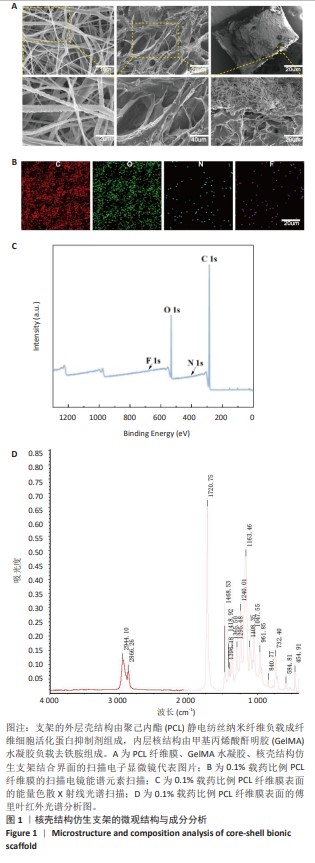
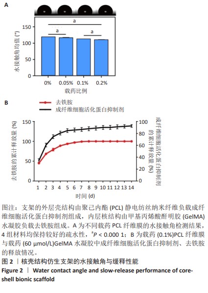
水接触角测量仪所拍摄的图像见图2A,载药比例0.2%聚己内酯纤维膜的水接触角[(113.0±0.8)°]较单纯聚己内酯纤维膜[(120.0±0.4)°]小,但整体改变幅度不大[14],仍然保持较好的疏水性。各载药聚己内酯纤维膜在2周之内可以有效释放出抗纤维化药物FAPI-4,各水凝胶中的去铁胺在溶胀效果下1周内便释放完毕,见图2B。"
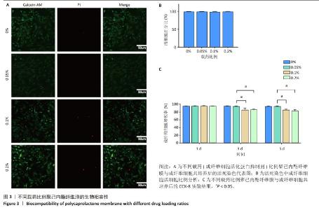
2.2 核壳结构仿生支架的生物相容性 活死染色与CCK-8实验结果显示,当成纤维细胞在不同载药比例的静电纺丝膜上共培养时,活细胞数量增加,死亡细胞数量少,随纤维膜中载药比例增加,死细胞增多;同样,随纤维膜中载药比例的增加,细胞增殖率受到抑制,见图3。"

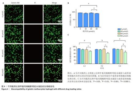
活死染色与CCK-8实验结果显示,当成骨前体细胞与不同载药比例甲基丙烯酸酐明胶水凝胶共培养时,同样可以观察到细胞存活性好,随着水凝胶中载药含量的增加,死细胞增加,同时细胞增殖率受到抑制,见图4。这证明了一定载药量范围内核壳结构仿生支架具有良好的生物相容性。为了在支架功能和生物相容性中取得一定平衡,后续实验均采取FAPI-1(0.1%)和去铁胺60 μmol/L的载药量进行。"


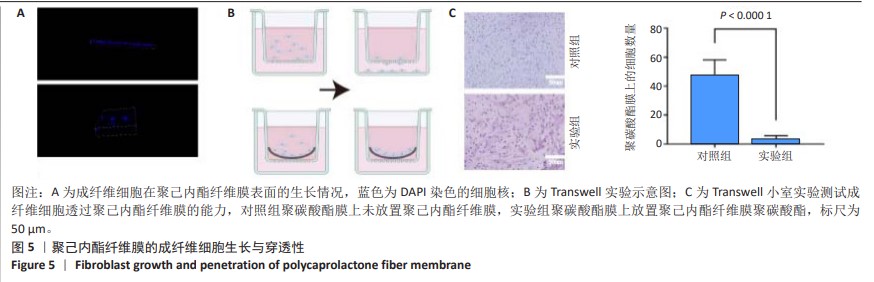
2.3 聚己内酯纤维膜的体外抗纤维化实验 2.3.1 聚己内酯纤维膜的物理阻隔作用 共聚焦激光扫描显微镜下可见成纤维细胞集中在纳米纤维膜的表面,难以进入更深层次的纤维膜内部(图5A)。如图5B示意图所示,在具有静电纺丝膜阻挡的Transwell小孔中,成纤维细胞很少穿过聚碳酸酯膜,几乎没有细胞可以穿越过聚己内酯纤维膜,这得益于纳米纤维微纳米的孔隙和致密的结构[14,36]。而在没有纳米纤维膜阻隔的Transwell小室中,成纤维细胞很容易就穿过小室的聚碳酸酯膜(图5C)。"
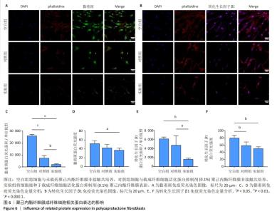
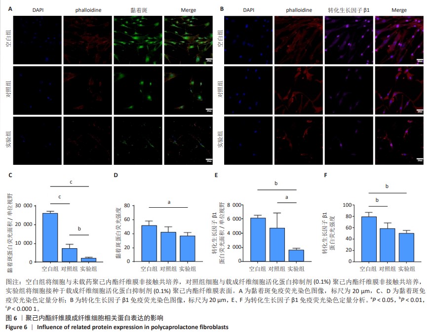
2.3.2 聚己内酯膜抑制成纤维相关蛋白表达 图6显示各组成纤维细胞黏着斑和转化生长因子β1的免疫荧光染色。空白组成纤维细胞增殖数量最多,细胞形态最好,黏着斑和转化生长因子β1的表达最丰富;对照组细胞形态变化不大,细胞数量明显抑制,这是由于FAPI-4抑制成纤维细胞生长增殖的作用;实验组细胞形态不规则,伸展性不佳,黏着斑和转化生长因子β1的表达最弱。"
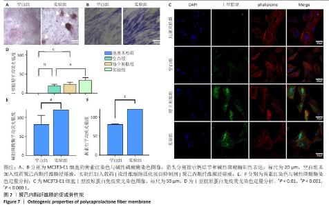
2.4 聚己内酯纤维膜的促成骨性能 茜素红染色与碱性磷酸酶染色显示,相较于空白组,实验组细胞钙沉积和碱性磷酸酶表达增强;Ⅰ型胶原蛋白免疫荧光染色结果显示,经地塞米松干预的细胞Ⅰ型胶原蛋白表达受到抑制,经特立帕肽和载药聚己内酯纤维膜浸提液干预的细胞明显表达更强的Ⅰ型胶原蛋白,并且均强于空白组,见图7。"


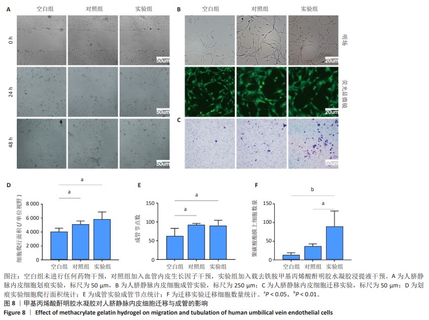
2.5 甲基丙烯酸酐明胶水凝胶的促血管形成作用 划痕实验、迁移实验与成骨实验结果显示,相较于空白组,血管内皮生长因子与载药甲基丙烯酸酐明胶水凝胶浸提液均可促进人脐静脉内皮细胞的迁移与成管,见图8。"
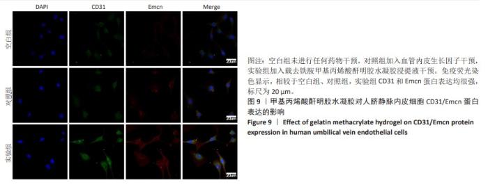
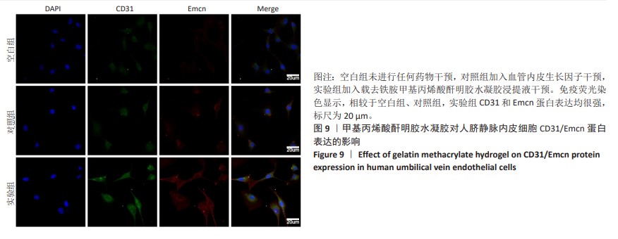
CD31/Emcn免疫荧光染色结果显示,空白组细胞CD31和Emcn蛋白表达均较弱;使用血管内皮生长因子干预细胞后,仅CD31蛋白表达较强,Emcn蛋白表达较弱;使用载药甲基丙烯酸酐明胶水凝胶浸提液干预细胞后,CD31和Emcn蛋白表达均很强,见图9。"
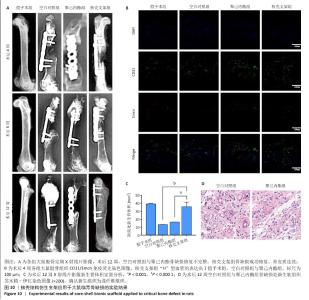
2.6 体内实验结果 2.6.1 实验动物数量分析 36只大鼠全部进入结果分析。 2.6.2 各组大鼠股骨X射线观察 术后4周,各组骨缺损部分都处于原始骨痂时期,新生的骨痂质地稍硬但在影像学图片上并不能显影;术后8周,核壳支架组可见骨折断端有部分连接,空白对照组和聚己内酯组无骨性连接;术后12周,空白对照组与聚己内酯骨缺损修复不完整,核壳支架组骨缺损成功修复,骨皮质连续,见图10。 2.6.3 各组大鼠股骨CD31/Emcn免疫荧光染色 术后4周,核壳支架组“H”型血管的表达高于假手术组、空白对照组与聚己内酯组,见图10。 2.6.4 大鼠股骨组织苏木精-伊红染色 为了确定空白对照组和聚己内酯组股骨修复是否为纤维化组织,对其进行苏木精-伊红染色分析。术后12周苏木精-伊红染色结果显示,对空白对照组和聚己内酯组骨缺损部分的非骨化组织为成纤维组织,见图10。"

| [1] HAK DJ, FITZPATRICK D, BISHOP JA, et al. Delayed union and nonunions: Epidemiology, clinical issues, and financial aspects. Injury. 2014;45: S3-S7. [2] SCHMITZ JP, HOLLINGER JO. The Critical Size Defect as an Experimental Model for Craniomandibulofacial Nonunions. Clin Orthop Relat Res. 1986:(205):299-308. [3] DALISSON B, CHARBONNIER B, AOUDE A, et al. Skeletal regeneration for segmental bone loss: Vascularised grafts, analogues and surrogates. Acta Biomater. 2021;136:37-55. [4] MA L, WANG X, ZHOU Y, et al. Biomimetic Ti–6Al–4V alloy/gelatin methacrylate hybrid scaffold with enhanced osteogenic and angiogenic capabilities for large bone defect restoration. Bioact Mater. 2021;6(10): 3437-3448. [5] BIGGEMANN J, PEZOLDT M, STUMPF M, et al. Modular ceramic scaffolds for individual implants. Acta Biomater. 2018;80: 390-400. [6] ZHANG Y, FAN Z, XING Y, et al. Effect of microtopography on osseointegration of implantable biomaterials and its modification strategies. Front Bioeng Biotechnol. 2022;10:981062. [7] WU L, GU Y, LIU L, et al. Hierarchical micro/nanofibrous membranes of sustained releasing VEGF for periosteal regeneration. Biomaterials. 2020;227:119555. [8] PENG F, ZHANG X, WANG Y, et al. Guided bone regeneration in long-bone defect with a bilayer mineralized collagen membrane. Collagen Leather. 2023;36(5). https://doi.org/10.1186/s42825-023-00144-4 [9] SALHOTRA A, SHAH HN, LEVI B, et al. Mechanisms of bone development and repair. Nat Rev Mol Cell Biol. 2020;21(11): 696-711. [10] PANTELI M, VUN JSH, POUNTOS I, et al. Biological and molecular profile of fracture non-union tissue: A systematic review and an update on current insights. J Cell Mol Med. 2022;26(3):601-623. [11] WANG L, TOWER RJ, CHANDRA A, et al. Periosteal Mesenchymal Progenitor Dysfunction and Extraskeletally-Derived Fibrosis Contribute to Atrophic Fracture Nonunion. J Bone Miner Res. 2019;34(3):520-532. [12] YU HS, PARK J, LEE HS, et al. Feasibility of Polycaprolactone Scaffolds Fabricated by Three-Dimensional Printing for Tissue Engineering of Tunica Albuginea. World J Mens Health. 2018;36(1):66-72. [13] ZHOU X, HE X, SHI K, et al. Injectable Thermosensitive Hydrogel Containing Erlotinib-Loaded Hollow Mesoporous Silica Nanoparticles as a Localized Drug Delivery System for NSCLC Therapy. Adv Sci (Weinh). 2020;7(23):2001442. [14] NIRWAN VP, KOWALCZYK T, BAR J, et al. Advances in Electrospun Hybrid Nanofibers for Biomedical Applications. Nanomaterials (Basel). 2022;12(11):1829. [15] WANG W, ZHAO J, YAO Z, et al. Oriented inner fabrication of bi-layer biomimetic tendon sheath for anti-adhesion and tendon healing. Ther Adv Chronic Dis. 2020;11:2040622320944779. [16] ZHANG X, REAGAN MR, KAPLAN DL. Electrospun silk biomaterial scaffolds for regenerative medicine. Adv Drug Deliv Rev. 2009;61(12): 988-1006. [17] LOKTEV A, LINDNER T, BURGER EM, et al. Development of Fibroblast Activation Protein-Targeted Radiotracers with Improved Tumor Retention. J Nucl Med. 2019;60(10):1421-1429. [18] GIESEL FL, KRATOCHWIL C, LINDNER T, et al. (68)Ga-FAPI PET/CT: Biodistribution and Preliminary Dosimetry Estimate of 2 DOTA-Containing FAP-Targeting Agents in Patients with Various Cancers. J Nucl Med. 2019;60(3):386-392. [19] CHEN Q, LI J, HAN F, et al. A Multifunctional Composite Hydrogel That Rescues the ROS Microenvironment and Guides the Immune Response for Repair of Osteoporotic Bone Defects. Adv Funct Mater. 2022;32(27):2201067.1-2201067.18. [20] WEI Z, XIONG C, LIU Z, et al. Release characteristics and processing-structure-performance relationship of electro-spinning curcumin-loaded polyethersulfone based porous ultrafine fibers. J Biomater Sci Polym Ed. 2018;29(15):1825-1838. [21] PATTNAIK S, SWAIN K, RAMAKRISHNA S. Optimal delivery of poorly soluble drugs using electrospun nanofiber technology: Challenges, state of the art, and future directions. Wiley Interdiscip Rev Nanomed Nanobiotechnol. 2023;15(2):e1859. [22] PENG Y, WU S, LI Y, et al. Type H blood vessels in bone modeling and remodeling. Theranostics. 2020;10(1):426-36. [23] XU R, YALLOWITZ A, QIN A, et al. Targeting skeletal endothelium to ameliorate bone loss. Nat Med. 2018;24(6):823-833. [24] RUAN Z, YIN H, WAN TF, et al. Metformin accelerates bone fracture healing by promoting type H vessel formation through inhibition of YAP1/TAZ expression. Bone Res. 2023;11(1):45. [25] YANG M, LI CJ, XIAO Y, et al. Ophiopogonin D promotes bone regeneration by stimulating CD31(hi) EMCN(hi) vessel formation. Cell Prolif. 2020;53(3):e12784. [26] LU W, ZENG M, LIU W, et al. Human urine-derived stem cell exosomes delivered via injectable GelMA templated hydrogel accelerate bone regeneration. Mater Today Bio. 2023;19:100569. [27] GHERASIM O, GRUMEZESCU AM, GRUMEZESCU V, et al. Bioactive Coatings Based on Hydroxyapatite, Kanamycin, and Growth Factor for Biofilm Modulation. Antibiotics (Basel). 2021;10(2):160. [28] ZENG Y, HUANG C, DUAN D, et al. Injectable temperature-sensitive hydrogel system incorporating deferoxamine-loaded microspheres promotes H-type blood vessel-related bone repair of a critical size femoral defect. Acta Biomater. 2022;153:108-123. [29] BAYANZAY K, ALZOEBIE L. Reducing the iron burden and improving survival in transfusion-dependent thalassemia patients: current perspectives. J Blood Med. 2016;7:159-169. [30] HADJIZADEH A, GHASEMKHAH F, GHASEMZAIE N. Polymeric Scaffold Based Gene Delivery Strategies to Improve Angiogenesis in Tissue Engineering: A Review. Polym Rev. 2017;57(3): 505-556. [31] CHEN S, YU Y, XIE S, et al. Local H2 release remodels senescence microenvironment for improved repair of injured bone. Nat Commun. 2023;14(1):7783. [32] YUE K, TRUJILLO-DE SANTIAGO G, ALVAREZ MM, et al. Synthesis, properties, and biomedical applications of gelatin methacryloyl (GelMA) hydrogels. Biomaterials. 2015;73:254-271. [33] CHEN YC, LIN RZ, QI H, et al. Functional Human Vascular Network Generated in Photocrosslinkable Gelatin Methacrylate Hydrogels. Adv Funct Mater. 2012;22(10):2027-2039. [34] KLOTZ BJ, GAWLITTA D, ROSENBERG AJWP, et al. Gelatin-Methacryloyl Hydrogels: Towards Biofabrication-Based Tissue Repair. Trends Biotechnol. 2016;34(5):394-407. [35] REICHERT JC, WULLSCHLEGER ME, CIPITRIA A, et al. Custom-made composite scaffolds for segmental defect repair in long bones. Int Orthop. 2010;35(8): 1229-1236. [36] CAI C, WANG W, LIANG J, et al. MMP-2 Responsive Unidirectional Hydrogel‐Electrospun Patch Loading TGF‐β1 siRNA Polyplexes for Peritendinous Anti‐Adhesion. Adv Funct Mater. 2020;31(6). doi: org/10.1002/adfm.202008364. [37] JOHNSON KA. Healing large bone defects. Vet Comp Orthop Traumatol. 2014;27(6): V.doi: 10.3415/VCOT-14-10-0162. [38] AGARWAL R, GARCÍA AJ. Biomaterial strategies for engineering implants for enhanced osseointegration and bone repair. Adv Drug Deliv Rev. 2015;94:53-62. [39] HO-SHUI-LING A, BOLANDER J, RUSTOM LE, et al. Bone regeneration strategies: Engineered scaffolds, bioactive molecules and stem cells current stage and future perspectives. Biomaterials. 2018;180:143-162. [40] CAO J, JIN L, YAN ZQ, et al. Reassessing endothelial-to-mesenchymal transition in mouse bone marrow: insights from lineage tracing models. Nat Commun. 2023;14(1):8461. |
| [1] | Sun Xianjuan, Wang Qiuhua, Zhang Jinyi, Yang Yangyang, Wang Wenshuang, Zhang Xiaoqing. Adhesion, proliferation, and vascular smooth muscle differentiation of bone marrow mesenchymal stem cells on different electrospinning membranes [J]. Chinese Journal of Tissue Engineering Research, 2025, 29(4): 661-669. |
| [2] | Zhao Zengbo, Li Chenxi, Dou Chenlei, Ma Na, Zhou Guanjun. Anti-inflammatory and osteogenic effects of chitosan/sodium glycerophosphate/sodium alginate/leonurine hydrogel [J]. Chinese Journal of Tissue Engineering Research, 2025, 29(4): 678-685. |
| [3] | Dong Meilin, Du Haiyu, Liu Yuan. Quercetin-loaded carboxymethyl chitosan hydrogel promotes wound healing in diabetic rats [J]. Chinese Journal of Tissue Engineering Research, 2025, 29(4): 692-699. |
| [4] |
Zhang Bo, Zhang Zhen, Jiang Dong.
Tannic acid modified interpenetrating network hydrogel promotes tissue remodeling of ruptured Achilles tendon after surgery#br#
#br#
[J]. Chinese Journal of Tissue Engineering Research, 2025, 29(4): 721-729.
|
| [5] | Wu Chen, Jiang Jiahui, Su Dou, Liu Chen, Ci Chao. Decellularized skin matrix/polyurethane blended fibrous scaffolds promote repair of skin defects in rats [J]. Chinese Journal of Tissue Engineering Research, 2025, 29(4): 745-751. |
| [6] | Wang Zilin, Mu Qiuju, Liu Hongjie, Shen Yuxue, Zhu Lili. Protective effects of platelet-rich plasma hydrogel on oxidative damage in L929 cells [J]. Chinese Journal of Tissue Engineering Research, 2025, 29(4): 771-779. |
| [7] | Zhao Hongxia, Sun Zhengwei, Han Yang, Wu Xuechao , Han Jing. Osteogenic properties of platelet-rich fibrin combined with gelatin methacryloyl hydrogel [J]. Chinese Journal of Tissue Engineering Research, 2025, 29(4): 809-817. |
| [8] | An Jiangru, Zhang Jinyi, Wang Qiuhua, Yang Yangyang, Wang Wenshuang, Zhang Xiaoqing. Mesenchymal stem cells combined with polycaprolactone-hyaluronic acid electrospinning membrane in repair of endometrial injury [J]. Chinese Journal of Tissue Engineering Research, 2025, 29(16): 3369-3379. |
| [9] | He Rui, Li Chongyi, Wang Ruiyao, Zeng Dan, Fan Daidi. Application of MXene-based hydrogels in wound repair [J]. Chinese Journal of Tissue Engineering Research, 2025, 29(16): 3486-3493. |
| [10] | Liu Zhongyu, Li Wenya, Fan Yonghong, Lyu Shuang, Pei Juan, Chen Yaqin, Liu Beiyu, Sun Hongyu. Methacrylated dermal extracellular matrix hydrogel promotes repair of abdominal wall defects [J]. Chinese Journal of Tissue Engineering Research, 2025, 29(10): 2074-2082. |
| [11] | Zhao Wenqi, Yu Haichi, Song Yiru, Yuan Tianyang, Liu Qinyi. Platelet-rich plasma and hydrogel for spinal cord injury [J]. Chinese Journal of Tissue Engineering Research, 2025, 29(10): 2189-2200. |
| [12] | Dang Yi, Du Chengyan, Yao Honglin, Yuan Nenghua, Cao Jin, Xiong Shan, Zhang Dingmei, Wang Xin. Hormonal osteonecrosis and oxidative stress [J]. Chinese Journal of Tissue Engineering Research, 2023, 27(9): 1469-1476. |
| [13] | Cheng Mingguang, Zhang Chaoyu, Zhuang Kangle, Ruan Peng, Zuo Yi, Zhou Zhengchun, Kong Xiang, Ge Jianjun, Cheng Guangcun. In vitro construction of Stanford type A aortic dissection 3D dynamic simulation diagram and individual tissue-engineered blood vessels [J]. Chinese Journal of Tissue Engineering Research, 2023, 27(3): 335-338. |
| [14] | Liu Liang, Hu Gaoquan, Wei Zhao, Chen Lin, Hong Feng. Potential of bacterial nanocellulose/polydopamine composite tubes as small-diameter artificial blood vessel [J]. Chinese Journal of Tissue Engineering Research, 2022, 26(22): 3535-3542. |
| [15] | Zhou Shicheng, Han Hongguang, Ji Fang, Xu Liying, Zhang Xiaohui, Sun Chang. Effect and histocompatibility of expended polytetrafluoroethylene in modified Blalock-Taussig shunt [J]. Chinese Journal of Tissue Engineering Research, 2022, 26(21): 3394-3400. |
| Viewed | ||||||
|
Full text |
|
|||||
|
Abstract |
|
|||||