Chinese Journal of Tissue Engineering Research ›› 2026, Vol. 30 ›› Issue (29): 7764-7772.doi: 10.12307/2026.269
Bibliometric analysis of research hotspots on mitochondria and spinal cord injury treatment
Wang Lei1, Hu Baoyang2, Fang Fang1
- 1Tongliao Clinical Medical College, Inner Mongolia Medical University, Tongliao 028000, Inner Mongolia Autonomous Region, China; 2Department of Spinal Surgery, Tongliao People’s Hospital, Tongliao 028000, Inner Mongolia Autonomous Region, China
Wang Lei, MS candidate, Tongliao Clinical Medical College, Inner Mongolia Medical University, Tongliao 028000, Inner Mongolia Autonomous Region, China
-
Received:2025-09-26Revised:2025-12-12Online:2026-10-18Published:2026-03-09 -
Contact:Fang Fang, PhD, Chief physician, Department of Spinal Surgery, Tongliao People’s Hospital, Tongliao 028000, Inner Mongolia Autonomous Region, China -
About author:Hu Baoyang, MS, Attending Physician, Department of Spinal Surgery, Tongliao People's Hospital, Tongliao 028000, Inner Mongolia Autonomous Region, China Wang Lei and Hu Baoyang contributed equally to this work.
CLC Number:
Cite this article
Wang Lei, Hu Baoyang, Fang Fang. Bibliometric analysis of research hotspots on mitochondria and spinal cord injury treatment[J]. Chinese Journal of Tissue Engineering Research, 2026, 30(29): 7764-7772.
share this article
Add to citation manager EndNote|Reference Manager|ProCite|BibTeX|RefWorks
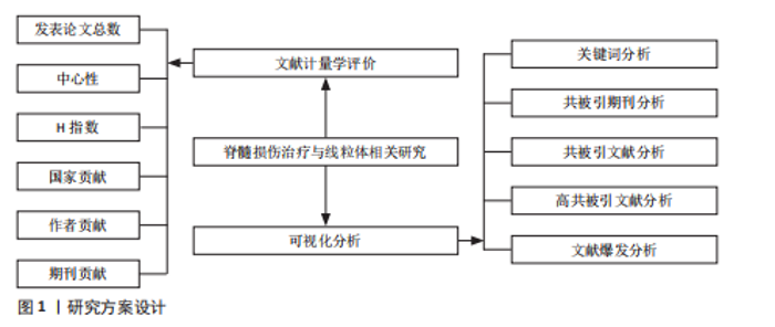
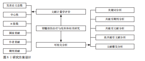
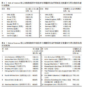
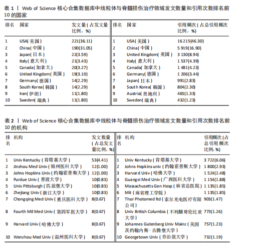
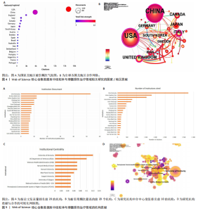
2.1 线粒体与脊髓损伤治疗领域数据和出版趋势 共纳入558篇文献进行文献计量学分析,涉及48个国家、3 036名作者、713家机构与252种期刊。近35年来,线粒体与脊髓损伤治疗领域的研究文献虽总量较少,但呈持续增长趋势,拟合曲线函数为“y=0.616 9x2- 6.643 2x+23.024,R2=0.998”,故线粒体与脊髓损伤治疗领域中研究增长曲线相对较好。文献筛选流程如图2所示。线粒体与脊髓损伤治疗领域年度发文趋势如图3所示。 2.2 线粒体与脊髓损伤治疗领域研究国家及地区分布 从国家和地区分布来看,48个国家及地区在线粒体脊髓损伤治疗领域进行研究。表1列出了基于发文量及引用频次排名前10的国家。数据显示,美国以221篇的发文量位居首位,约占总研究数量的36.11%;中国以190篇的发文量位列第二,约占总研究数量的31.05%;日本、意大利和加拿大的发文量位列前5。在论文引用频次上,美国以16 215次的引用量居首,约占总引用频次的46.3%;中国以5 919次紧随其后,约占总引用频次的16.9%;英国、意"
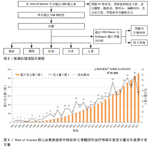
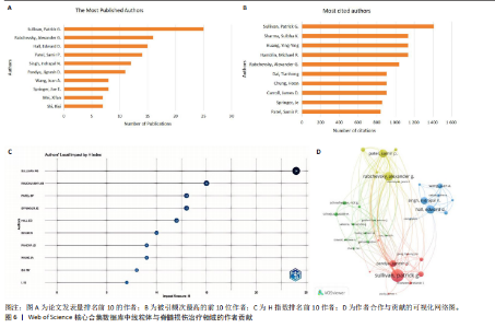
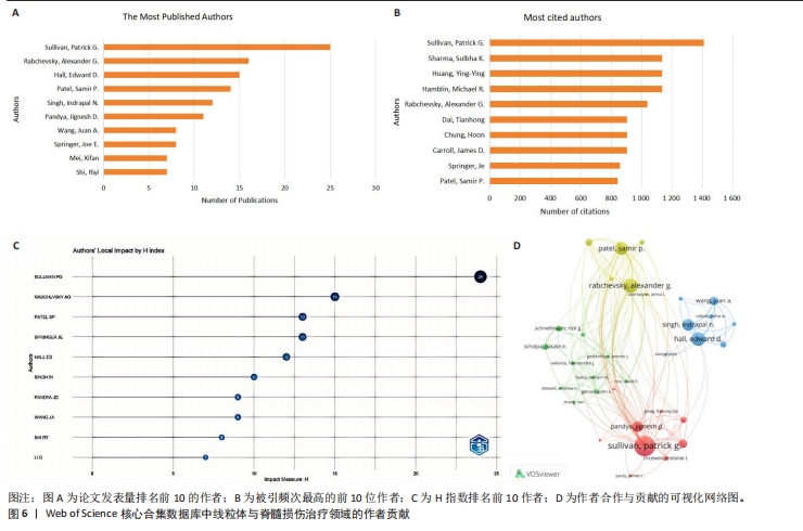
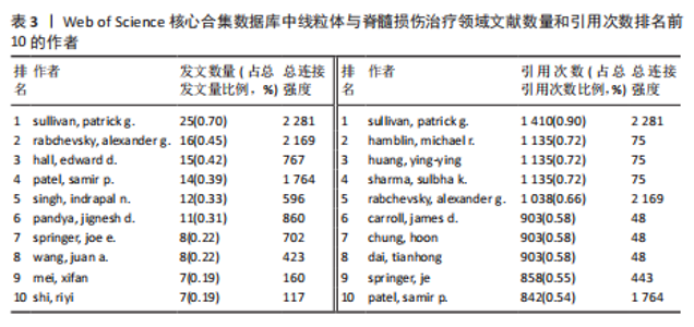
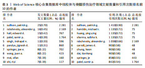
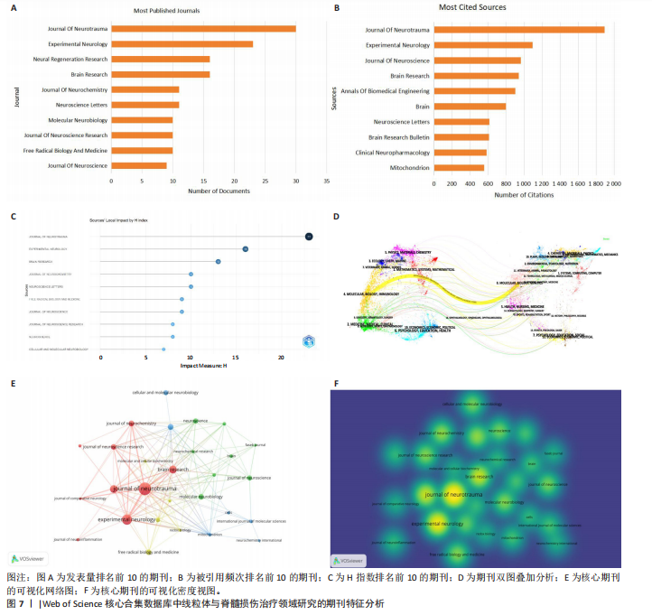
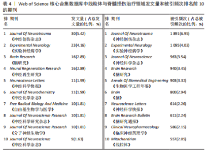
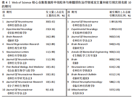
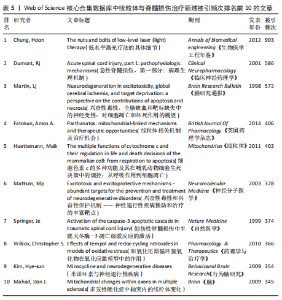
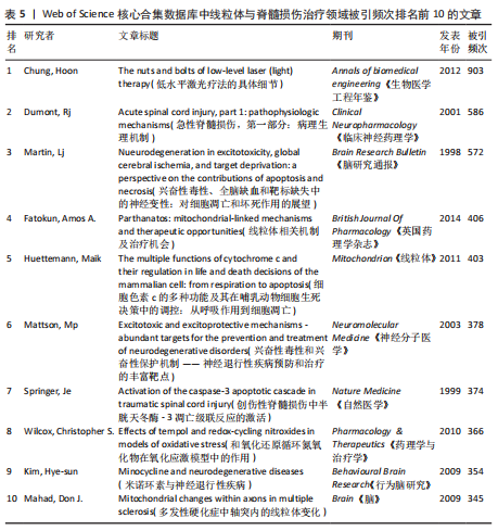
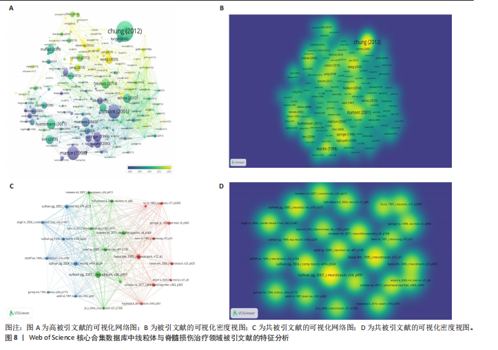
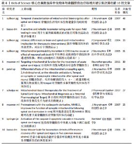
大利和加拿大也位列引用频次前5(图4A)。图4B展示了各国及地区间的合作关系网络,节点大小与发文量呈正比,美国和中国因较高的发文量呈现出较大节点;此外,以英国、瑞典和意大利为代表的欧洲国家之间合作关系较为紧密。 2.3 线粒体与脊髓损伤治疗领域研究机构与学者 表2列出了该领域基于发文量和引用频次排名前10的机构。其中,发文量最多的机构是肯塔基大学(53篇,4.41%),其次是锦州医科大学(12篇,1.00%)、约翰斯·霍普金斯大学(12篇,1.00%)。总引用频次最多的机构是肯塔基大学(n=3 722,6.06%),其次是约翰斯·霍普金斯大学(n=1 800,2.93%)、哈佛大学(n=1 524,2.48%)(图5A,B)。从研究机构贡献与合作的可视化网络图及研究机构中介中心度值看出,肯塔基大学处于该领域研究的前沿,该机构对该领域相关研究的认可度很高(图5C,D)。 共有3 036位学者在该领域发表相关文章。作者发文量及其文章引用频次可反映他们在该领域的影响力。通过分析作者与引用作者的合作网络能够识别出具有影响力的学者,为研究人员寻找潜在合作对象提供参考[17]。其中,Sullivan, Patrick G.发表文章最多,为25篇,约占总发文量的0.7%;Rabchevsky, Alexander G.发表文章16篇,约占总发文量的0.45%;Hall, Edward D.发表文章15篇,约占总发文量的0.42%(图6A)。被引用频次最高的作者为Sullivan, Patrick G.,共被引1 410次,其次是Hamblin, Michael R.、Huang, Ying-Ying、Sharma, Sulbha K.等,被引1 135次(图6B)。表3列出了该领域文献数量和引用次数排名的前10位作者。H指数(一个人在一定期间内发表的论文至少有H篇的被引频次不低于H次)前4位的作者分别为Sullivan, Patrick G.、Hamblin, Michael R.、Patel, Samir P.、Springer, Joe E(图6C)。Sullivan, Patrick G.在该领领域占据领先地位。作者共现网络显示该领域内有4个合作团体,各组内部作者共现联系密切,组间作者也存在合作关系(图6D)。 2.4 线粒体与脊髓损伤治疗领域期刊、被引用文章和共同引用文章 该领域相关研究发表于252种期刊,其中发文量排名前10的期刊累计发表146篇文章,发文量排名前2位的期刊为《Journal Of Neurotrauma》和《Experimental Neurology》,其被引频次也位居前2位(图7A,B),表明《Journal of Neurotrauma》和《Experimental Neurology》在该领域研究中具有重要影响力。表4列出了该领域发文量与被引频次排名前10的期刊。H指数排名前3的期刊分别为《Journal of Neurotrauma》《Experimental Neurology》和《Brain》(图7C)。图7D为Web of Science核心合集数据库该领域研究期刊双图叠加分析。期刊网络可视化分析结果显示,被引频次最高的期刊为《Journal of Neurotrauma》《Experimental Neurology》和《Brain Research》(图7E,F)。 表5列出了被引频次最多的10篇文章。其中,CHUNG 等[18]于2012年发表的《The "

"
"

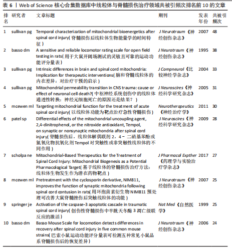
nuts and bolts of low-level laser (light) therapy》被引高达903次,位居第一,该文献明确指出低水平激光疗法的核心作用机制与线粒体密切相关,低水平激光疗法通过调节线粒体能量代谢、信号分子释放实现了对炎症、损伤等有治疗作用。排名第二的是DUMONT等[19]于2001年发表的《Acute spinal cord injury, part 1: pathophysiologic mechanisms》,被引586次,该文献明确指出通过保护线粒体功能可以减轻脊髓损伤进展、减少神经细胞的死亡,为后续脊髓损伤临床治疗提供了理论靶点和方向。MARTIN等[20]于1998年发表的《Nueurodegeneration in excitotoxicity, global cerebral ischemia, and target deprivation: a perspective on the contributions of apoptosis and necrosis》被引572次,该文献对兴奋性毒性、全脑缺血等导致神经退行性变机制进行研究,为脊髓损伤研究提供了思路,比如可以从抑制兴奋性毒性、调节细胞死亡形式、保护线粒体功能等方向去探索治疗脊髓损伤的新方法。在高被引文献中,Chung于2012年发表的文章被引频次最高(图8A,B)。 表6列出了共引用频次排名前10的文章。其中,SULLIVAN等[21]于2007年发表的《Temporal characterization of mitochondrial bioenergetics after spinal cord injury》共被引频次最高(共被引频次48),文献主要研究了脊髓损伤后线粒体生物能量学的变化,阐述了线粒体在脊髓损伤病理过程中的重要作用。排名第二的是BASSO等[22]1995年发表的《A sensitive and reliable locomotor rating scale for open field testing in rats》,共被引38次,该文献指出从肢体运动、尾巴位置、关节运动、躯干姿势、步幅和体质量支撑能力等多个方面对大鼠后肢功能进行评估,为研究人员提供了一种更具辨别力的行为结果测量方法,以评估脊髓损伤后的治疗效果。此外,SULLIVAN等[23]2004年发表的《Intrinsic differences in brain and spinal cord mitochondria: Implication for therapeutic "
"

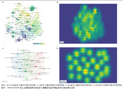
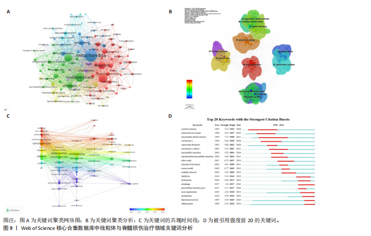
interventions》共被引33次,该文献为理解脊髓损伤的病理机制以及寻找有效治疗策略提供了重要理论依据。从共被引文献可视化网络与密度视图看,Sullivan, Patrick G.在2007年的研究成果在学术界获得广泛认可,在其他文献中多次被引用(图8C,D)。 2.5 线粒体与脊髓损伤治疗领域关键词分析 关键词共现分析基于文献内容的核心词来分析并反映文献的主要内容和研究方向,关键词共现分析可以识别领域的热点及研究前沿,有助于研究人员了解某一特定时期的热点词[24]。线粒体与脊髓损伤治疗领域的研究热点主要与线粒体、凋亡、氧化应激相关联(图9A)。共现网络将相关关键词划分为6大聚类,叠加可视化结果显示多数关键词集中产生于2010-2018年间(图9B,C)。图9D展示了被引强度排名前20的关键词,被引强度最高的5个关键词分别是肌萎缩侧索硬化、小鼠模型、一氧化氮、细胞色素C和超氧化物歧化酶。总体来看,这20个关键词的被引用度在4.17-7.17之间,持续时长为3-13年。研究表明,自噬、轴突再生、机制、功能恢复及炎症等关键词在2024年前仍将是该领域的研究热点,这意味着这些关键词不仅是当前该领域的热门话题,也会持续成为未来的研究重点。"
"
"
"

"
"
| [1] BIE F, WANG KY, XU T, et al. The potential roles of circular RNAs as modulators in traumatic spinal cord injury. Biomed Pharmacother. 2021;141:111826. [2] FISCHER I, DULIN JN, LANE MA. Transplanting neural progenitor cells to restore connectivity after spinal cord injury. Nat Rev Neurosci. 2020;21(7):366-383. [3] KARSY M, HAWRYLUK G. Modern Medical Management of Spinal Cord Injury. Curr Neurol Neurosci. 2019;19(9):65 [4] LIN JL, XIONG ZC, GU JH, et al. Sirtuins: Potential Therapeutic Targets for Defense against Oxidative Stress in Spinal Cord Injury. Oxid Med Cell Longev. 2021;2021:7207692. [5] XIANG XN, ZONG HY, OU Y, et al. Exoskeleton-assisted walking improves pulmonary function and walking parameters among individuals with spinal cord injury: a randomized controlled pilot study. J Neuroeng Rehabil. 2021;18(1):120. [6] FLACK JA, SHARMA KD, XIE JYH. Delving into the recent advancements of spinal cord injury treatment: a review of recent progress. Neural Regen Res. 2022;17(2):283-291. [7] QUADRI SA, FAROOQUI M, IKRAM A, et al. Recent update on basic mechanisms of spinal cord injury. Neurosurg Rev. 2020;43(2):425-441. [8] SONG YH, AGRAWAL NK, GRIFFIN JM, et al. Recent advances in nanotherapeutic strategies for spinal cord injury repair. Adv Drug Deliver Rev. 2019;148:38-59. [9] BADHIWALA JH, WILSON JR, FEHLINGS MG. Global burden of traumatic brain and spinal cord injury. Lancet Neurol. 2019;18(1):24-25. [10] BARONCINI A, MAFFULLI N, ESCHWEILER J, et al. Pharmacological management of secondary spinal cord injury. Expert Opin Pharmaco. 2021; 22(13):1793-1800. [11] YING YB, ZHANG YF, TU YR, et al. Hypoxia Response Element-Directed Expression of aFGF in Neural Stem Cells Promotes the Recovery of Spinal Cord Injury and Attenuates SCI-Induced Apoptosis. Front Cell Dev Biol. 2021;9:693694. [12] MORCIANO G, PEDRIALI G, SBANO L, et al. Intersection of mitochondrial fission and fusion machinery with apoptotic pathways: Role of Mcl-1. Biol Cell. 2016;108(10):279-293. [13] PATEL VA, MASSENBURG D, VUJICIC S, et al. Apoptotic Cells Activate AMP-activated Protein Kinase (AMPK) and Inhibit Epithelial Cell Growth without Change in Intracellular Energy Stores. J Biol Chem. 2015;290(37):22352-22369. [14] NEGANOVA ME, ALEKSANDROVA YR, NEBOGATIKOV VO, et al. Promising Molecular Targets for Pharmacological Therapy of Neurodegenerative Pathologies. Acta Naturae. 2020;12(3):60-80. [15] YIN MC, XU CQ, MA JM, et al. A Bibliometric Analysis and Visualization of Current Research Trends in the Treatment of Cervical Spondylotic Myelopathy. Glob Spine J. 2021;11(6):988-998. [16] LIN GX, KOTHEERANURAK V, MAHATTHANATRAKUL A, et al. Worldwide research productivity in the field of full-endoscopic spine surgery: a bibliometric study. Eur Spine J. 2020;29(1):153-160. [17] ZHENG KY, DAI GY, LAN Y, et al. Trends of Repetitive Transcranial Magnetic Stimulation From 2009 to 2018: A Bibliometric Analysis. Front Neurosci-Switz. 2020;14:586314. [18] CHUNG H, DAI TH, SHARMA SK, et al. The Nuts and Bolts of Low-level Laser (Light) Therapy. Ann Biomed Eng. 2012;40(2):516-533. [19] DUMONT RJ, OKONKWO DO, VERMA S, et al. Acute spinal cord injury, part I: pathophysiologic mechanisms. Clin Neuropharmacol. 2001;24(5): 254-264. [20] MARTIN LJ, AL-ABDULLA NA, BRAMBRINK AM, et al. Neurodegeneration in excitotoxicity, global cerebral ischemia, and target deprivation: A perspective on the contributions of apoptosis and necrosis. Brain Res Bull. 1998;46(4):281-309. [21] SULLIVAN PG, KRISHNAMURTHY S, PATEL SP, et al. Temporal characterization of mitochondrial bioenergetics after spinal cord injury. J Neurotrauma. 2007;24(6):991-999. [22] BASSO DM, BEATTIE MS, BRESNAHAN JC. A sensitive and reliable locomotor rating scale for open field testing in rats. J Neurotrauma. 1995; 12(1):1-21. [23] SULLIVAN PG, RABCHEVSKY AG, KELLER JN, et al. Intrinsic differences in brain and spinal cord mitochondria: Implication for therapeutic interventions. J Comp Neurol. 2004;474(4):524-534. [24] ZHOU K, ZHOU Y, ZENG Y, et al. Research Hotspots and Global Trends of Transcranial Direct Current Stimulation in Stroke: A Bibliometric Analysis. Neuropsychiatr Dis Treat. 2023;19:601-613. [25] ELI I, LERNER DP, GHOGAWALA Z. Acute Traumatic Spinal Cord Injury. Neurol Clin. 2021;39(2):471-488. [26] AHUJA CS, NORI S, TETREAULT L, et al. Traumatic Spinal Cord Injury-Repair and Regeneration. Neurosurgery. 2017;80(3):S9-S22. [27] SLATER PG, DOMíNGUEZ-ROMERO ME, VILLARREAL M, et al. Mitochondrial function in spinal cord injury and regeneration. Cell Mol Life Sci. 2022;79(5):233.DOI:10.1007/s00018-022-04211-7 [28] SHANG ZZ, WANYAN P, WANG MC, et al. Bibliometric analysis of stem cells for spinal cord injury: current status and emerging frontiers. Front Pharmacol. 2023;14:1235324 [29] CAJIGAS I, VEDANTAM A. Brain-Computer Interface, Neuromodulation, and Neurorehabilitation Strategies for Spinal Cord Injury. Neurosurg Clin N Am. 2021;32(3):407-417. [30] AHUJA CS, MOTHE A, KHAZAEI M, et al. The leading edge: Emerging neuroprotective and neuroregenerative cell-based therapies for spinal cord injury. Stem Cell Transl Med. 2020;9(12): 1509-1530. [31] YAMAZAKI K, KAWABORI M, SEKI T, et al. Clinical Trials of Stem Cell Treatment for Spinal Cord Injury. Int J Mol Sci. 2020;21(11):6234.DOI:10.3390/ijms21116234 [32] CERVERA MA, SOEKADAR SR, USHIBA J, et al. Brain-computer interfaces for post-stroke motor rehabilitation: a meta-analysis. Ann Clin Transl Neur. 2018;5(5):651-663. [33] HUANG LY, ZHANG Q, FU CY, et al. Effects of hyperbaric oxygen therapy on patients with spinal cord injury: A systematic review and meta-analysis of Randomized Controlled Trials. J Back Musculoskelet. 2021;34(6):905-913. [34] ZHANG LG, FISHER JP, LEONG KW, et al. 3D bioprinting and nanotechnology in tissue engineering and regenerative medicine. London, England: Academic Press, 2015. [35] MCEWEN ML, SULLIVAN PG, RABCHEVSKY AG, et al. Targeting Mitochondrial Function for the Treatment of Acute Spinal Cord Injury. Neurotherapeutics. 2011;8(2):168-179. [36] SPRINGER JE, PRAJAPATI P, SULLIVAN PG. Targeting the mitochondrial permeability transition pore in traumatic central nervous system injury. Neural Regen Res. 2018;13(8):1338-1341. [37] KIM JW, MAHAPATRA C, HONG JY, et al. Functional Recovery of Contused Spinal Cord in Rat with the Injection of Optimal-Dosed Cerium Oxide Nanoparticles. Adv Sci. 2017;4(10):1700034. [38] LUO WQ, WANG YM, LIN F, et al. Selenium-Doped Carbon Quantum Dots Efficiently Ameliorate Secondary Spinal Cord Injury via Scavenging Reactive Oxygen Species. Int J Nanomed. 2020;15: 10113-10125. |
| [1] | Xu Canli, He Wenxing, Wang Yuping, Ba Yinying, Chi Li, Wang Wenjuan, Wang Jiajia. Research context and trend of TBK1 in autoimmunity, signaling pathways, gene expression, tumor prevention and treatment [J]. Chinese Journal of Tissue Engineering Research, 2026, 30(在线): 1-11. |
| [2] | Zhu Xiaolong, Zhang Wei, Yang Yang. Visualization analysis of research hotspots and cutting-edge information in the field of intervertebral disc regeneration and repair [J]. Chinese Journal of Tissue Engineering Research, 2026, 30(9): 2391-2402. |
| [3] | Wen Fayan, Li Yan, Qiang Tianming, Yang Chen, Shen Linming, Li Yadong, Liu Yongming. Unilateral biportal endoscopic technology for treatment of lumbar degenerative diseases: global research status and changing trends [J]. Chinese Journal of Tissue Engineering Research, 2026, 30(9): 2380-2390. |
| [4] | Lai Yu, Chen Yueping, Zhang Xiaoyun. Research hotspots and frontier trends of bioactive materials in treating bone infections [J]. Chinese Journal of Tissue Engineering Research, 2026, 30(8): 2132-2144. |
| [5] | Liu Anting, Lu Jiangtao, Zhang Wenjie, He Ling, Tang Zongsheng, Chen Xiaoling. Regulation of AMP-activated protein kinase by platelet lysate inhibits cadmium-induced neuronal apoptosis [J]. Chinese Journal of Tissue Engineering Research, 2026, 30(7): 1800-1807. |
| [6] | Hou Chaowen, Li Zhaojin, Kong Jianda, Zhang Shuli. Main physiological changes in skeletal muscle aging and the multimechanism regulatory role of exercise [J]. Chinese Journal of Tissue Engineering Research, 2026, 30(6): 1464-1475. |
| [7] | Lyu Xiaofan, Huang Yi, Ding Liucheng . Mitochondrial mechanism and intervention therapy in diabetic cystopathy [J]. Chinese Journal of Tissue Engineering Research, 2026, 30(6): 1508-1515. |
| [8] | Huang Jie, Zeng Hao, Wang Wenchi, Lyu Zhucheng, Cui Wei. Visualization analysis of literature on the effect of lipid metabolism on osteoporosis [J]. Chinese Journal of Tissue Engineering Research, 2026, 30(6): 1558-1568. |
| [9] | Yin Yongcheng, Zhao Xiangrui, Yang Zhijie, Li Zheng, Li Fang, Ning Bin. Effect and mechanism of peroxiredoxin 1 in microglial inflammation after spinal cord injury [J]. Chinese Journal of Tissue Engineering Research, 2026, 30(5): 1106-1113. |
| [10] | Yang Zeyu, Zhi Liang, Wang Jia, Zhang Jingyi, Zhang Qingfang, Wang Yulong, Long Jianjun. A visualized analysis of research hotspots in high-frequency repetitive transcranial magnetic stimulation from the macroscopic perspective [J]. Chinese Journal of Tissue Engineering Research, 2026, 30(5): 1320-1330. |
| [11] | Zhang Zhilong, Wang Haiying, Ma Fenghua, Hou Yanjie. Regulating mitochondrial dynamics balance in nucleus pulposus cells inhibits cell apoptosis [J]. Chinese Journal of Tissue Engineering Research, 2026, 30(29): 7520-7528. |
| [12] | Li Wenhao, Yang Xi, Du Xinran, Bai Shi, Li Zhongshan. Effect of magnetic field mitochondrial regulation technology combined with low-load blood flow restriction on the strength of lower limb muscle groups [J]. Chinese Journal of Tissue Engineering Research, 2026, 30(29): 7555-7564. |
| [13] | Wang Degang, Mei Junhua, Wang Junli, Zheng Li, Chen Guohua. Bibliometric and visualization analysis of the mechanism of osteogenic factors and neurotransmitters in the bone-brain axis [J]. Chinese Journal of Tissue Engineering Research, 2026, 30(29): 7724-7731. |
| [14] | Xia Tiange, Zhou Yi, Li Shaoshuo, Wang Jianwei, Shao Yang. Signaling pathways related to active ingredients of ginseng in the treatment of musculoskeletal degenerative diseases [J]. Chinese Journal of Tissue Engineering Research, 2026, 30(29): 7639-7647. |
| [15] | Liu Yan, Zuo Qingchun, Li Weiying, Wu Xubo. Research hotspots and trends of optogenetics in behavioral neuroscience [J]. Chinese Journal of Tissue Engineering Research, 2026, 30(28): 7396-7403. |
| Viewed | ||||||
|
Full text |
|
|||||
|
Abstract |
|
|||||