Chinese Journal of Tissue Engineering Research ›› 2023, Vol. 27 ›› Issue (18): 2831-2836.doi: 10.12307/2023.317
Previous Articles Next Articles
Effect of elastin degradation of patellar tendon on the quasi-static tensile mechanical properties
Liu Xiaoyun1, Deng Yuping1, 2, 3, Li Feifei1, Zhao Dongliang3, 4, Yang Yang1, Huang Tao1, 5, Tan Wenchang3, 4, Wu Yaobin1, Huang Wenhua1, 6, Li Yanbing1
- 1Guangdong Engineering Research Center for Translation of Medical 3D Printing Application; 2Department of Orthopedics and Traumatology, Integrated Hospital of Traditional Chinese Medicine and Western Medicine, Southern Medical University; 3Institute of Biomedical Engineering, Shenzhen Bay Laboratory; 4School of Chemical Biology and Biotechnology; 5The Third Affiliated Hospital of Guangzhou University of Chinese Medicine; 6The Third Affiliated Hospital of Southern Medical University
-
Received:2022-05-05Accepted:2022-06-06Online:2023-06-28Published:2022-09-17 -
Contact:Li Yanbing, MD, Professor, Master’s supervisor, Guangdong Engineering Research Center for Translation of Medical 3D Printing Application, Guangdong Provincial Key Laboratory of Medical Biomechanics, National Key Discipline of Human Anatomy, School of Basic Medical Sciences, Southern Medical University, Guangzhou 510515, Guangdong Province, China -
About author:Liu Xiaoyun, Master candidate, Guangdong Engineering Research Center for Translation of Medical 3D Printing Application, Guangdong Provincial Key Laboratory of Medical Biomechanics, National Key Discipline of Human Anatomy, School of Basic Medical Sciences, Southern Medical University, Guangzhou 510515, Guangdong Province, China -
Supported by:Guangdong Science and Technology Program Project, No. 2018B090944002 (to LYB); Guangdong Provincial Basic and Applied Basic Research Fund, No. 2020B1515120001 (to HWH); Shenzhen Science and Technology Plan, No. JCYJ20210324130401005 (to ZDL)
CLC Number:
Cite this article
Liu Xiaoyun, Deng Yuping, Li Feifei, Zhao Dongliang, Yang Yang, Huang Tao, Tan Wenchang, Wu Yaobin, Huang Wenhua, Li Yanbing. Effect of elastin degradation of patellar tendon on the quasi-static tensile mechanical properties[J]. Chinese Journal of Tissue Engineering Research, 2023, 27(18): 2831-2836.
share this article
Add to citation manager EndNote|Reference Manager|ProCite|BibTeX|RefWorks
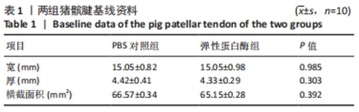
2.1 标本数量分析 所有23个髌腱标本均完成实验。在应力松弛实验过程中,PBS对照组有2个标本发生轻微抖动,为了保持一致性,PBS对照组和弹性蛋白酶处理组(弹性蛋白酶组)各选择8个应力松弛实验数据进行应力-时间分析。 2.2 两组基线资料比较 两组猪髌腱的宽、厚、横截面积差异均无显著性意义,具有可比性,见表1。"
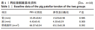
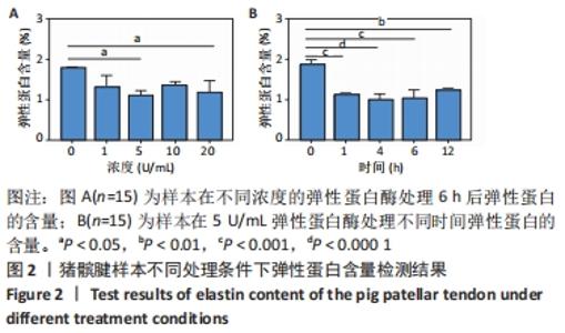
2.3 髌腱弹性蛋白含量检测结果 处理后,髌腱中残留的弹性蛋白含量与弹性蛋白酶的浓度成反比,见图2A,随着酶浓度的增加,弹性蛋白含量表示一致的相对下降趋势。对于不同的处理时间,随着时间的增加,弹性蛋白含量也显示一致的下降趋势,但在4-6 h后基本趋于平稳,见图2B。用于测试的2个髌腱具有不同的总弹性蛋白含量,因此用于评估不同浓度及不同时间的弹性蛋白含量会有略微差异。由于弹性蛋白酶的成本高,且弹性蛋白酶浓度在5 U/mL时的含量最低,酶浓度从5-20 U/mL增加时,弹性蛋白含量波动幅度不大,统计学分析没有显著性意义,因此选择5 U/mL弹性蛋白酶孵育8 h作为弹性蛋白酶处理方案。"
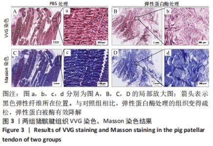
2.4 组织学染色观察结果 组织学染色分析弹性蛋白酶对髌腱中弹性蛋白纤维和胶原蛋白纤维的影响。VVG染色将弹性蛋白纤维染成深黑色,将胶原纤维染成红色,与PBS处理组相比,弹性蛋白酶处理的组织清楚显示弹性蛋白降解效果,见图3A,B。Masson染色将胶原蛋白纤维染成蓝色,显示弹性蛋白酶处理前后的胶原纤维方向不受酶处理的影响,显示了弹性蛋白沿着胶原纤维分布,胶原纤维呈波浪状,卷曲清晰可见,见图3C,D。在弹性蛋白酶处理之前,组织中的胶原纤维和弹性纤维排列整齐紧凑;弹性蛋白酶处理后,组织变得疏松,弹性蛋白在组织中零碎存在,胶原纤维波浪状的卷曲度减少。除此之外,还可见弹性蛋白酶从外表面开始向内降解弹性蛋白。"

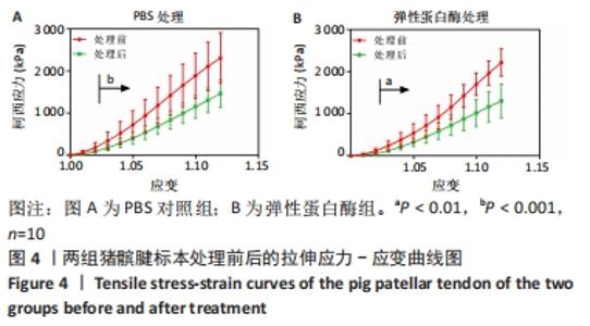
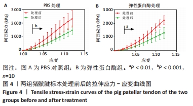
2.5 弹性蛋白对力学性能的影响 2.5.1 两组髌腱处理前后拉伸柯西应力变化结果 PBS组和弹性蛋白酶组都显示了处理前的柯西应力要比处理后的大,差异有显著性意义,两组应力降低百分比约40%(PBS对照组P=0.000 3,见图4A;弹性蛋白酶组P=0.001 3,见图4B)。但是PBS处理前后的柯西应力差值与弹性蛋白酶处理前后柯西应力差值比较,差异无显著性意义(P=0.595)。"
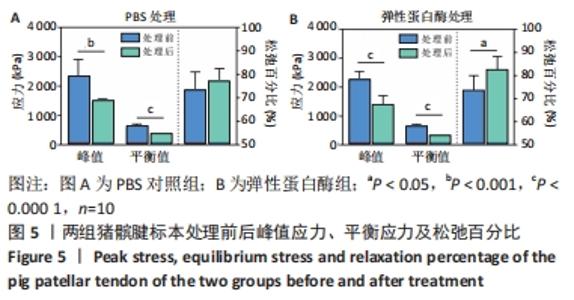
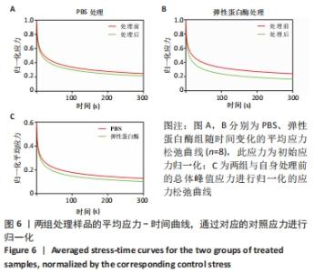
2.5.2 两组髌腱处理前后应力松弛 图5显示了PBS对照组和弹性蛋白酶组处理前后的峰值应力、平衡应力、松弛百分比。PBS对照组处理后的峰值应力和平衡应力比处理前的小,峰值应力降低约37%,平衡应力降低约43%,差异有显著性意义(P=0.001,P < 0.000 1),应力下降幅度差异无显著性意义(P=0.241),见图5A。弹性蛋白酶组处理后的峰值应力和平衡应力比处理前的小,峰值应力降低约41%,平衡应力降低约61%,差异有显著性意义(P < 0.000 1),应力下降幅度增加约12%,差异有显著性意义(P=0.007),见图5B。两组应力随时间的变化趋势显示,与处理前相比,组织经过处理后表现出明显下降趋势,应力值差异有显著性意义(P值均< 0.000 1),见图6A,B。通过对相应组内所有样品的归一化应力取平均值绘制平均归一化应力-时间曲线,见图6C,显示了弹性蛋白酶组比PBS对照组引起的应力降低值更大(P < 0.000 1)。"

"
| [1] HUTCHISON MK, HOUCK J, CUDDEFORD T, et al. Prevalence of Patellar Tendinopathy and Patellar Tendon Abnormality in Male Collegiate Basketball Players: A Cross-Sectional Study. J Athl Train. 2019;54(9):953-958. [2] BREDA SJ, OEI E, ZWERVER J, et al. Effectiveness of progressive tendon-loading exercise therapy in patients with patellar tendinopathy: a randomised clinical trial. Br J Sports Med. 2021;55(9):501-509. [3] BURTON I. Interventions for prevention and in-season management of patellar tendinopathy in athletes: A scoping review. Phys Ther Sport. 2022;55:80-89. [4] FIRMINGER CR, EDWARDS WB. Effects of cyclic loading on the mechanical properties and failure of human patellar tendon. J Biomech. 2021;120:110345. [5] FIRMINGER CR, EDWARDS WB. A biomechanical study of clamping technique on patellar tendon surface strain and material properties using digital image correlation. J Mech Behav Biomed Mater. 2021;113:104156. [6] GOLMAN M, WRIGHT ML, WONG TT, et al. Rethinking Patellar Tendinopathy and Partial Patellar Tendon Tears: A Novel Classification System. Am J Sports Med. 2020;48(2):359-369. [7] RUMIAN AP, DRAPER ER, WALLACE AL, et al. The influence of the mechanical environment on remodelling of the patellar tendon. J Bone Joint Surg Br. 2009; 91(4):557-564. [8] ZHANG C, COUPPÉ C, SCHEIJEN J, et al. Regional collagen turnover and composition of the human patellar tendon. J Appl Physiol (1985). 2020;128(4): 884-891. [9] HELLAND C, BOJSEN-MØLLER J, RAASTAD T, et al. Mechanical properties of the patellar tendon in elite volleyball players with and without patellar tendinopathy. Br J Sports Med. 2013;47(13):862-868. [10] 陈一言, 陆阿明, 齐亚楠, 等. 大强度跳跃负荷下p38MAPK对兔髌腱起止点微细结构影响[J]. 体育科研,2021,42(6):86-92. [11] TORNIAINEN J, RISTANIEMI A, SARIN JK, et al. Near infrared spectroscopic evaluation of biochemical and crimp properties of knee joint ligaments and patellar tendon. PLoS One. 2022;17(2):e263280. [12] RISTANIEMI A, TORNIAINEN J, STENROTH L, et al. Comparison of water, hydroxyproline, uronic acid and elastin contents of bovine knee ligaments and patellar tendon and their relationships with biomechanical properties. J Mech Behav Biomed Mater. 2020;104: 103639. [13] DE MOUDT S, LELOUP A, FRANSEN P. Aortic Stiffness Hysteresis in Isolated Mouse Aortic Segments Is Intensified by Contractile Stimuli, Attenuated by Age, and Reversed by Elastin Degradation. Front Physiol. 2021;12:723972. [14] UGRADAR S, KARLIN J, LE A, et al. Photochemical Collagen Cross-Linking Reverses Elastase-Induced Mechanical Degradation of Upper Eyelid Tarsus. Ophthalmic Plast Reconstr Surg. 2020;36(6):562-565. [15] HILL JR, EEKHOFF JD, BROPHY RH, et al. Elastic fibers in orthopedics: Form and function in tendons and ligaments, clinical implications, and future directions. J Orthop Res. 2020;38(11):2305-2317. [16] GRANT TM, YAPP C, CHEN Q, et al. The Mechanical, Structural, and Compositional Changes of Tendon Exposed to Elastase. Ann Biomed Eng. 2015;43(10):2477-2486. [17] HENNINGER HB, UNDERWOOD CJ, ROMNEY SJ, et al. Effect of elastin digestion on the quasi-static tensile response of medial collateral ligament. J Orthop Res. 2013;31(8):1226-1233. [18] AKINTUNDE A, ROBISON KM, CAPONE D, et al. Effects of elastase digestion on the murine vaginal wall biaxial mechanical response. J Biomech Eng. 2018;141(2): 210111-2101111. [19] LEVILLAIN A, ORHANT M, TURQUIER F, et al. Contribution of collagen and elastin fibers to the mechanical behavior of an abdominal connective tissue. J Mech Behav Biomed Mater. 2016;61:308-317. [20] YANG F, DAS D, CHASIOTIS I. Strain rate induced toughening of individual collagen fibrils. Appl Phys Lett. 2022;120(11):114101. [21] KIM DK, AHN J, KIM SA, et al. Improved Healing of Rabbit Patellar Tendon Defects After an Atelocollagen Injection. Am J Sports Med. 2021;49(11):2924-2932. [22] SVENSSON RB, ERIKSEN CS, TRAN P, et al. Mechanical properties of human patellar tendon collagen fibrils. An exploratory study of aging and sex. J Mech Behav Biomed Mater. 2021;124:104864. [23] LIU Y, BALLARINI R, EPPELL SJ. Tension tests on mammalian collagen fibrils. Interface Focus. 2016;6(1):20150080. [24] BLOOM ET, LEE AH, ELLIOTT DM. Tendon Multiscale Structure, Mechanics, and Damage Are Affected by Osmolarity of Bath Solution. Ann Biomed Eng. 2021; 49(3):1058-1068. [25] LEE AH, ELLIOTT DM. Freezing does not alter multiscale tendon mechanics and damage mechanisms in tension. Ann N Y Acad Sci. 2017;1409(1):85-94. [26] FANG F, LAKE SP. Multiscale mechanical integrity of human supraspinatus tendon in shear after elastin depletion. J Mech Behav Biomed Mater. 2016;63:443-455. [27] 胡凯, 陈维明, 骆井万, 等. 过氧乙酸/乙醇对异体皮性能的影响[J]. 中国组织工程研究,2022,26(10):1537-1543. [28] DUENWALD SE, VANDERBY RJ, LAKES RS. Viscoelastic relaxation and recovery of tendon. Ann Biomed Eng. 2009;37(6):1131-1140. [29] LEE AH, SZCZESNY SE, SANTARE MH, et al. Investigating mechanisms of tendon damage by measuring multi-scale recovery following tensile loading. Acta Biomater. 2017;57:363-372. [30] LAKE SP, MILLER KS, ELLIOTT DM, et al. Effect of fiber distribution and realignment on the nonlinear and inhomogeneous mechanical properties of human supraspinatus tendon under longitudinal tensile loading. J Orthop Res. 2009;27(12):1596-1602. [31] 黄涛, 刘晓云, 邓羽平, 等. 实时动态拉伸下成人肱二头肌、肱三头肌的生物力学特性[J]. 中国组织工程研究,2022,26(12):1828-1833. [32] CASTILE RM, SKELLEY NW, BABAEI B, et al. Microstructural properties and mechanics vary between bundles of the human anterior cruciate ligament during stress-relaxation. J Biomech. 2016,49(1):87-93. [33] HIRANO E, KNUTSEN RH, SUGITANI H, et al. Functional rescue of elastin insufficiency in mice by the human elastin gene: implications for mouse models of human disease. Circ Res. 2007;101(5):523-531. [34] KIM SH, MONTICONE RE, MCGRAW KR, et al. Age-associated proinflammatory elastic fiber remodeling in large arteries. Mech Ageing Dev. 2021;196:111490. [35] HENNINGER HB, VALDEZ WR, SCOTT SA, et al. Elastin governs the mechanical response of medial collateral ligament under shear and transverse tensile loading. Acta Biomater. 2015;25:304-312. [36] GODINHO MS, THORPE CT, GREENWALD SE, et al. Elastase treatment of tendon specifically impacts the mechanical properties of the interfascicular matrix. Acta Biomater. 2021;123:187-196. [37] JACOBS NT, SMITH LJ, HAN WM, et al. Effect of orientation and targeted extracellular matrix degradation on the shear mechanical properties of the annulus fibrosus. J Mech Behav Biomed Mater. 2011;4(8):1611-1619. [38] FAZAELI S, MIRAHMADI F, EVERTS V, et al. Alteration of structural and mechanical properties of the temporomandibular joint disc following elastase digestion. J Biomed Mater Res B Appl Biomater. 2020;108(8):3228-3240. [39] EEKHOFF JD, ABRAHAM JA, SCHOTT HR, et al. Fascicular elastin within tendon contributes to the magnitude and modulus gradient of the elastic stress response across tendon type and species. Acta Biomater. 2022;S1742-7061(22)00156-8. [40] PATEL D, ZAMBOULIS DE, SPIESZ EM, et al. Structure-function specialisation of the interfascicular matrix in the human achilles tendon. Acta Biomater. 2021;131: 381-390. [41] HILL JR, EEKHOFF JD, BROPHY RH, et al. Elastic fibers in orthopedics: Form and function in tendons and ligaments, clinical implications, and future directions.J Orthop Res. 2020;38(11):2305-2317. [42] CHAUDHURI O, COOPER-WHITE J, JANMEY PA, et al. Effects of extracellular matrix viscoelasticity on cellular behaviour. Nature. 2020;584(7822):535-546. [43] ROSS CJ, LAURENCE DW, ECHOLS AL, et al. Effects of enzyme-based removal of collagen and elastin constituents on the biaxial mechanical responses of porcine atrioventricular heart valve anterior leaflets. Acta Biomater. 2021;135:425-440. [44] EEKHOFF JD, STEENBOCK H, BERKE IM, et al. Dysregulated assembly of elastic fibers in fibulin-5 knockout mice results in a tendon-specific increase in elastic modulus. J Mech Behav Biomed Mater. 2021;113:104134. |
| [1] | Zhong Yizheng, Huang Peizhen, Cai Qunbin, Zheng Liqin, He Xingpeng, Dong Hang. Microstructural indexes that determine the trabecular bone maximum stress of micro-finite element models [J]. Chinese Journal of Tissue Engineering Research, 2023, 27(9): 1313-1318. |
| [2] | Wu Taoguang, Nie Shaobo, Chen Hua, Zhu Zhengguo, Qi Lin, Tang Peifu. Biomechanical characteristics of a new multi-dimensional cross locking plate in the treatment of subtrochanteric nonunion [J]. Chinese Journal of Tissue Engineering Research, 2023, 27(9): 1330-1334. |
| [3] | Peng Zhixin, Yan Wengang, Wang Kun, Zhang Zhenjiang. Finite element analysis and structural optimization design of 3D printed forearm braces [J]. Chinese Journal of Tissue Engineering Research, 2023, 27(9): 1340-1345. |
| [4] | Liu Jinyu, Zhang Hanshuo, Cui Hongpeng, Pan Lingzhi, Zhao Boran, Li Fei, Ding Yu. Finite element biomechanical analysis of minimally invasive treatment of cervical spondylotic myelopathy and accurate exercise rehabilitation [J]. Chinese Journal of Tissue Engineering Research, 2023, 27(9): 1359-1364. |
| [5] | Wen Xinghua, Ding Huanwen, Cheng Kai, Yan Xiaonan, Peng Yuanhao, Wang Yuning, Liu Kang, Zhang Huiwu. Three-dimensional finite element model analysis of intramedullary nailing fixation design for large femoral defects in Beagle dogs [J]. Chinese Journal of Tissue Engineering Research, 2023, 27(9): 1371-1376. |
| [6] | Liu Jiaxin, Jia Peng, Men Yutao, Liu Lu, Wang Yeming, Ye Jinduo. Design and optimization of bone trabecular structure with triply periodic minimal surfaces [J]. Chinese Journal of Tissue Engineering Research, 2023, 27(7): 992-997. |
| [7] | Li Huiqin, Wang Chunjuan, Wang Yu, Wang Weifeng, Chen Dinggen, Li Na. Clear aligner orthodontic therapy of rotated mandibular teeth with different shapes: a three-dimensional finite element analysis [J]. Chinese Journal of Tissue Engineering Research, 2023, 27(7): 1050-1054. |
| [8] | Li Yaping, Liu Hong, Gao Zhen, Chen Xiaolin, Huang Wujie, Jiang Zheng. Three-dimensional motion analysis of lower limb biomechanical performance in Tai Chi practitioners accompanied by knee joint pain [J]. Chinese Journal of Tissue Engineering Research, 2023, 27(4): 520-526. |
| [9] | Li Shihao, Li Qi, Li Zhen, Zhang Yuanyuan, Liu Miaomiao, Ouyang Yi, Xu Weiguo. Plantar pressure and gait analysis in patients with anterior cruciate ligament injury and reconstruction [J]. Chinese Journal of Tissue Engineering Research, 2023, 27(4): 626-631. |
| [10] | Zhan Yi, Wang Biao, Ma Yuli, He Simin, Sun Honghui, Hao Dingjun. Biomechanical comparison between a novel bone cement screw system and common surgical methods for the treatment of Kummell’s disease [J]. Chinese Journal of Tissue Engineering Research, 2023, 27(3): 385-390. |
| [11] | Lu Hui, Wu Qimei, Liu Rong. Finite element analysis and application of unilateral and bilateral bone-filling mesh container in treatment of osteoporotic vertebral compression fracture [J]. Chinese Journal of Tissue Engineering Research, 2023, 27(3): 391-397. |
| [12] | Gao Xu, Xing Wenhua. Application of finite element analysis in spine surgery [J]. Chinese Journal of Tissue Engineering Research, 2023, 27(18): 2921-2927. |
| [13] | Hao Yunteng, Shi Jun, Liu Yuhang, Li Kun, Ma Yuan, Zhang Shaojie, Wang Chaoqun, Chen Jie, Zhang Zhifeng, Zheng Leigang, Wang Xing, Li Zhijun. Comparative finite element analysis of the cervical articular process after resection of different ranges of the uncinate processes [J]. Chinese Journal of Tissue Engineering Research, 2023, 27(18): 2789-2796. |
| [14] | Huang Dingan, Liu Chen. Three-dimensional finite element analysis of a new height adjustable cervical fusion cage [J]. Chinese Journal of Tissue Engineering Research, 2023, 27(18): 2797-2803. |
| [15] | Liu Yan, Ge Hongqing, Chen Wenzhi, Liang Zeqian, Li Junye, He Jielong. Finite element analysis of different fibular support methods to reconstruct the poor medial column of humeral proximal fractures [J]. Chinese Journal of Tissue Engineering Research, 2023, 27(18): 2804-2808. |
| Viewed | ||||||
|
Full text |
|
|||||
|
Abstract |
|
|||||