Chinese Journal of Tissue Engineering Research ›› 2021, Vol. 25 ›› Issue (4): 537-541.doi: 10.3969/j.issn.2095-4344.2365

Previous Articles     Next Articles

Stress of three-dimensional finite element models of E-MAX porcelain inlay

Zhang Guomei1, Zhu Jun1, Hu Yang2, Jiao Hongwei3   

  1. 1Department of Stomatology, Luodian Hospital of Baoshan District of Shanghai, Shanghai 201908, China; 2Department of Prosthodontics, First Affiliated Hospital of Xinjiang Medical University, Urumqi 830054, Xinjiang Uygur Autonomous Region, China; 3Cavity Prevention Center in Shanghai, Shanghai Stomatological Hospital, Yongjia Road Special Need Clinic, Shanghai 200001, China
  • Received:2019-12-24 Revised:2019-12-27 Accepted:2020-04-22 Online:2021-02-08 Published:2020-11-21
  • Contact: Jiao Hongwei, Associate chief physician, Cavity Prevention Center in Shanghai, Shanghai Stomatological Hospital, Yongjia Road Special Need Clinic, Shanghai 200001, China
  • About author:Zhang Guomei, Master, Department of Stomatology, Luodian Hospital of Baoshan District of Shanghai, Shanghai 201908, China
  • Supported by:
    the Special Fund for Key Research & Development Tasks of Xinjiang Uygur Autonomous Region, No. 2016B03049-2

Abstract: BACKGROUND: E-Max porcelain inlay has good aesthetic, bonding and mechanical properties, and has a broad application prospect in the field of tooth defect repair. 
OBJECTIVE: To build the model of mesio-occluso-distal cavity E-Max porcelain inlay with different adhesives and different depths of holes, and to explore the stress distribution and regional law of different data models.
METHODS: Micro-CT was used to scan the human mandibular third molar model. Medical modeling software mimics 20, reverse engineering software Geomagic Studio 2014, and three-dimensional mechanical drawing software NX 10 were utilized to construct the three-dimensional finite element models of mesio-occluso-distal cavity E-Max porcelain inlay with different adhesives and different depths of holes: Group A with a hole depth of 2 mm, using 3M RelyX™ Unicem adhesive; group B with a hole depth of 3 mm, using 3M RelyX™ Unicem adhesive; group C with a hole depth of 4 mm, using 3M RelyX™ Unicem adhesive; group D with a hole depth of 2 mm, using vario-link N adhesive; group E with a hole depth of 3 mm, using vario-link N adhesive; group F with a hole depth of 4 mm, using vario-link N adhesive. Finite element analysis software ANSYS workbench 18.2 was used for meshing. Stress distribution of each model at 10 N•mm torque, 45° loading 175 N and 90° loading 600 N was analyzed.  
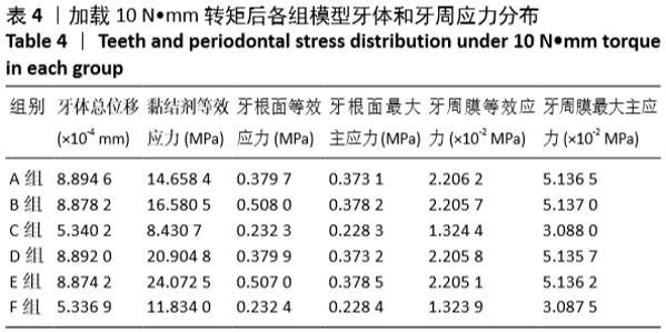
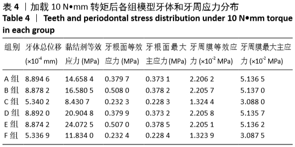
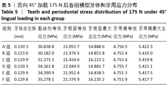
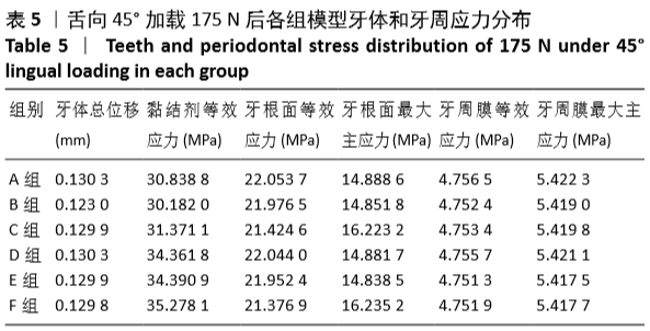
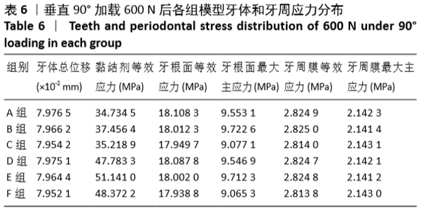
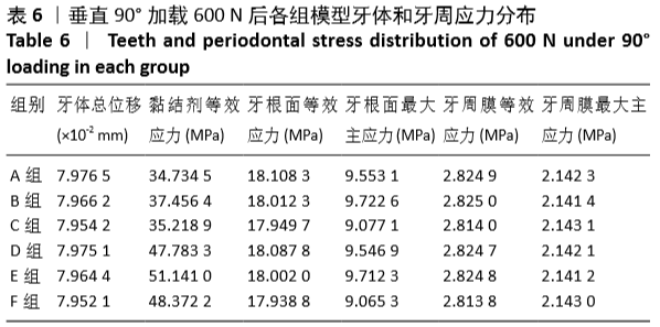
RESULTS AND CONCLUSION: (1) After 10 N•mm torque loading, with the increase of cavity depth, total displacement of the teeth and the equivalent stress of the periodontal ligament decreased with the same adhesive; when the cavity depth was 3 mm, the root surface equivalent stress and the adhesive equivalent stress were largest. Under the same cavity depth, the equivalent stress and the maximum principal stress of the root surface were larger when using vario link N adhesive. (2) When 175 N was applied at 45° lingual direction and the same adhesive was used, the equivalent stress on the root surface decreased with the increase of the cavity depth. When the depth of the cavity was 4 mm, the total displacement of the tooth and the equivalent stress of the adhesive were largest. When the cavity depth was 2 mm, the equivalent stress and the maximum principal stress of periodontal ligament were largest. At the same cavity depth, the maximum principal stress, equivalent stress of root surface and equivalent stress of periodontal membrane of models using 3M RelyX™ T Unicem adhesive were higher and equivalent stress of the adhesive smaller than those of other models. (3) When 600 N was applied at 90° lingual direction and the same adhesive was used, with the increase of the cavity depth, the total displacement of the tooth and the equivalent stress of the periodontal membrane decreased. When the cavity depth was 3 mm, the maximum principal stress of the root surface and the equivalent stress of the adhesive were maximum; when the cavity depth was 4 mm, the maximum principal stress of periodontal ligament was largest. At the same cavity depth, the equivalent stress of the root surface, the total displacement of the tooth, the maximum principal stress and the maximum principal stress of the root surface were high, while the equivalent stress of the adhesive was small in models using 3M RelyX™ Unicem adhesive. (4) Results indicate that stress concentration areas are the root bifurcation area, the inlay edge line, the pulp chamber top, and the gingival wall; and key areas of stress concentration and destruction are the bonding interface, the gingival wall and the root bifurcation area in the three-dimensional finite element models of mesio-occluso-distal cavity E-Max porcelain inlay.

Key words: denture, material, three-dimensional finite element, porcelain inlay, adhesive, stress distribution, tooth defect

CLC Number: