Chinese Journal of Tissue Engineering Research ›› 2017, Vol. 21 ›› Issue (12): 1805-1812.doi: 10.3969/j.issn.2095-4344.2017.12.001
Effect of different resting time on patella-patellar tendon healing after acute injury
Liang Xiao-tian1, Wang Bo1, Zeng Xiao-hui1, Tang Yi-ni1, Xiao Fang-xin1, Li Hai-wei2, Wang Lin3
- 1Beijing Sport University, Beijing 100084, China; 2School of Physical Education, Shanxi Normal University, Linfen 041000, Shanxi Province, China; 3Department of Sports Medicine, Beijing Sport University, Beijing 10084, China
-
Received:2017-03-10Online:2017-04-28Published:2017-05-16 -
Contact:Wang Lin, M.D., Professor, Department of Sports Medicine, Beijing Sport University, Beijing 10084, China -
About author:Liang Xiao-tian, M.D., Beijing Sport University, Beijing 100084, China -
Supported by:the Scientific and Technologic Development Project of the Educational Ministry of China, No. 20111112110005
CLC Number:
Cite this article
Liang Xiao-tian1, Wang Bo1, Zeng Xiao-hui1, Tang Yi-ni1, Xiao Fang-xin1, Li Hai-wei2, Wang Lin3. Effect of different resting time on patella-patellar tendon healing after acute injury[J]. Chinese Journal of Tissue Engineering Research, 2017, 21(12): 1805-1812.
share this article

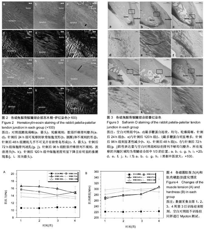
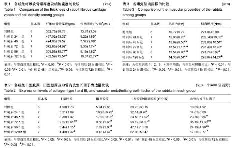
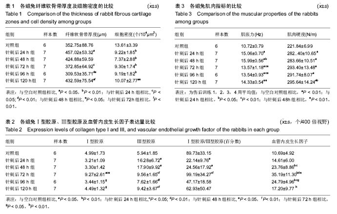
2.1 实验动物数量分析 实验选用新西兰大白兔40只,分为6组,实验过程无脱失,全部进入结果分析。 2.2 组织形态学描述 2.2.1 苏木精-伊红染色 图2显示:空白对照组细胞形态正常,轮廓清晰,有明显的“潮线”,胶原纤维整齐有序、呈波浪状排列(图2a,d);与空白对照组相比,针刺后24 h组细胞轮廓不清,软骨细胞变形,胶原纤维分离、变直(图2b,e);针刺后48 h组潮线消失,出现明显的软骨岛,胶原排列变直消融(图2c,f);针刺后72 h组潮线周围模糊不清,细胞排列零散,胶原纤维不连续(图2g,j);针刺后96 h组潮线虽然可见,但仍能观察到明显的细胞排列紊乱与波浪状排列消失(图2h,k);针刺后120 h组呈现出较规则的骨腱结合部轮廓,但是细胞密度显著低于空白对照组,潮线明显向肌腱方向上涨(图2i,l)。 2.2.2 番红染色 图3显示:空白对照组中黏多糖蛋白沿骨腱结合部均匀分布(图3a,d);与空白对照组相比,针刺后24 h组(图3b,e)与针刺后120h组(图3i,l)明显增多;针刺后48 h组(图3c,f)和针刺后72 h组(图3g,j)结构完整,黏多糖蛋白含量也较为接近空白对照组,针刺后96 h组阳性染色面积则明显减少(图3h,k)。 2.3 组织学/免疫组织化学定量指标 与空白对照组的纤维软骨带厚度相比,针刺后24 h组和针刺后120 h组显著增加(P < 0.01),针刺后72 h组显著低于针刺后24 h组(P < 0.05)但与其他各训练组无显著性差异。最低值出现在针刺后96 h组,其显著低于针刺后24 h组,针刺后48 h和针刺后120 h组(P < 0.01),见表1。 空白对照组软骨细胞密度显著高于针刺后24 h组(P < 0.01),针刺后48 h(P < 0.01),针刺后72 h(P < 0.01),针刺后96 h (P < 0.01)和针刺后120 h组(P < 0.05)。在各实验组中,仅针刺后48 h组低于针刺后120 h组有统计学上的意义(P < 0.05),见表1。 针刺后72 h组的Ⅰ型胶原表达显著高于其他各组(P < 0.01),且其他各组之间无显著性差异。针刺后48 h组的Ⅲ型胶原表达量达到峰值,分别是空白对照组的301%(P < 0.01),针刺后72 h组的187%(P < 0.01),针刺后96 h组的235%(P < 0.01)和针刺后120 h组的190%(P < 0.01)。第二高值出现在针刺后24 h组,也显著高于空白对照组(P < 0.01),针刺后72 h组(P < 0.05),针刺后96 h组(P < 0.01)和针刺后120 h组(P < 0.05)。与空白对照组相比,针刺后24 h组和针刺后48 h组的Ⅰ型胶原与Ⅲ型胶原的比值(后文简述为“胶原比值”)明显偏低(P < 0.05)。针刺后72 h组的胶原比值分别是针刺后24 h组的448%(P < 0.05)及针刺后48 h组的404%(P < 0.05)。针刺后48 h,针刺后72 h和针刺后96 h组的血管内皮生长因子表达量分别是空白对照组的222%,329%和232%(P < 0.01),针刺后72 h组显著高于其他所有组(P < 0.05),见表2。 2.4 肌张力及肌肉硬度 重复测量方差分析结果显示伤后不同时间恢复训练与时间并无交互关系(P > 0.05)。主体间比较显示肌张力随时间变化呈线性特征(P < 0.05),见图4A,主体内比较显示肌张力在各组之间的差异有统计学意义(P < 0.05)。 LSD检验显示针刺后24 h,针刺后48 h,针刺后72 h,针刺后96 h,针刺后120 h组的肌张力分别为空白对照组的140%,149%,127%,126%和134%(P < 0.01)。球形检验及Greenhouse-Geisser校正测试显示肌肉硬度在4周测试中不全相同,在各组间存在显著性差异(P < 0.05)。在伤后训练1周时相对于针刺后24 h和针刺后48 h组,针刺后 96 h和针刺后120 h组的肌肉硬度有明显的升高;但训练4周结束时各伤后训练组之间则未见统计学意义上的差异 (P > 0.05),见表3。 4周训练的肌张力平均值与Ⅲ型胶原存在中等相关关系(r=0.501;P < 0.01),与细胞密度(r=-0.570;P < 0.01)及胶原比值(r=-0.538;P < 0.01)存在负相关,而肌肉硬度与血管内皮生长因子表达量存在中等相关关系(r=0.613; P < 0.01),见图4B。"
"
| [1] Zwerver J,Bredeweg SW, van den Akker-Scheek I. Prevalence of Jumper's knee among nonelite athletes from different sports: a cross-sectional survey.Am J Sports Med. 2011;39:1984-1988.[2] Cassel M,Baur H,Hirschmuller A,et al.Prevalence of Achilles and patellar tendinopathy and their association to intratendinous changes in adolescent athletes. Scand.J Med Sci Sports.2015;25:e310-318.[3] Fisher B,Nyland J,Brand E,et al. Medial patellofemoral ligament reconstruction for recurrent patellar dislocation: a systematic review including rehabilitation and return-to-sports efficacy.Arthroscopy.2010;26:1384-1394.[4] Killian ML,Cavinatto L,Galatz LM,et al. The role of mechanobiology in tendon healing.J Shoulder Elbow Surg. 2012;21:228-237.[5] Frizziero A, Salamanna F, Della Bella E, et al. The Role of Detraining in Tendon Mechanobiology. Front.Aging Neurosci. 016;8:43.[6] Kubo K, Ikebukuro T, Yata H,et al. Time course of changes in muscle and tendon properties during strength training and detraining.J Strength Cond Res. 2010;24:322-331.[7] Nakama LH, King KB, Abrahamsson S, Rempel DM. Evidence of tendon microtears due to cyclical loading in an in vivo tendinopathy model. J Orthop Res. 2005;23: 1199-1205.[8] Deol RS, Roche A, Calder JD. Return to Training and Playing After Acute Lisfranc Injuries in Elite Professional Soccer and Rugby Players.Am J Sports Med. 2016;44:166-170.[9] Calder JD, Sexton SA, Pearce CJ. Return to training and playing after posterior ankle arthroscopy for posterior impingement in elite professional soccer. Am J Sports Med. 2010;38:120-124.[10] Berry CC, Shelton JC, Bader DL,et al. Influence of external uniaxial cyclic strain on oriented fibroblast-seeded collagen gels. Tissue Eng.2003;9:613-624.[11] Thomopoulos S,Kim HM,Rothermich SY,et al.Decreased muscle loading delays maturation of the tendon enthesis during postnatal development.J Orthop Res. 2007;25: 1154-1163.[12] Thomopoulos S, Genin GM, Galatz LM.The development and morphogenesis of the tendon-to-bone insertion - what development can teach us about healing. J. Musculoskelet. Neuronal Interact.2010;10:35-45.[13] Lu H, Chen C, Qu J, et al. Initiation Timing of Low-Intensity Pulsed Ultrasound Stimulation for Tendon-Bone Healing in a Rabbit Model.Am J Sports Med. Am J Sports Med. 2016; 44(10):2706-2715.[14] Dakin SG, Dudhia J, Smith RK. Resolving an inflammatory concept: the importance of inflammation and resolution in tendinopathy.Vet Immunol Immunopathol. 2014;158:121-127.[15] Lin TW, Cardenas L, Soslowsky LJ.Biomechanics of tendon injury and repair. J. Biomech.2004;37:865-877.[16] Thomopoulos S, Williams GR, Soslowsky LJ.Tendon to bone healing: differences in biomechanical, structural, and compositional properties due to a range of activity levels.J Biomech Eng.2003;125:106-113.[17] Esteve-Lanao J, Foster C, Seiler S,et al.Impact of training intensity distribution on performance in endurance athletes.J Strength Cond Res. 2007;21:943-949.[18] Vetter RE, Symonds ML.Correlations between injury, training intensity, and physical and mental exhaustion among college athletes.J Strength Cond Res. 2010;24:587-596.[19] Frizziero A, Fini M, Salamanna F, et al. Effect of training and sudden detraining on the patellar tendon and its enthesis in rats. BMC Musculoskelet.Disord. 2011;12:20.[20] Zhang ZJ, Ng GY, Lee WC, et al. Increase in passive muscle tension of the quadriceps muscle heads in jumping athletes with patellar tendinopathy. Scand J Med Sci Sports. 2016 Aug 19. doi: 10.1111/sms.12749. [21] Silldorff MD, Choo AD, Choi AJ, et al.Effect of supraspinatus tendon injury on supraspinatus and infraspinatus muscle passive tension and associated biochemistry.J Bone Joint Surg Am.2014;96:e175.[22] Wang L, Gao W, Xiong K,et al. The effects of an early return to training on the bone-tendon junction post-acute micro-injury healing.J Sports Sci Med. 2012;11:238-244.[23] Wang L,Gao W,Xiong K,et al.VEGF and BFGF Expression and Histological Characteristics of the Bone-Tendon Junction during Acute Injury Healing. J Sports Sci Med.2014;13:15-21.[24] Wang L, Qin L, Lu HB, et al.Extracorporeal shock wave therapy in treatment of delayed bone-tendon healing. Am J Sports Med.2008;36:340-347.[25] Fu SC, Shum WT, Hung LK, et al. Low-intensity pulsed ultrasound on tendon healing: a study of the effect of treatment duration and treatment initiation. Am J Sports Med. 2008;36:1742-1749.[26] Aird L,Samuel D,Stokes M.Quadriceps muscle tone, elasticity and stiffness in older males: reliability and symmetry using the MyotonPRO. Arch. Gerontol Geriatr. 2012;55:e31-39.[27] Lian OB, Engebretsen L, Bahr R. Prevalence of jumper's knee among elite athletes from different sports: a cross-sectional study. Am J Sports Med.2005;33:561-567.[28] Archambault JM, Hart DA, Herzog W. Response of rabbit Achilles tendon to chronic repetitive loading. Connect Tissue Res.2001;42:13-23.[29] Zhao J, Zhang P, Qin L,et al. Hypoxia is essential for bone-tendon junction healing: the molecular biological evidence.Int Orthop.2011;35:925-928.[30] Krivic A,Majerovic M,Jelic I,et al. Modulation of early functional recovery of Achilles tendon to bone unit after transection by BPC 157 and methylprednisolone. Inflamm Res.2008;57:205-210.[31] Thorpe CT, Chaudhry S, Lei, II, et al.Tendon overload results in alterations in cell shape and increased markers of inflammation and matrix degradation. Scand. J Med Sci Sports.2015;25:e381-391.[32] Farnsworth NL,Antunez LR,Bryant SJ.Influence of chondrocyte maturation on acute response to impact injury in PEG hydrogels.J Biomech.2012;45:2556-2563.[33] Chen CT, Burton-Wurster N, Borden C,et al.Chondrocyte necrosis and apoptosis in impact damaged articular cartilage. J Orthop Res.2001;19:703-711.[34] Sakane M, Mutsuzaki H, Nakajima H, et al. Anterior cruciate ligament insertion after partial tear: histological changes and chondrocyte turnover.Knee Surg Sports Traumatol Arthrosc. 2012;20:102-108.[35] Thomopoulos S, Fomovsky GM, Chandran PL,et al. Collagen fiber alignment does not explain mechanical anisotropy in fibroblast populated collagen gels. J Biomech Eng.2007; 129: 642-650.[36] Tabuchi K, Soejima T, Kanazawa T,et al.Chronological changes in the collagen-type composition at tendon-bone interface in rabbits.Bone Joint Res. 2012;1:218-224.[37] Sahin H, Tholema N, Petersen W,et al. Impaired biomechanical properties correlate with neoangiogenesis as well as VEGF and MMP-3 expression during rat patellar tendon healing.J Orthop Res.2012;30:1952-1957.[38] Molloy T, Wang Y, Murrell G. The roles of growth factors in tendon and ligament healing.Sports Med.2003;33:381-394.[39] Kubo K,Kanehisa H,Kawakami Y,et al.Influences of repetitive muscle contractions with different modes on tendon elasticity in vivo. J ApplPhysiol (1985). 2001;91:277-282.[40] Parent G, Huppe N, Langelier E.Low stress tendon fatigue is a relatively rapid process in the context of overuse injuries. Ann. Biomed. Eng.2011;39:1535-1545.[41] Ditroilo M, Hunter AM, Haslam S,et al. The effectiveness of two novel techniques in establishing the mechanical and contractile responses of biceps femoris. Physiol Meas. 2011;32:1315-1326.[42] Williams HB.The value of continuous electrical muscle stimulation using a completely implantable system in the preservation of muscle function following motor nerve injury and repair:an experimental study.Microsurgery. 1996;17: 589-596. |
| [1] | Tan Xinfang, Guo Yanxing, Qin Xiaofei, Zhang Binqing, Zhao Dongliang, Pan Kunkun, Li Yuzhuo, Chen Haoyu. Effect of uniaxial fatigue exercise on patellofemoral cartilage injury in a rabbit [J]. Chinese Journal of Tissue Engineering Research, 2022, 26(在线): 1-6. |
| [2] | Liu Feng, Feng Yi. Finite element analysis of different Kirschner wire tension bands on transverse patella fractures during gait cycle [J]. Chinese Journal of Tissue Engineering Research, 2022, 26(9): 1367-1371. |
| [3] | Zhang Jinglin, Leng Min, Zhu Boheng, Wang Hong. Mechanism and application of stem cell-derived exosomes in promoting diabetic wound healing [J]. Chinese Journal of Tissue Engineering Research, 2022, 26(7): 1113-1118. |
| [4] | Duan Chao, Shang Xiaoqiang, Duan Xianglin, Yang Ping, Tao Shengxiang. Stability of patellar claw versus loop plate combined with patellar claw for the treatment of comminuted fractures of the lower pole of the patella [J]. Chinese Journal of Tissue Engineering Research, 2022, 26(6): 934-937. |
| [5] | Yang Kuangyang, Wang Changbing. MRI evaluation of graft maturity and knee function after anterior cruciate ligament reconstruction with autogenous bone-patellar tendon-bone and quadriceps tendon [J]. Chinese Journal of Tissue Engineering Research, 2022, 26(6): 963-968. |
| [6] | Li Jie, Zhang Haitao, Chen Jinlun, Ye Pengcheng, Zhang Hua, Zhou Bengen, Zhao Changqing, Sun Youqiang, Chen Jianfa, Xiang Xiaobing, Zeng Yirong. Anterior cruciate ligament rupture and patellofemoral joint stability before sagittal and axial measurement using MRI [J]. Chinese Journal of Tissue Engineering Research, 2022, 26(6): 969-972. |
| [7] | Li Weiming, Xu Qingwen, Li Yijun, Sun Yanbo, Cui Jin, Xu Pengyuan . Deep seawater promotes wound healing in diabetic mice by activating PI3K/Akt pathway [J]. Chinese Journal of Tissue Engineering Research, 2022, 26(5): 724-729. |
| [8] | Zhao Tianyu, Jin Song, Zhang Di, Liu Xiaoxiao, Ma Jiang, Wang Ju. Baduanjin training for patellar tendinopathy in a randomized controlled trial: improving pain, muscle flexibility and lower limb balance stability [J]. Chinese Journal of Tissue Engineering Research, 2022, 26(11): 1662-1668. |
| [9] | Zhao Zixi, Xu Jun, Ding Min, Li Xiwen, Zhang Jinghang, Wang Penghua. Changes of type I and III collagen and matrix metalloproteinase 2 and 9 on the wound of diabetic foot ulcer with external application of medical collagen dressing [J]. Chinese Journal of Tissue Engineering Research, 2022, 26(10): 1544-1550. |
| [10] | Zhou Jihui, Li Xinzhi, Zhou You, Huang Wei, Chen Wenyao. Multiple problems in the selection of implants for patellar fracture [J]. Chinese Journal of Tissue Engineering Research, 2021, 25(9): 1440-1445. |
| [11] | Zhang Zhenkun, Li Zhe, Li Ya, Wang Yingying, Wang Yaping, Zhou Xinkui, Ma Shanshan, Guan Fangxia. Application of alginate based hydrogels/dressings in wound healing: sustained, dynamic and sequential release [J]. Chinese Journal of Tissue Engineering Research, 2021, 25(4): 638-643. |
| [12] | Chen Yu, Yu Zhongxiang, Kuang Yong, Wang Shuqiang, Cai Yuwei, Fang Lei, Xu Shengming. Double-rows technique with suture anchors in eight patients with fracture of distal patellar pole: reliable initial fixation [J]. Chinese Journal of Tissue Engineering Research, 2021, 25(33): 5341-5344. |
| [13] | Lin Xiaodong, Liu Wengang, Xu Xuemeng, Liu Xin, Lu Chao, Song Min, Li Congcong. Unicompartmental knee arthroplasty superior to open-wedge high tibial osteotomy: differences of mechanical parameters and knee function [J]. Chinese Journal of Tissue Engineering Research, 2021, 25(30): 4793-4798. |
| [14] | Wen Xu, Shen Jing. Application of nano-silver antibacterial hydrogel dressing combined with recombinant human basic fibroblast growth factor on wound healing after debridement of open ankle fracture [J]. Chinese Journal of Tissue Engineering Research, 2021, 25(29): 4638-4643. |
| [15] | Xiong Binglang, Lin Tianye, Yang Peng, Xu Jingli, Zou Qizhao, Lai Qizhong, Zhang Qingwen. Visualization of global status and trends in anterior cruciate ligament reconstruction [J]. Chinese Journal of Tissue Engineering Research, 2021, 25(29): 4656-4663. |
| Viewed | ||||||
|
Full text |
|
|||||
|
Abstract |
|
|||||