Chinese Journal of Tissue Engineering Research ›› 2013, Vol. 17 ›› Issue (16): 2859-2868.doi: 10.3969/j.issn.2095-4344.2013.16.002
Previous Articles Next Articles
Bone marrow mesenchymal stem cells seeded into an allogeneic scaffold repair critical-sized iliac defects in sheep
Yang Nan, He Hui-yu, Hu Yang, Yang Chuan-bo
- Department of Prosthodontics, the First Affiliated Hospital of Xinjiang Medical University, Urumqi 830054, Xinjiang Uygur Autonomous Region, China
-
Received:2013-01-14Revised:2013-01-22Online:2013-04-16Published:2013-05-24 -
Contact:He Hui-yu, Master’s supervisor, Professor, Department of Prosthodontics, the First Affiliated Hospital of Xinjiang Medical University, Urumqi 830054, Xinjiang Uygur Autonomous Region, China hehuiyu02@sina.com -
About author:Yang Nan★, Studying for master’s degree, Department of Prosthodontics, the First Affiliated Hospital of Xinjiang Medical University, Urumqi 830054, Xinjiang Uygur Autonomous Region, China yangnan8513@sina.com -
Supported by:National Natural Science Foundation of China, No. 81060088*; Natural Science Foundation of Xinjiang Uygur Autonomous Region, No. 2011211A073*

CLC Number:
Cite this article
Yang Nan, He Hui-yu, Hu Yang, Yang Chuan-bo. Bone marrow mesenchymal stem cells seeded into an allogeneic scaffold repair critical-sized iliac defects in sheep[J]. Chinese Journal of Tissue Engineering Research, 2013, 17(16): 2859-2868.
share this article
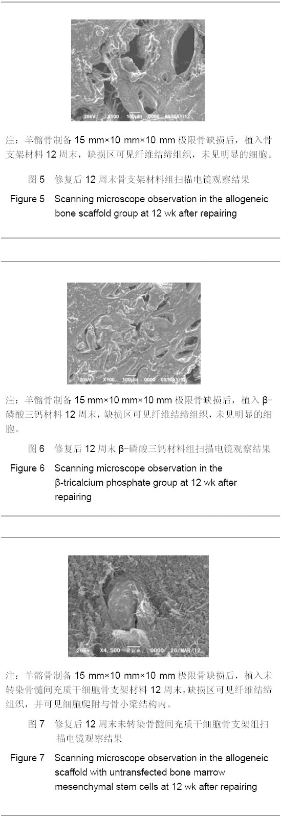
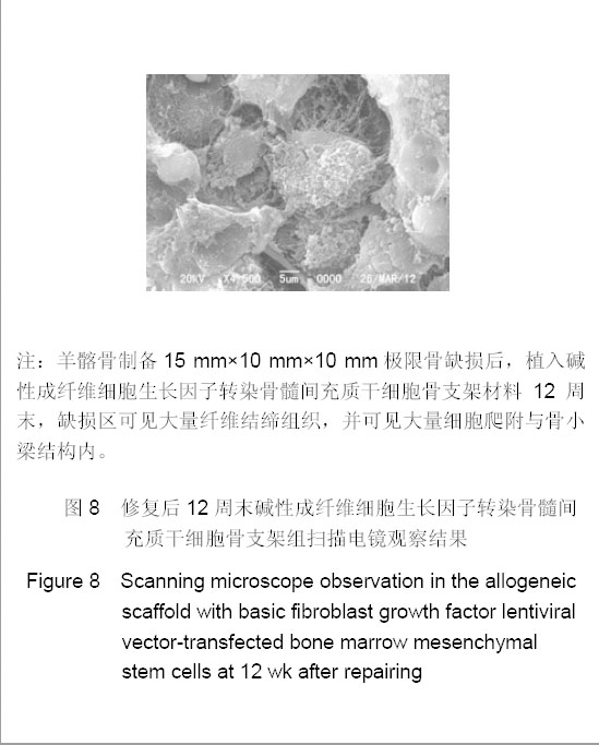
2.1 实验动物数量分析 羊双侧髂骨各制造2处缺损,将4组材料同时植入,9只羊于实验4,8,12周末每次处死3只,全部进入结果分析,无脱失。 2.2 大体观察结果 术后4,8,12周观察动物,无一跛行,感染或死亡。 4,8,12周末随机处死3只动物,将植骨区扩大5 mm电钻锯断取出,去尽软组织,观察标本发现12周末骨支架组材料完全被骨膜覆盖,术区充满结缔组织生长,植骨区硬度明显下降;β-磷酸三钙材料完全被骨膜覆盖,术区不平整,缺损凸起表面修复,硬度变化小;骨髓间充质干细胞复合骨支架组标本完全被骨膜覆盖,表面充满结缔组织,植骨区特别是中央部位硬度也有明显下降;碱性成纤维细胞生长因子转染组术区完全被骨膜覆盖,术区充满结缔组织,缺损修复区硬度与周围基本一致,骨皮质表面平整无凹陷或凸起。 2.3 扫描电镜结果 4,8,12周末处死动物,将植骨区扩大5 mm电钻锯断取出,去尽软组织,扫描各组不同材料植骨区。发现骨支架组结合区有纤维化,基本无细胞爬入支架骨小梁内,类似死骨结构。β-磷酸三钙材料组无明显支架结构存在,在结合区有大量纤维化,中央区无细胞及纤维结缔组织爬入。骨髓间充质干细胞复合骨支架组骨小梁结构内有少量细胞爬入,但中央区仍为死骨结构。碱性成纤维细胞生长因子转染组植骨区有大量细胞,细胞形态饱满,细胞伸出伪足,还可见纤维结缔组织覆盖骨支架空隙,见图5-8。"
"
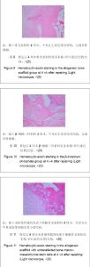
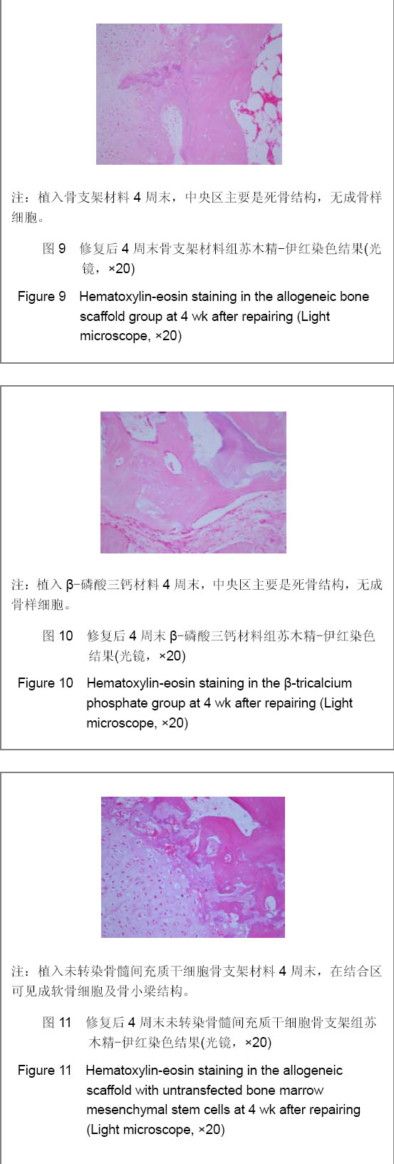
2.4 苏木精-伊红染色结果 12周末处死动物,将植骨区扩大5 mm电钻锯出,去尽软组织,将标本置于体积分数4%甲醛溶液中(pH=7.4,4 ℃)固定24 h,2%EDTA脱钙2-4周,梯度乙醇脱水,石蜡包埋后切片,切片厚度5 μm,进行苏木精-伊红染色,光镜下观察。 4周末,骨支架组、β-磷酸三钙材料组主要为死骨结构,术区材料周围有成纤维细胞包绕;未转染细胞组结合区可见成软骨样结构,中央区为死骨结构,见图9-12。 8周末,骨支架组、β-磷酸三钙材料组主要为死骨结构,术区结合部位有成软骨化表现;未转染细胞组在手术结合区有成软骨样结构及成骨样细胞出现,中央区为死骨结构;而碱性成纤维细胞生长因子转染组可见手术结合区及支架空隙周围有成软骨样结构及成骨样细胞出现,内有成骨样细胞排列。 12周末,骨支架组、β-磷酸三钙材料组术区可见成纤维细胞,结合区为成软骨化表现;未转染细胞组结合区有成软骨样结构及骨小梁出现,中央区少量成纤维细胞;而碱性成纤维细胞生长因子转染组可见手术结合区及支架空隙周围有大量成软骨样结构及成骨样细胞出现,结合区可见骨小梁结构。 "

"

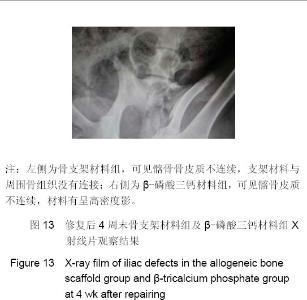
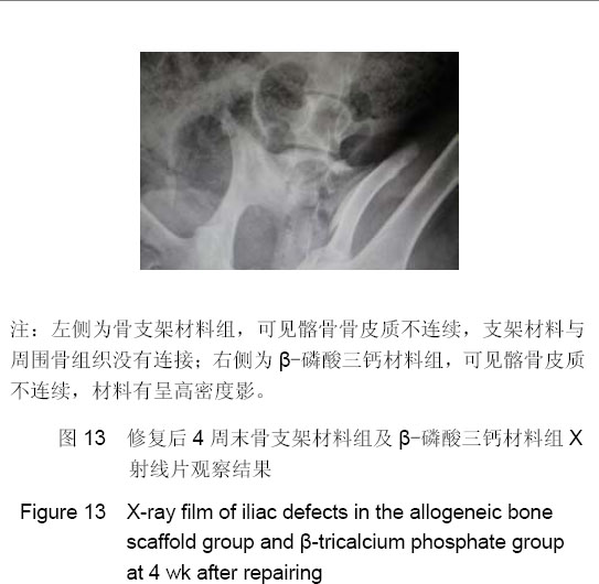
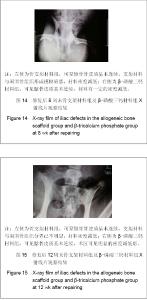
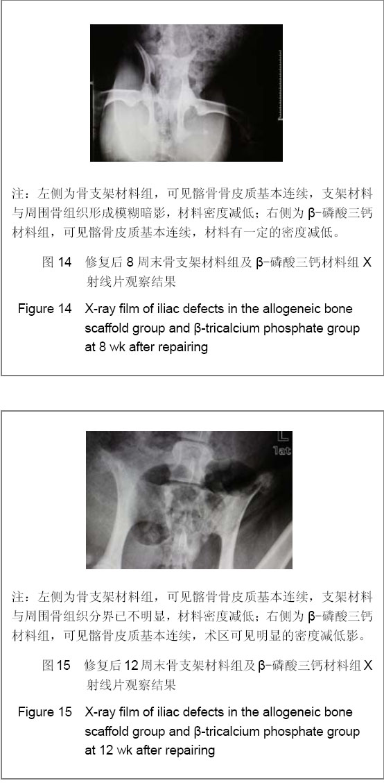
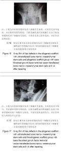
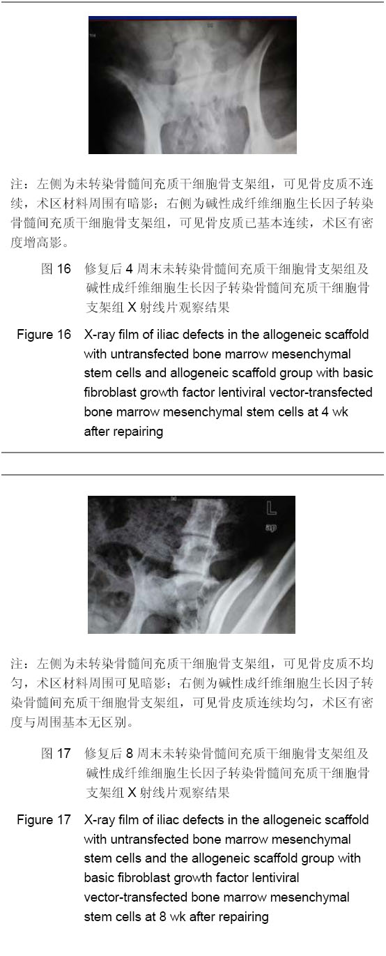
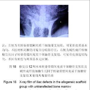
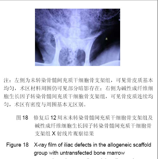
2.5 X射线片观察结果 在4,8,12周末处死动物前,将动物人工固定,进行数字化DR片检查,采用荷兰 PhilipsDigital diagnost 床式数字图像增强器,配备电脑DAP显示,采用自动曝光控制中心电离室技术,400速自动曝光。 操作参考文献[22]进行。动物摄片体位为骨盆正位。采用西门子PACS影像信息管理系统,分辨率为4 096× 2 560,见图13-18。 由X射线摄片检查结果显示: 4周时,转染组术区影密度最高,骨皮质基本有增生影,术区骨密度均匀,并高于其他3组;未转染细胞组相比单纯骨支架组、β-磷酸三钙材料组术区密度较均匀;单纯骨支架材料组、β-磷酸三钙材料组可见明显的材料植入区与周边缺损影形成灰度差,说明材料仍未降解及吸收。 8周时,术区影密度:转染组>未转染细胞组>骨支架组>β-磷酸三钙材料组,转染组术区骨皮质已基本连续;而骨支架材料组与β-磷酸三钙材料组术区骨皮质仍有不平,单纯骨支架材料组、β-磷酸三钙材料组可见明显的材料植入区与周边缺损影仍有灰度差,说明材料开始降解但还未充分吸收。 12周时,术区影密度:转染组>未转染细胞组>骨支架组>β-磷酸三钙材料组;转染组骨皮质连续,均匀;而细胞组密度影像基本均匀,骨皮质连续;骨支架材料组、β-磷酸三钙材料组出现明显术区骨密度影减低,证明在12周末,转染组良好地修复了缺损区,而骨支架材料组材料出现了明显降解和吸收,大于β-磷酸三钙材料组的降解和吸收。 "
"
"
"
| [1]Vacanti JP, Langer R. Tissue engineering: The design and fabrication of living replacement devices for surgical reconstruction and transplantation. Lancet. 1999;354(Suppl 1):SI32-SI34. [2]Rozalia D, George I M, Giorgio MC, et al. The role of barrier membranes for guided bone regeneration and restoration of large bone defects: current experimental and clinical evidence. BMC Medicin. 2012; 10: 81. [3]Aronson J. Limb-lengthening, skeletal reconstruction, and bone transport with the Ilizarov method. J Bone Joint Surg Am. 1997;79(8):1243-1258.[4]Muschler GF, Nakamoto C, Griffith LG. Engineering principles of clinical cell-based tissue engineering. J Bone Joint Surg Am. 2004;86-A:1541-1558.[5]Li F, Wang X, Niyibizi C. Distribution of single-cell expanded marrow derived progenitors in a developing mouse model of osteogenesis imperfecta following systemic transplantation. Stem Cells. 2007;25:3183-3193.[6]Mankani MH, Kuznetsov SA, Wolfe RM, et al. In vivo bone formation by human bone marrow stromal cells: reconstruction of the mouse calvarium and mandible. Stem Cells. 2006;24:2140-2149.[7]Arthur A, Zannettino A, Gronthos S. The therapeutic applications of multipotential mesenchymal/stromal stem cells in skeletal tissue repair. J Cell Physiol. 2009;218:237-245. [8]Enzmann GU, Benton RL, Talbott JF, et al. Whittemore SR. Functional considerations of stem cell transplantation therapy for spinal cord repair. J Neurotrauma. 2006;23:479-495. [9]Chen J, Li Y, Katakowski M, et al. Intravenous bone marrow stromal cell therapy reduces apoptosis and promotes endogenous cell proliferation after stroke in female rat. J Neurosci Res. 2003;73:778-786.[10]Widenfalk J, Lundströmer K, Jubran M, et al. Neurotrophic factors and receptors in the immature and adult spinal cord after mechanical injury or kainic acid. J Neurosci. 2001;21: 3457-3475. [11]Mocchetti I, Wrathall JR. Neurotrophic factors in central nervous system trauma. J Neurotrauma. 1995;12:853–870. [12]Bennett J. Immune response following intraocular delivery of recombinant viral vectors. Gene Ther. 2003;10(11):977-982.[13]Xu HF, He HY, Tang XX, et al. Zhongguo Zuzhigongcheng yanjiu yu Linchuang Kangfu. 2011;15(47): 8749-8752.许慧芬,何惠宇,唐小雪,等. 不同方法制备异种骨支架材料的生物相容性评价[J].中国组织工程研究与临床康复,2011,15(47): 8749-8752.[14]Berger MG, Veyrat-Masson R, Rapatel C, et al.Cell culture medium composition and translational adult bone marrow-derived stem cell research. Stem Cells. 2006;24(12):2888-2990.[15]Dimarakis I,Levicar N. Cell culture medium composition and translational adult bone marrow-derived stem cell research. Stem Cells. 2006;24(5):1407-1408.[16]Tang XX, He HY, Xu HF, et al. Zhongguo Zuzhi Gongcheng Yanjiu. 2011;15(49): 9137-9140.唐小雪,何惠宇,许慧芬,等.两种不同分离方法对羊骨髓间充质干细胞生物活性的影响[J].中国组织工程研究与临床康复.2011; 15(49): 9137-9140.[17]Hu Y, Ma Y, He HY. Zhongguo Zuzhi Gongcheng Yanjiu yu Linchuang Kangfu. 2011;15(20): 3653-3656.胡杨,马莹,何惠宇. 兔下颌骨前牙区剩余牙槽嵴模型的建立[J].中国组织工程研究与临床康复,2011,15(20): 3653-3656.[18]Itaka K,Ohba S,Miyata K,et al. Bone regeneration by regulated invivo gene transfer using biocompatible polyplex nanomicelles. Mol Ther. 2007;15(9):1655-1662. [19]Ivkovic A,Pascher A,Hudetz D,et al. Current concepts in gene therapy of the musculoskeletal system. Acta Chir Orthop Traumatol Cech. 2006;73(2):115-122.[20]Tsuda H,Wada T,Yamashita T,et al. Enhanced osteoinduction by mesenchymal stem cells transfected with a fiber-mutant adenoviral BMP2 gene. Gene Med. 2005;7(10):1322-1334.[21]Xu HF, He HY, Tang XX. Zhongguo Zuzhi Gongcheng Yanjiu. 2012;16(6):958-962.许慧芬,何惠宇,唐小雪.物理联合化学或化学方法处理去抗原异种松质骨支架与骨髓间充质干细胞的细胞相容性[J].中国组织工程研究,2012,16(6):958-962.[22]Guo H, Zhang TL, Nuer, et al. Zhongguo Xunzheng Yixue Zazhi. 2011;11(10):1129-1132.郭辉,张铁亮,努尔,等. 优化婴幼儿胸部DR图像质量和辐射剂量的前瞻性临床研究[J].中国循证医学杂志,2011,11(10): 1129-1132.[23]Chen T,Li NY. Shandong Yiyao. 2009;49(7):27-29.陈涛,李宁毅. 血管化复合组织工程骨的构建及在实验性下颌骨缺损修复中的应用[J].山东医药,2009,49(7):27-29.[24]Xing H,Chen XM,Zhang HQ. Shengwu Guke Cailiao yu Linchuang Yanjiu. 2004;1(5):35-39.邢辉,陈晓明,张宏泉.骨组织工程支架材料[J].生物骨科材料与临床研究,2004,1(5):35-39.[25]Mastrogiacomo M, Muraglia A, Komlev V, et al. Tissue engineering of bone:search for a better scaffold. Ortho Craniofac Res. 2005;8(4):277-284.[26]Zheng L,Wang Q,Pei GX. Zhonghua Waike Zazhi. 2000; 38(10): 745-748.郑磊,王前,裴国献.骨组织工程中理想细胞外基质材料的选择[J].中华外科杂志,2000,38(10):745-748.[27]Simon CG,Khatri CA,Wight SA,et al. Preliminary report on the biocompatibility of a moldable,resorbable,composite bone graft consisting of calcium phosphate cement and poly(lactide-co-glycolide) microspheres. Orthop Res. 2002; 20:473-482.[28]Ni S,Chang J.In vitro degradation,bioactivity,and cytocompatibility of calcium silicate,dimagnesium silicate,and tricalcium phosphate bioceramics. J Biomater Appl. 2009; 24(2):139-158.[29]Erdemli O,Captug O,Bilgili H,et al.In vitro and in vivo evaluation of the effects of demineralized bone matrix or calcium sulfate addition to polycaprolactone bioglass composites.J Mater Sci Mater Med. 2010;21(1):295-308.[30]Jahn K,Braunstein V,Furlong PI,et al.A rapid method for the generation of uniform acellular bone explants.J Orthop Surg Res. 2010;10(5):28-32.[31]Connolly JF. Injectable bone marrow preparations to stimulate osteogenic repair. Clin Orthop. 1995; (313):8-18.[32]Joyner CJ, Bennett A, Triffitt JT. Identification and enrichment of human osteoprogenitor cells by using differentiation stage - specific monoclonal antibodies. Bone. 1997;21:1-6.[33]Li CM,Wang X,Liu BL,et al. Shiyong Kouqiang Yixue Zazhi. 2009;25(1):26-30.李春明,王鑫,刘宝林,等.种植体+ bFGF基因修饰骨髓基质细胞复合Bio2Oss胶原修复犬颌骨缺损的实验研究[J].实用口腔医学杂志,2009,25(1):26-30.[34]Wang L, Zou D, Zhang S, et al. Repair of bone defects around dental implants with bone morphogenetic protein/fibroblast growth factor-loaded porous calcium phosphate cement: a pilot study in a canine model. Clin Oral Impl Res. 2011; 22: 173-181.[35]Fan XQ.Xiao CW,Zhou HF,et al. Zhonghua Yanke Zazhi. 2009; 45(1):66-72.范先群,肖彩雯,周慧芳,等.基因修饰的组织工程骨修复眶骨缺损的实验研究[J].中华眼科杂志,2009,45(1):66-72.[36]Cheng T,Li NY. Shandong Yiyao. 2009;49(7):27-29.陈涛,李宁毅. 血管化复合组织工程骨的构建及在实验性下颌骨缺损修复中的应用[J].山东医药,2009,49(7):27-29.[37]Bonab MM, Alimoghaddam K, Talebian F, et al.Aging of mesenchymal stem cell in vitro. BMC Cell Biology. 2006;7:14. [38]Kotev-Emeth S, Pitaru S, Pri-Chen S, et al. Establishment of a rat long-term culture expressing the osteogenic phenotype: dependence on dexamethasone and FGF-2. Connect Tissue Res. 2002;43(4):606-612. |
| [1] | Tao Daiju, Su Haiyu, Wang Yuqi, Shen Zhiqiang, He Bo . Construction and identification of stable PC12 cell lines with high/low expression of miR-122-5p [J]. Chinese Journal of Tissue Engineering Research, 2026, 30(7): 1790-1799. |
| [2] | Yu Shiyu, Yu Sutong, Xu Yang, Zhen Xiangyan, Han Fengxuan. Advances in research and application of tissue engineering therapeutic strategies in oral submucous fibrosis [J]. Chinese Journal of Tissue Engineering Research, 2026, 30(4): 936-948. |
| [3] | Xu Hongtao, Wang Jianping, Xu Yuehong, Li Qin, Qi Qihua, Xia Qipeng. Asiaticoside promotes osteogenic differentiation in osteoporotic rats [J]. Chinese Journal of Tissue Engineering Research, 2026, 30(35): 9113-9119. |
| [4] | Chen Yida, Cheng Xinyi, Zhao Huan, Zhou Xichao, Gu Qiaoli, Lin Xiao, Shi Qin. Effects of physiological osmotic pressure on chondrocyte differentiation and extracellular matrix metabolism [J]. Chinese Journal of Tissue Engineering Research, 2026, 30(35): 9143-9150. |
| [5] | Wang Quan, Tang Zhenglong. Visualization analysis of literature on medication-related osteonecrosis of the jaw [J]. Chinese Journal of Tissue Engineering Research, 2026, 30(35): 9327-9335. |
| [6] | Liu Hongwei, Chang Lungji. Lentivirus-mediated gene therapy in a beta-thalassemia mouse model [J]. Chinese Journal of Tissue Engineering Research, 2026, 30(34): 8978-8985. |
| [7] | Li Xuanze, Fang Hanhong, Xu Zhe. Preparation and cytocompatibility of sodium alginate-hydroxyapatite-graphene oxide hydrogels [J]. Chinese Journal of Tissue Engineering Research, 2026, 30(32): 8420-8426. |
| [8] | Jin Yuan, Zhou Jiabing. Applications and advances of hydrogels in bone tissue engineering repair related to sports injuries [J]. Chinese Journal of Tissue Engineering Research, 2026, 30(32): 8512-8520. |
| [9] | Xia Jinyan, Su Meifang, Yang Wenyu, Hu Qilan, Gong Li. Application of novel materials in tissue repair of diabetic foot wounds [J]. Chinese Journal of Tissue Engineering Research, 2026, 30(32): 8563-8568. |
| [10] | Yang Zishu, Yang Jian, Huang Cuiyuan, Cheng Chen, Wu Di, Zhang Jing. Construction and identification of RNA interference lentiviral vector for rat disruptor of telomeric silencing 1-like gene [J]. Chinese Journal of Tissue Engineering Research, 2026, 30(31): 8197-8204. |
| [11] | Liu Lei, Mao Wen, Qi Fu, Li Hong, Zhang Minghuan. Hypoxia preconditioning of bone marrow mesenchymal stem cells promotes angiogenesis in human umbilical vein endothelial cells through paracrine cytokines [J]. Chinese Journal of Tissue Engineering Research, 2026, 30(31): 8047-8053. |
| [12] | Zhou Zixiang, Zhao Baoxiang. Research progress in the relationship between nontraumatic necrosis of the femoral head and lipid metabolism and its treatment [J]. Chinese Journal of Tissue Engineering Research, 2026, 30(3): 680-690. |
| [13] | Yang Hu, Zheng Yu, Jia Chengming, Wang Tong, Zhang Guangfei, Ji Yaoyao. Immune microenvironment regulates bone regeneration [J]. Chinese Journal of Tissue Engineering Research, 2026, 30(3): 701-710. |
| [14] | Hu Yingnan, Shi Wanwan, Wang Datao. Blood cells and the occurrence and progression of osteoporosis: biomarkers and emerging therapeutic strategies [J]. Chinese Journal of Tissue Engineering Research, 2026, 30(29): 7632-7638. |
| [15] | Yang Guang, Yin Zhitao, Xu Yan. Three-dimensional printed isoniazid liposome photothermal composite scaffolds and their performance evaluation [J]. Chinese Journal of Tissue Engineering Research, 2026, 30(26): 6701-6709. |
| Viewed | ||||||
|
Full text |
|
|||||
|
Abstract |
|
|||||