Chinese Journal of Tissue Engineering Research ›› 2013, Vol. 17 ›› Issue (12): 2091-2099.doi: 10.3969/j.issn.2095-4344.2013.12.001
Self-made Fe3O4 versus gelatin sponge embolic agents in rabbit pulmonary microcirculation
Yang Qing, Tong Yu-yun, Guo Li, Wang Jia-ping, Li Ying-chun, Jiang Hua
- Department of Interventional Radiology, Second Hospital of Kunming Medical University, Kunming 650101, Yunnan Province, China
-
Received:2012-12-12Revised:2013-02-05Online:2013-03-19Published:2013-04-27 -
Contact:Tong Yu-yun, Master, Attending physician, Department of Interventional Radiology, Second Hospital of Kunming Medical University, Kunming 650101, Yunnan Province, China 1012580@qq.com -
About author:Yang Qing☆, Doctor, Attending physician, Department of Interventional Radiology, Second Hospital of Kunming Medical University, Kunming 650101, Yunnan Province, China yq7119@hotmail.com

CLC Number:
Cite this article
Yang Qing, Tong Yu-yun, Guo Li, Wang Jia-ping, Li Ying-chun, Jiang Hua. Self-made Fe3O4 versus gelatin sponge embolic agents in rabbit pulmonary microcirculation[J]. Chinese Journal of Tissue Engineering Research, 2013, 17(12): 2091-2099.
share this article
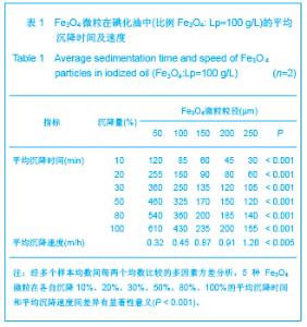
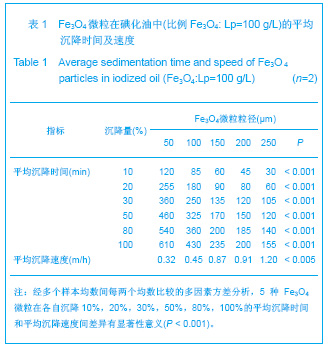
2.1 实验动物数量分析 实验共用兔21只,其中1只行肺血管铸型,实验过程中死亡12只,死亡兔经解剖排除与异位栓塞相关,死因与麻醉过量、感染相关,及时补充实验兔达实验组规定数目,栓塞成功,最终计入结果分析兔共20只。 2.2 Fe3O4微粒体外形态 Fe3O4微粒加入碘化油中,初期微粒聚集呈大小不等团块沉于碘化油底部。充分振荡后,磁微粒团块离散,微粒呈均匀、分散状悬浮于碘化油中,然后缓慢沉降。用注射器推注可顺利通过直径为3-6F的动脉导管。5种规格及不同比例的Fe3O4微粒在碘化油中的悬浮情况见表1。5种Fe3O4微粒在各自沉降10%,20%,30%,50%,80%,100%的平均沉降时间和平均沉降速度间差异有显著性意义(P < 0.001)。"
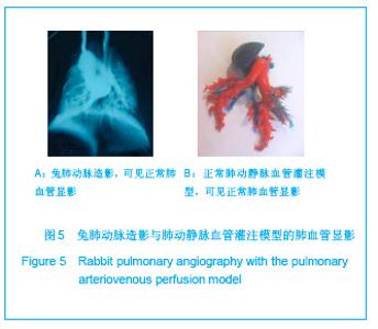
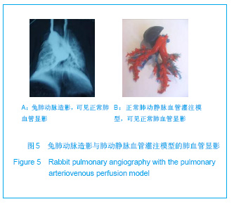
2.3 正常兔肾动脉造影表现及血管铸型 每只实验用兔在栓塞前,经兔耳缘静脉行右心室造影,双侧肺动脉均无狭窄、闭塞、畸形等异常表现,血管铸型在解剖显微镜下能观察到细小血管,见图5。应用计算机病理图像定量分析同样方法,测量兔肺动脉第四五级分支直径在[368±42.5E (SEM=0.23)] μm兔肺静脉第四五级分支直径在[467±62.5E (SEM=0.26)] μm。"
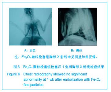
2.4 实验兔栓塞后胸部X射线平片、数字减影血管造影复查结果 兔栓塞后1,2,4周,胸部X射线平片未见异常征像,见图6。"
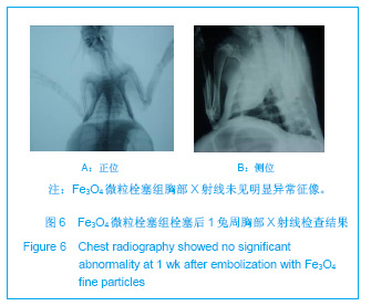
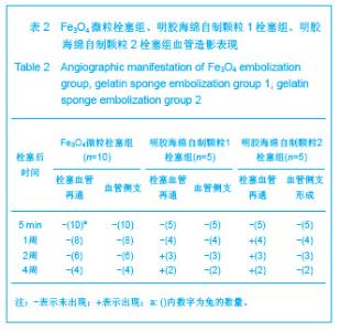
数字减影血管造影复查:Fe3O4微粒栓塞组:栓塞后1,2,4周,栓塞后血管缺损区未见恢复,未见血管侧支形成。明胶海绵自制颗粒1栓塞组:栓塞后1,2周栓塞后血管缺损区未见恢复,未见血管侧支形成。第4周栓塞后血管缺损区部分再通,未见血管侧支形成。明胶海绵自制颗粒2栓塞组:栓塞后第1周栓塞后血管缺损区未见恢复,未见血管侧支形成。第2周栓塞后血管缺损区部分再通,部分血管侧支形成。第4周栓塞后血管缺损大部分再通。见表2。"
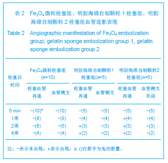
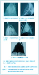
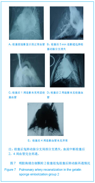
栓塞后5 min数字减影血管造影造影结果显示,Fe3O4微粒栓塞组均显示实验动物兔肺动脉主干存留,部分肺动脉分支消失,血流中断,兔无明显异常表现。明胶海绵自制颗粒1栓塞组均显示实验动物兔肺动脉主干存留,超选择栓塞肺动脉分支消失,血流中断,兔无明显异常表现。明胶海绵自制颗粒2栓塞组显示实验动物兔肺动脉主干存留,超选择栓塞动脉分支消失,血流中断,兔无明显异常表现,栓塞后1-4周造影复查,见图7。"
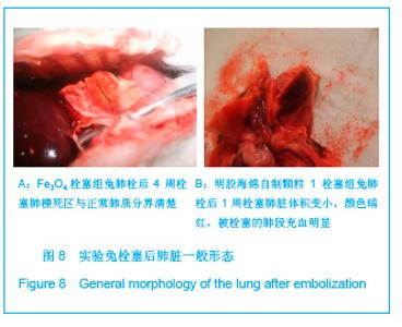
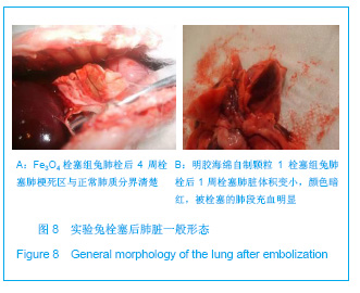
2.5 实验兔栓塞后1,2,4周肺组织病理分析结果 大体观察结果:Fe3O4微粒栓塞组兔肺栓塞后1周(2只),栓塞肺脏体积变小,颜色暗红,被栓塞的肺段充血明显。2周(2只)、4周(6只),栓塞肺脏,表面有散在出血点,质地变韧,实质出现梗死区域,梗死区与正常肺质分界清楚。明胶海绵自制颗粒1栓塞组兔栓塞肺动脉后1周(1只) ,栓塞肺体积稍增大,颜色深红,2周时(1只),栓塞肺体积肺恢复正常,未见有明显梗死区域。4周时(2只),仍未见有明显梗死改变。明胶海绵自制颗粒2栓塞组兔栓塞肺动脉后1周(1只)栓塞肺体积稍增大,颜色正常,2周时(1只),栓塞肺体积肺恢复正常,未见有明显梗死区域。4周时(2只),仍未见有明显梗死改变。见图8。"
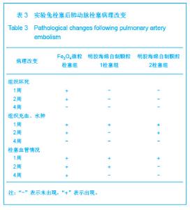
光镜下兔肺栓塞组织病理改变:见表3。"
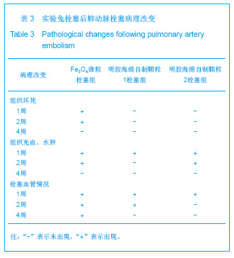
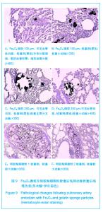
Fe3O4微粒栓塞组兔肺栓塞后1,2,4周,苏木精-伊红染色示50-250 μm的Fe3O4微粒:黑色Fe3O4微粒栓塞肺细小血管内,在较大血管中颗粒互相集聚成团,较小血管中偶可见单颗粒,各组颗粒栓塞血管程度不同,50 μm组栓塞可见少量位于肺泡周毛细血管及毛细血管前小动脉,多数集聚于小动脉内,栓塞彻底。100 μm粒径很少栓塞毛细血管,常五六个视野内未见栓塞毛细血管,毛细血管前小动脉见栓塞,多数集聚于5级分支小动脉内,150-250 μm栓塞范围在小动脉四五级小动脉内。明胶海绵自制颗粒1栓塞组兔栓塞肺动脉后1周(1只),可见3级大动脉及4级小动脉内栓塞剂栓塞,远端小动脉未见栓塞。明胶海绵自制颗粒2栓塞组基本与明胶海绵自制颗粒1栓塞组相同。Fe3O4微粒栓塞组可见局部组织坏死,其他两组未见异常。见图9。"
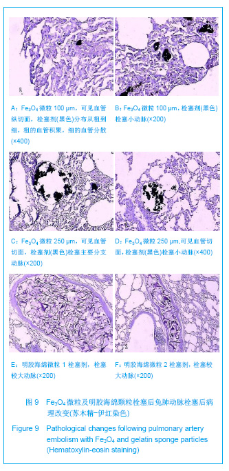

50 μm粒径Fe3O4微粒能栓塞直径50-500 μm的毛细血管及毛细血管前小动脉,多集中在直径100- 200 μm血管范围里;100-200 μm Fe3O4微粒能栓塞直径150-700 μm毛细血管前小动脉,多集中在直径200-400 μm的毛细血管前小动脉范围里,250 μm Fe3O4能栓塞直径250-1 500 μm小动脉,多集中在350-700 μm的小动脉范围里。明胶海绵颗粒1 能栓塞(1 300±348) μm小动脉,明胶海绵颗粒2能栓塞 (1 263±436) μm小动脉。各种粒径Fe3O4微粒间差距有显著性意义(P < 0.05)。"

| [1] Bilbao JI, Martínez-Cuesta A, Urtasun F. Complications of embolization. Semin Intervent Radiol. 2006;23(2):126-142. [2] Walsh KP. Advanced embolization techniques. Pediatr Cardiol. 2005;26(3):275-288.[3] Al-Shahi R, Warlow C. Arteriovenous malformations of the brain: ready to randomisel. J Neurol Neurosurg Psychiatry. 2005;76(10):1327-1329[4] White RI Jr. Pulmonary arteriovenous malformations: how do I embolize? Tech Vasc Interv Radiol. 2007;10(4):283-290. [5] Kjeldsen AD, Andersen PE, Oxhøj H, et al. Percutaneous transluminal embolization of pulmonary arteriovenous malformations. Ugeskr Laeger. 1998;160(10):1465-1469.[6] Vaidya S, Kathleen R. An overview of embolic agents. Semin Intervent Radiol. 2008;25(3):204-215. [7] Becker TA, Preul MC, Bichard WD, et al. Calcium alginate gel as a biocompatible material for endovascular arteriovenous malformation embolization: six-month results in an animal model. Neurosurgery. 2005;56:793-801. [8] Becker TA, Preul MC, Bichard WD, et al. Preliminary investigation of calcium alginate gel as a biocompatible material for endovascular aneurysm embolization in vivo. Neurosurgery. 2007;60:1119-1128. [9] Vanninen R L, Manninen I. Onyx, a new liquid embolic material for peripheral interventions: preliminary experience in aneurysm, pseudoaneurysm, and pulmonary arteriovenous malformation embolization. Cardiovasc Intervent Radiol. 2007; 30(2):196-200. [10] Raffi L, Simonetti L, Cenni P, et al. Use of Glubran 2 acrylic glue in interventional neuroradiology. Neuroradiology. 2007; 49:829-836. [11] Pollak JS, White RI. The use of cyanoacrylate adhesives in peripheral embolization. J Vasc Interv Radiol. 2001;12: 907-913. [12] Osuga K, Mikami K, Higashihara H, et al. Principles and techniques of transcatheter embolotherapy for peripheral vascular lesions. Radiat Med. 2006;24(4):309-314. [13] Spies J B, Cornell C, Worthington-Kirsch R, et al. Long-term outcome from uterine fibroid embolization with tris-acryl gelatin microspheres: results of a multicenter study. J Vasc Interv Radiol. 2007;18(2):203-207. [14] Becker TA, Kipke DR, Brandon T. Calcium alginate gel: a biocompatible and mechanically stable polymer for endovascular embolization. J Biomed Mater Res. 2001; 54(1):76-86. [15] López-Benítez R, Richter GM, Kauczor HU, et al. Analysis of nontarget embolization mechanisms during embolization and chemoembolization procedures. Cardiovasc Intervent Radiol. 2009;32(4):615-622. [16] Hidaka K, Nakamura M, Osuga K, et al. Elastic characteristics of microspherical embolic agents used for vascular interventional radiology. 2010;3(7):497-503.[17] Hidaka K, Moine L, Collin G, et al. Elasticity and viscoelasticity of embolization microspheres. J Mech Behav Biomed Mater. 2011;4(8):2161-2167.[18] Yoon W. Embolic agents used for bronchial artery embolisation in massive haemoptysis. Expert Opin Pharmacother. 2004;5(2):361-367.[19] Loffroy R, Guiu B, Cercueil JP, et al. Endovascular therapeutic embolisation: an overview of occluding agents and their effects on embolised tissues. Curr Vasc Pharmacol. 2009;7(2):250-263.[20] Lewis AL, Adams C, Busby W etal.Comparative in vitro evaluation of microspherical embolisation agents. J Mater Sci Mater Med. 2006;17(12):1193-1204.[21] Stampfl S, Bellemann N, Stampfl U, et al. Arterial distribution characteristics of Embozene particles and comparison with other spherical embolic agents in the porcine acute embolization model. J Vasc Interv Radiol. 2009;20(12): 1597-607. [22] Stampfl S, Bellemann N, Stampfl U, et al. Inflammation and recanalization of four different spherical embolization agents in the porcine kidney model. J Vasc Interv Radiol. 2008;19(4): 577-586. [23] Bilbao JI, de Luis E, García de Jalón JA, et al. Comparative study of four different spherical embolic particles in an animal model: a morphologic and histologic evaluation. J Vasc Interv Radiol. 2008;19(11):1625-1638. [24] Abada HT, Golzarian J. Gelatine sponge particles: handling characteristics for endovascular use. Tech Vasc Interv Radiol. 2007;10(4):257-260. [25] White RI Jr, Pollak JS, Wirth JA. Pulmonary arteriovenous malformations: diagnosis and transcatheter embolotherapy. J Vasc Interv Radiol. 1996;7(6):787-804.[26] Gulati MS, Srinivasan A, Paul SB, et al. Uterine restoration following fibroid expulsion after uterine artery embolisation using gelfoam. J Postgrad Med. 2004;50(1):80.[27] Katsumori T, Kasahara T, Akazawa K. Long-term outcomes of uterine artery embolization using gelatin sponge particles alone for symptomatic fibroids. AJR Am J Roentgenol. 2006; 186(3):848-854.[28] The Ministry of Science and Technology of the People’s Republic of China. Guidance Suggestions for the Care and Use of Laboratory Animals. 2006-09-30. |
| [1] | Sun Yaotian, Xu Kai, Wang Peiyun. Potential mechanisms by which exercise regulates iron metabolism in immune inflammatory diseases [J]. Chinese Journal of Tissue Engineering Research, 2026, 30(6): 1486-1498. |
| [2] | Yu Shiyu, Yu Sutong, Xu Yang, Zhen Xiangyan, Han Fengxuan. Advances in research and application of tissue engineering therapeutic strategies in oral submucous fibrosis [J]. Chinese Journal of Tissue Engineering Research, 2026, 30(4): 936-948. |
| [3] | Li Lei, Zhao Qisheng. Effects of exercise intervention on cortical excitability and motor performance in healthy populations: a meta-analysis based on transcranial magnetic stimulation measurements [J]. Chinese Journal of Tissue Engineering Research, 2026, 30(35): 9302-9308. |
| [4] | Wang Quan, Tang Zhenglong. Visualization analysis of literature on medication-related osteonecrosis of the jaw [J]. Chinese Journal of Tissue Engineering Research, 2026, 30(35): 9327-9335. |
| [5] | Jin Yuan, Zhou Jiabing. Applications and advances of hydrogels in bone tissue engineering repair related to sports injuries [J]. Chinese Journal of Tissue Engineering Research, 2026, 30(32): 8512-8520. |
| [6] | Xia Jinyan, Su Meifang, Yang Wenyu, Hu Qilan, Gong Li. Application of novel materials in tissue repair of diabetic foot wounds [J]. Chinese Journal of Tissue Engineering Research, 2026, 30(32): 8563-8568. |
| [7] | Li Xuanze, Fang Hanhong, Xu Zhe. Preparation and cytocompatibility of sodium alginate-hydroxyapatite-graphene oxide hydrogels [J]. Chinese Journal of Tissue Engineering Research, 2026, 30(32): 8420-8426. |
| [8] | Yang Hu, Zheng Yu, Jia Chengming, Wang Tong, Zhang Guangfei, Ji Yaoyao. Immune microenvironment regulates bone regeneration [J]. Chinese Journal of Tissue Engineering Research, 2026, 30(3): 701-710. |
| [9] | Zhao Ke, Zhang Ping, Xi Yongping, Zeng Fanzhi, Zhang Liru. Optimal exercise prescription for chronic low back pain in adults: a network meta-analysis [J]. Chinese Journal of Tissue Engineering Research, 2026, 30(29): 7688-7695. |
| [10] | Yang Guang, Yin Zhitao, Xu Yan. Three-dimensional printed isoniazid liposome photothermal composite scaffolds and their performance evaluation [J]. Chinese Journal of Tissue Engineering Research, 2026, 30(26): 6701-6709. |
| [11] | Chen Gang, Ge Caijun, Chen Jianpeng, Wang Yuanbin, Wang Qianliang. Mechanism of ferrostatin-1 hydrogel in treatment of lumbar disc herniation [J]. Chinese Journal of Tissue Engineering Research, 2026, 30(26): 6807-6813. |
| [12] | Wei Xinyi, Zheng Yan, Chen Qian, Ren Jiajia, Li Jian. Molecular dynamic characteristics of rat gastrocnemius muscle under acute and short-term exercise intervention during the subacute phase of spinal cord injury [J]. Chinese Journal of Tissue Engineering Research, 2026, 30(24): 6196-6206. |
| [13] | Wang Yinfeng, Yao Lijuan, Ma Zhennan, Chen Leqin. Exercise-intestinal flora and aging [J]. Chinese Journal of Tissue Engineering Research, 2026, 30(24): 6337-6344. |
| [14] | Li Zijing, Chen Xuwu, Ouyang Xinye, Wang Maoyuan. Mitophagy impairment mediated muscular atrophy: insights from the Drosophila model [J]. Chinese Journal of Tissue Engineering Research, 2026, 30(23): 5897-5905. |
| [15] | Li Yiguang, Guo Haonan, Ding Xiaotao, Yuan Mengyao, Jiang Lijin, Fan Xinfeng, Feng Yan. Visual analysis of research hotspots in the field of gut microbiota in the elderly at home and abroad [J]. Chinese Journal of Tissue Engineering Research, 2026, 30(23): 6071-6080. |
| Viewed | ||||||
|
Full text |
|
|||||
|
Abstract |
|
|||||