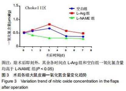
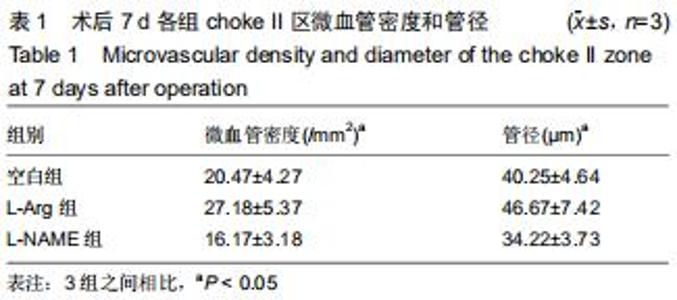
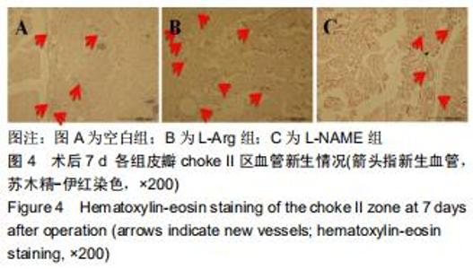
|
[1] TAYLOR GI,PALMER JH.The vascular territories (angiosomes) of the body experimental study and clinical applications.Br J Plast Surg.1987;40(2):113-141.
[2] TAYLOR GI, CORLETT RJ, DHAR SC, et al. The anatomical (angiosome) and clinical territories of cutaneous perforating arteries: development of the concept and designing safe flaps. Plast Reconstr Surg.2011;127(4):1447-1459.
[3] 李文波,贾丁丁,王飞,等.外源性L-精氨酸对大鼠背部跨区皮瓣成活的影响[J].浙江大学学报(医学版),2017,46(6):656-661.
[4] MONCADA S, HIGGS A. The L-arginine-nitric oxide pathway. N Engl J Med.1993;329(27):2002-2012.
[5] 任元鹏,杨鸣,姜柏生.互利与平衡:实验动物的伦理、福利与法律[J].中国医学伦理学,2016,29(6):1015-1017.
[6] 邓放,赵不非,马跃,等.超氧化物歧化酶与L-精氨酸联合预处理对大鼠肝缺血再灌注损伤的保护作用[J].中国老年学杂志,2015, 35(23):6710-6712.
[7] 赵林,丰昀.左旋精氨酸甲酯对脑缺血再灌注期脑组织炎症反应的影响[J].湖南师范大学学报(医学版),2009,6(3):16-18.
[8] INNOCENTI M, SANTINI M, DREASSI E, et al. Effects of cutaneous negative pressure application on perforator artery flow in healthy volunteers: a preliminary study.J Reconstr Microsurg.2019; 35(3):189-193.
[9] 刘燕玲,高磊,王江宁.前列地尔改善大鼠缺血皮瓣微循环[J].微循环学杂志,2018,28(1):1-6.
[10] 蔡洁云,林博杰,潘新元,等.天然水蛭素联合高压氧治疗对大鼠随意皮瓣成活的影响研究[J].中国修复重建外科杂志,2018,32(4): 484-490.
[11] GAO ZM, LIN DM, WANG Y, et al. Role of the NO/cGMP pathway in postoperative vasodilation in perforator flaps.J Reconstr Microsurg.2015;31(2):107-121.
[12] 白永铎.L-精氨酸影响动脉内皮功能的荟萃分析及脑卒中的遗传易感性研究[D].北京:北京协和医学院,2009.
[13] 晏泽,刘春利,苑凯华,等.皮瓣掀起后内皮素、一氧化氮变化与微循环关系的实验研究[J].中华整形外科杂志,2000,16(3):157-160.
[14] MCDONALD WS,LO TP J,THURMOND M,et al.Role of nitric oxide in skin flap delay.Plast Reconstr Surg.2004;113(3): 927-931.
[15] GRIBBE O,SAMUELSON UE,WIKLUND NP.Effects of nitric oxide synthase inhibition on blood flow and survival in experimental skin flaps.J Plast Reconstr Aesthet Surg. 2007; 60(3):287-293.
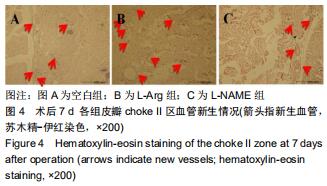
[16] 李文波,张超,石杰,等.穿支皮瓣中choke vessels的新生过程研究及“血管体”概念[J].中国组织工程研究,2018,22(8):1261-1266.
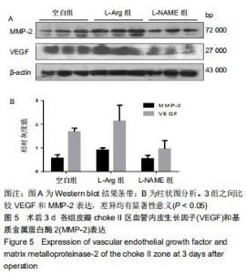
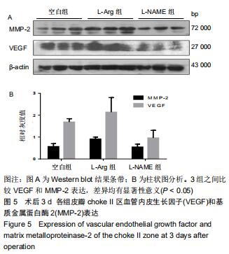
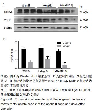
[17] FERRARA N.Vascular endothelial growth factor. Arterioscler Thromb Vasc Biol.2009;29(6):789-791.
[18] ZOELLER JJ,WHITELOCK JM,IOZZO RV. Perlecan regulates developmental angiogenesis by modulating the VEGF-VEGF R2 axis.Matrix Biology.2009;28(5):284-291.
[19] PHILLIPS TM,FADIA M,LEA-HENRY TN,et al.MMP2 and MMP9 associate with crescentic glomerulonephritis.Clin Kidney J.2017;10(2):215-220.
|