[1] FARRIS AB, COLVIN RB. Renal interstitial fibrosis: mechanisms and evaluation. Curr Opin Nephrol Hypertens. 2012;21(3):289-300.
[2] 马园园,刘成海,陶艳艳.肾纤维化动物模型特点与研究进展[J].中国实验动物学报,2018,26(3):398-403.
[3] 吴锋,何立群.单侧输尿管梗阻模型肾纤维化的中医药研究进展[J].辽宁中医杂志,2010,37(6):1168-1171.
[4] 吴燕升,万强,史丽强,等.高尿酸血症肾纤维化动物模型研究进展[J].成都医学院学报,2016,11(6):747-750,756.
[5] KLAHR S, MORRISSEY J. Obstructive nephropathy and renal fibrosis. Am J Physiol Renal Physiol. 2002;283(5):F861-875.
[6] CHEVALIER RL, FORBES MS, THORNHILL BA. Ureteral obstruction as a model of renal interstitial fibrosis and obstructive nephropathy. Kidney Int. 2009;75(11):1145-1152.
[7] BECKER GJ, HEWITSON TD. Animal models of chronic kidney disease: useful but not perfect. Nephrol Dial Transplant. 2013;28(10):2432-2438.
[8] LAWSON J, ELLIOTT J, WHEELER-JONES C, et al. Renal fibrosis in feline chronic kidney disease: known mediators and mechanisms of injury. Vet J. 2015;203(1):18-26.
[9] LI RX, YIU WH, TANG SC. Role of bone morphogenetic protein-7 in renal fibrosis. Front Physiol. 2015;6:114.
[10] MENG XM, TANG PM, LI J, et al. TGF-β/Smad signaling in renal fibrosis. Front Physiol. 2015;6:82.
[11] LI H, PENG X, WANG Y, et al. Atg5-mediated autophagy deficiency in proximal tubules promotes cell cycle G2/M arrest and renal fibrosis. Autophagy. 2016;12(9):1472-1486.
[12] FEDERICO G, MEISTER M, MATHOW D, et al. Tubular Dickkopf-3 promotes the development of renal atrophy and fibrosis. JCI Insight. 2016;1(1):e84916.
[13] KIM J, IMIG JD, YANG J, et al. Inhibition of soluble epoxide hydrolase prevents renal interstitial fibrosis and inflammation. Am J Physiol Renal Physiol. 2014;307(8):F971-980.
[14] AN HJ, KIM JY, KIM WH, et al. The Protective Effect of Melittin on Renal Fibrosis in an Animal Model of Unilateral Ureteral Obstruction. Molecules. 2016;21(9): E1137.
[15] 韩红,孙东,麻艳艳,等.单侧输尿管梗阻模型小鼠肾脏微血管损伤与肾间质纤维化的关系研究[J].中国病理生理杂志, 2010, 26(12):2478-2481,2486.
[16] HE P, LI D, ZHANG B. Losartan attenuates renal interstitial fibrosis and tubular cell apoptosis in a rat model of obstructive nephropathy. Mol Med Rep. 2014;10(2):638-644.
[17] ZHANG D, SUN L, XIAN W, et al. Low-dose paclitaxel ameliorates renal fibrosis in rat UUO model by inhibition of TGF-beta/Smad activity. Lab Invest. 2010;90(3):436-447.
[18] 谢盛彬,王伟铭,陈楠.UUO模型大鼠肾间质纤维化动态进展及α-SMA、TGF-β1 和VDR表达变化[J].上海交通大学学报(医学版),2010,30(7): 752-757,796.
[19] 张选明,杨百京,叶学锋,等.补阳还五汤对肾间质纤维化大鼠模型胶原纤维和TGF-β1的作用[J].中药药理与临床, 2011, 27(6):3-6.
[20] 陆敏,周娟,王飞,等.川芎嗪对肾间质纤维化模型大鼠Smad7和SnoN蛋白表达的影响[J].中国中药杂志, 2009, 34(1):84-88.
[21] 汪燕,王显,沈雯雯,等.树突状细胞在多柔比星诱导的大鼠肾纤维化模型中的作用[J].安徽医科大学学报,2011,46(1):15-19.
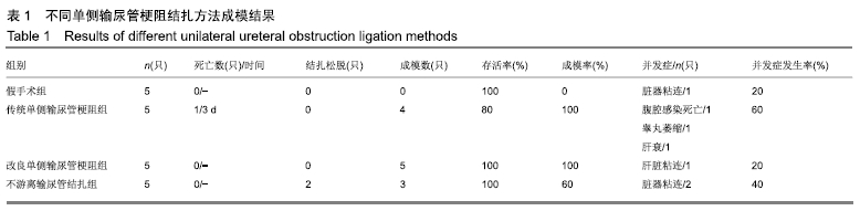
[22] 刘克剑,张悦,李靖,等.单侧输尿管梗阻法制作大鼠肾间质纤维化模型的改进[J].中国实验动物学报, 2007, 15(6):410-412.
[23] 王艳非,常晶,高云,等.老年性肾间质纤维化动物模型研究新进展[J].中华老年多器官疾病杂志,2018,17(12):952-955.
[24] 刘言振,赵婷婷,李佳霖,等.中药治疗肾间质纤维化研究进展[J].中日友好医院学报,2018,32(6):366-369.
[25] 李丹,郭玉莹,邓昊,等.实验动物麻醉剂使用的福利伦理问题研究进展[J].中国比较医学杂志,2017,27(9):87-91.
[26] 冯定浩,李均,徐静雅,等.丹参酮IIA联合DAPT对UUO模型大鼠肾组织Notch/Jagged通路相关蛋白表达的影响[J].中国中西医结合杂志,2019, 29(7):826-831.
[27] 柴亚男,郝娟,马雪莲,等.益气活血解毒中药对梗阻性肾病细胞自噬的调控作用[J].新中医, 2018, 50(12):8-11.
[28] HESKETH EE, VERNON MA, DING P, et al. A murine model of irreversible and reversible unilateral ureteric obstruction. J Vis Exp. 2014;(94):e52559.
|