Chinese Journal of Tissue Engineering Research ›› 2020, Vol. 24 ›› Issue (19): 3004-3009.doi: 10.3969/j.issn.2095-4344.2038
Previous Articles Next Articles
The role of PI3K/Akt pathway in hypoxia-induced cell proliferation and endothelial cell differentiation of adipose-derived stem cells
Yin Lingni, Chen Dexuan
- Department of Geriatrics, Jiading Central Hospital, Shanghai University of Medicine & Health Sciences, Shanghai 201800, China
-
Received:2019-07-04Revised:2019-07-09Accepted:2019-08-09Online:2020-07-08Published:2020-04-08 -
Contact:Chen Dexuan, Master, Chief physician, Department of Geriatrics, Jiading Central Hospital, Shanghai University of Medicine & Health Sciences, Shanghai 201800, China -
About author:Yin Lingni, Master, Physician, Department of Geriatrics, Jiading Central Hospital, Shanghai University of Medicine & Health Sciences, Shanghai 201800, China
CLC Number:
Cite this article
Yin Lingni, Chen Dexuan. The role of PI3K/Akt pathway in hypoxia-induced cell proliferation and endothelial cell differentiation of adipose-derived stem cells[J]. Chinese Journal of Tissue Engineering Research, 2020, 24(19): 3004-3009.
share this article

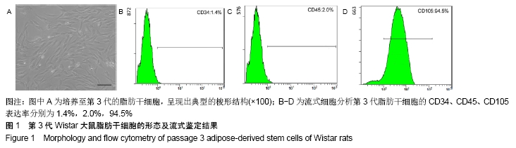
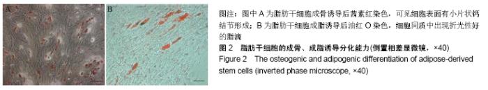
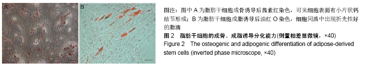
2.1 脂肪干细胞的形态及鉴定结果 体外培养至第 3代的脂肪干细胞呈现出典型的梭形结构。经流式细胞分析鉴定后发现间充质细胞表面标志物CD105表达阳性,而造血细胞表面标志物CD34和CD45表达阴性,说明培养至第3代的脂肪干细胞具有干细胞特性,是一群均一的未分化干细胞,见图1。加入成骨诱导液培养2周后,显微镜下可见细胞表面有小片状钙结节形成,茜素红染色阳性,说明脂肪干细胞有向骨细胞分化的能力。加入成脂诱导液培养2周后,细胞间质中出现折光性好的脂滴,油红O染色显示脂质变红,表明脂肪干细胞有向脂肪细胞分化的能力,见图2。 "
"

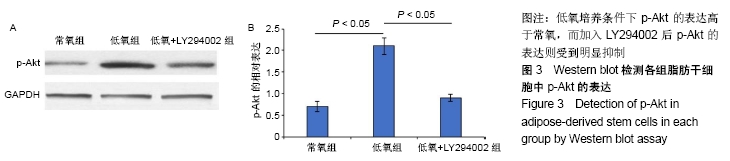
2.2 Western blot检测低氧培养条件下PI3K/Akt通路的激活 低氧培养24 h时,Western blot检测发现p-Akt的表达明显高于常氧组,加入LY294002后,p-Akt的表达受到明显抑制,说明LY294002可以有效抑制PI3K/Akt通路的激活,见图3。 "
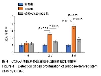
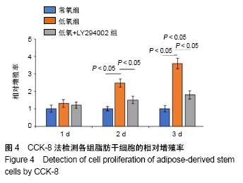
2.3 CCK-8检测细胞增殖 在培养第1天时3组之间差异无显著性意义,第2天时低氧组的细胞增殖率明显高于常氧组、低氧+LY294002组(P < 0.05),常氧组、低氧+LY294002组之间差异无显著性意义(P > 0.05),而培养第3天时,3组之间两两比较差异均有显著性意义(P < 0.05),说明加入LY294002后抑制了低氧诱导的脂肪干细胞增殖,见图4。 "

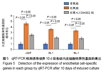
2.4 qRT-PCR检测内皮细胞特异性标志物的表达 经过低氧培养后细胞vWF、flt-1及flk-1的表达明显高于常氧组(P < 0.05),低氧+LY294002组vWF、flt-1及flk-1的表达则明显低于低氧组(P < 0.05),且与常氧组比较差异均有显著性意义(P < 0.05),见图5。 "

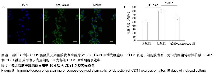
2.5 抗CD31免疫荧光染色结果 免疫荧光染色CD31抗原,显微镜下观察可见CD31抗原表达于细胞膜表面,低氧组CD31阳性细胞表达率远远高于常氧组(P < 0.05),低氧+ LY294002组CD31阳性细胞表达率低于低氧组(P < 0.05),但仍高于常氧组(P < 0.05),见图6。 "
| [1] WANG Z, XING H, HU H, et al. Intraglandular transplantation of adipose-derived stem cells combined with platelet-rich fibrin extract for the treatment of irradiation-induced salivary gland damage. Exp Ther Med. 2018;15(1):795-805. [2] CHAN TM, HARN HJ, LIN HP, et al. The use of ADSCs as a treatment for chronic stroke. Cell Transplant. 2014;23(4-5): 541-547. [3] GADELKARIM M, ABUSHOUK AI, GHANEM E, et al. Adipose-derived stem cells: Effectiveness and advances in delivery in diabetic wound healing. Biomed Pharmacother. 2018; 107:625-633. [4] SHANG T, LI S, ZHANG Y, et al. Hypoxia promotes differentiation of adipose-derived stem cells into endothelial cells through demethylation of ephrinB2. Stem Cell Res Ther. 2019;10(1):133. [5] DENG Y, HUANG G, CHEN F, et al. Hypoxia enhances buffalo adipose-derived mesenchymal stem cells proliferation, stemness, and reprogramming into induced pluripotent stem cells. J Cell Physiol. 2019;234(10):17254-17268. [6] FAN LJ, XIAO QR, LIN KS, et al. Comparison of endothelial differentiation capacity of adipose-derived stem cells and bone marrow mesenchymal stem cells from rats. Nan Fang Yi Ke Da Xue Xue Bao. 2016;36(9):1247-1254. [7] CHEN J, YUE C, XU J, et al. Downregulation of receptor tyrosine kinase-like orphan receptor 1 in preeclampsia placenta inhibits human trophoblast cell proliferation, migration, and invasion by PI3K/AKT/mTOR pathway accommodation. Placenta. 2019;82: 17-24. [8] SHENG L, MAO X, YU Q, et al. Effect of the PI3K/AKT signaling pathway on hypoxia-induced proliferation and differentiation of bone marrow-derived mesenchymal stem cells. Exp Ther Med. 2017;13(1):55-62. [9] CHEN H, LIU Y, JIANG CJ, et al. Calcium-Activated Chloride Channel A4 (CLCA4) Plays Inhibitory Roles in Invasion and Migration Through Suppressing Epithelial-Mesenchymal Transition via PI3K/AKT Signaling in Colorectal Cancer. Med Sci Monit. 2019;25:4176-4185. [10] TONG X, PELLING JC. Targeting the PI3K/Akt/mTOR axis by apigenin for cancer prevention. Anticancer Agents Med Chem. 2013;13(7):971-978. [11] HONG SJ, TRAKTUEV DO, MARCH KL. Therapeutic potential of adipose-derived stem cells in vascular growth and tissue repair. Curr Opin Organ Transplant. 2010;15(1):86-91. [12] ZUK PA, ZHU M, ASHJIAN P, et al. Human adipose tissue is a source of multipotent stem cells. Mol Biol Cell. 2002;13(12): 4279-4295. [13] FRESE L, DIJKMAN PE, HOERSTRUP SP. Adipose Tissue-Derived Stem Cells in Regenerative Medicine. Transfus Med Hemother. 2016;43(4):268-274. [14] WYSTRYCHOWSKI W, PATLOLLA B, ZHUGE Y, et al. Multipotency and cardiomyogenic potential of human adipose-derived stem cells from epicardium, pericardium, and omentum. Stem Cell Res Ther. 2016;7(1):84. [15] ZHAO K, LI R, BI S, et al. Combination of mild therapeutic hypothermia and adipose-derived stem cells for ischemic brain injury. Neural Regen Res. 2018;13(10):1759-1770. [16] BUERK DG, SHONAT RD, RIVA CE, et al. O2 gradients and countercurrent exchange in the cat vitreous humor near retinal arterioles and venules. Microvasc Res. 1993;45(2):134-148. [17] SHANG T, LI S, ZHANG Y, et al. Hypoxia promotes differentiation of adipose-derived stem cells into endothelial cells through demethylation of ephrinB2. Stem Cell Res Ther. 2019;10(1):133. [18] BEKHITE MM, FINKENSIEPER A, REBHAN J, et al. Hypoxia, leptin, and vascular endothelial growth factor stimulate vascular endothelial cell differentiation of human adipose tissue-derived stem cells. Stem Cells Dev. 2014;23(4):333-351. [19] WANG F, ZACHAR V, PENNISI CP, et al. Hypoxia Enhances Differentiation of Adipose Tissue-Derived Stem Cells toward the Smooth Muscle Phenotype. Int J Mol Sci. 2018;19(2): E517. [20] HERS I, VINCENT EE, TAVARÉ JM. Akt signalling in health and disease. Cell Signal. 2011;23(10):1515-1527. [21] KHAN S, VILLALOBOS MA, CHORON RL, et al. Fibroblast growth factor and vascular endothelial growth factor play a critical role in endotheliogenesis from human adipose-derived stem cells. J Vasc Surg. 2017;65(5):1483-1492. [22] LI D, XIE K, ZHANG L, et al. Dual blockade of vascular endothelial growth factor (VEGF) and basic fibroblast growth factor (FGF-2) exhibits potent anti-angiogenic effects. Cancer Lett. 2016;377(2):164-173. [23] LEE EY, XIA Y, KIM WS, et al. Hypoxia-enhanced wound-healing function of adipose-derived stem cells: increase in stem cell proliferation and up-regulation of VEGF and bFGF. Wound Repair Regen. 2009;17(4):540-547. [24] SONG SY, CHUNG HM, SUNG JH. The pivotal role of VEGF in adipose-derived-stem-cell-mediated regeneration. Expert Opin Biol Ther. 2010;10(11):1529-1537. [25] WATANABE H, SAITO H, UEDA J, et al. Regulation of pancreatic duct cell differentiation by phosphatidylinositol-3 kinase. Biochem Biophys Res Commun. 2008;370(1):33-37. [26] OUYANG N, ZHANG P, FU R, et al. Mechanical strain promotes osteogenic differentiation of bone mesenchymal stem cells from ovariectomized rats via the phosphoinositide 3‑kinase/Akt signaling pathway. Mol Med Rep. 2018;17(1):1855-1862. [27] XIAO X, WANG W, LIU D, et al. The promotion of angiogenesis induced by three-dimensional porous beta-tricalcium phosphate scaffold with different interconnection sizes via activation of PI3K/Akt pathways. Sci Rep. 2015;5:9409. [28] MANGI AA, NOISEUX N, KONG D, et al. Mesenchymal stem cells modified with Akt prevent remodeling and restore performance of infarcted hearts. Nat Med. 2003;9(9):1195-1201. [29] YUAN L, FAN L, LI Q, et al. Inhibition of miR-181b-5p protects cardiomyocytes against ischemia/reperfusion injury by targeting AKT3 and PI3KR3. J Cell Biochem. 2019 Jul 11. doi: 10.1002/jcb.29271. [Epub ahead of print] [30] DATTA SR, DUDEK H, TAO X, et al. Akt phosphorylation of BAD couples survival signals to the cell-intrinsic death machinery. Cell. 1997;91(2):231-241. [31] CARDONE MH, ROY N, STENNICKE HR, et al. Regulation of cell death protease caspase-9 by phosphorylation. Science. 1998;282 (5392):1318-1321. |
| [1] | Lin Qingfan, Xie Yixin, Chen Wanqing, Ye Zhenzhong, Chen Youfang. Human placenta-derived mesenchymal stem cell conditioned medium can upregulate BeWo cell viability and zonula occludens expression under hypoxia [J]. Chinese Journal of Tissue Engineering Research, 2021, 25(在线): 4970-4975. |
| [2] | Gu Xia, Zhao Min, Wang Pingyi, Li Yimei, Li Wenhua. Relationship between hypoxia inducible factor 1 alpha and hypoxia signaling pathway [J]. Chinese Journal of Tissue Engineering Research, 2021, 25(8): 1284-1289. |
| [3] | Hou Jingying, Yu Menglei, Guo Tianzhu, Long Huibao, Wu Hao. Hypoxia preconditioning promotes bone marrow mesenchymal stem cells survival and vascularization through the activation of HIF-1α/MALAT1/VEGFA pathway [J]. Chinese Journal of Tissue Engineering Research, 2021, 25(7): 985-990. |
| [4] | Fan Quanbao, Luo Huina, Wang Bingyun, Chen Shengfeng, Cui Lianxu, Jiang Wenkang, Zhao Mingming, Wang Jingjing, Luo Dongzhang, Chen Zhisheng, Bai Yinshan, Liu Canying, Zhang Hui. Biological characteristics of canine adipose-derived mesenchymal stem cells cultured in hypoxia [J]. Chinese Journal of Tissue Engineering Research, 2021, 25(7): 1002-1007. |
| [5] | Li Cai, Zhao Ting, Tan Ge, Zheng Yulin, Zhang Ruonan, Wu Yan, Tang Junming. Platelet-derived growth factor-BB promotes proliferation, differentiation and migration of skeletal muscle myoblast [J]. Chinese Journal of Tissue Engineering Research, 2021, 25(7): 1050-1055. |
| [6] | Liu Cong, Liu Su. Molecular mechanism of miR-17-5p regulation of hypoxia inducible factor-1α mediated adipocyte differentiation and angiogenesis [J]. Chinese Journal of Tissue Engineering Research, 2021, 25(7): 1069-1074. |
| [7] | Zhao Min, Feng Liuxiang, Chen Yao, Gu Xia, Wang Pingyi, Li Yimei, Li Wenhua. Exosomes as a disease marker under hypoxic conditions [J]. Chinese Journal of Tissue Engineering Research, 2021, 25(7): 1104-1108. |
| [8] | Wang Shiqi, Zhang Jinsheng. Effects of Chinese medicine on proliferation, differentiation and aging of bone marrow mesenchymal stem cells regulating ischemia-hypoxia microenvironment [J]. Chinese Journal of Tissue Engineering Research, 2021, 25(7): 1129-1134. |
| [9] | Ma Zetao, Zeng Hui, Wang Deli, Weng Jian, Feng Song. MicroRNA-138-5p regulates chondrocyte proliferation and autophagy [J]. Chinese Journal of Tissue Engineering Research, 2021, 25(5): 674-678. |
| [10] | Wang Yujiao, Liu Dan, Sun Song, Sun Yong. Biphasic calcium phosphate loaded with advanced platelet rich fibrin can promote the activity of rabbit bone marrow mesenchymal stem cells [J]. Chinese Journal of Tissue Engineering Research, 2021, 25(4): 504-509. |
| [11] | Zhou Jihui, Yao Meng, Wang Yansong, Li Xinzhi, Zhou You, Huang Wei, Chen Wenyao. Influence of novel nanoscaffolds on biological behaviors of neural stem cells and the related gene expression [J]. Chinese Journal of Tissue Engineering Research, 2021, 25(4): 532-536. |
| [12] | Chen Yang, Huang Denggao, Gao Yuanhui, Wang Shunlan, Cao Hui, Zheng Linlin, He Haowei, Luo Siqin, Xiao Jingchuan, Zhang Yingai, Zhang Shufang. Low-intensity pulsed ultrasound promotes the proliferation and adhesion of human adipose-derived mesenchymal stem cells [J]. Chinese Journal of Tissue Engineering Research, 2021, 25(25): 3949-3955. |
| [13] | Zhou Wu, Wang Binping, Wang Yawen, Cheng Yanan, Huang Xieshan. Transforming growth factor beta combined with bone morphogenetic protein-2 induces the proliferation and differentiation of mouse MC3T3-E1 cells [J]. Chinese Journal of Tissue Engineering Research, 2021, 25(23): 3630-3635. |
| [14] | Yu Chengshuai, Du Gang, Pang Shenning, Lao Shan. Chemerin, a pro-inflammatory adipokine, regulates chondrocyte proliferation and metabolism by increasing production of nitric oxide [J]. Chinese Journal of Tissue Engineering Research, 2021, 25(2): 258-263. |
| [15] | Dai Yaling, Chen Lewen, He Xiaojun, Lin Huawei, Jia Weiwei, Chen Lidian, Tao Jing, Liu Weilin. Construction of miR-146b overexpression lentiviral vector and the effect on the proliferation of hippocampal neural stem cells [J]. Chinese Journal of Tissue Engineering Research, 2021, 25(19): 3024-3030. |
| Viewed | ||||||
|
Full text |
|
|||||
|
Abstract |
|
|||||