Chinese Journal of Tissue Engineering Research ›› 2015, Vol. 19 ›› Issue (3): 488-492.doi: 10.3969/j.issn.2095-4344.2015.03.029
Design, fabrication and clinical application of surgical implant guides
Xiang Mei, Zhang Yu
- Department of Dental Implantation, Nanfang Hospital of Southern Medical University, Stomatological School of Southern Medical University, Guangzhou 510515, Guangdong Province, China
-
Online:2015-01-15Published:2015-01-15 -
Contact:Zhang Yu, Associate chief physician, Master’s supervisor, Department of Dental Implantation, Nanfang Hospital of Southern Medical University, Stomatological School of Southern Medical University, Guangzhou 510515, Guangdong Province, China -
About author:Xiang Mei, Studying for master’s degree, Department of Dental Implantation, Nanfang Hospital of Southern Medical University, Stomatological School of Southern Medical University, Guangzhou 510515, Guangdong Province, China -
Supported by:the Science and Technology Plan of Guangdong Province, No. 2010B010800023
CLC Number:
Cite this article
Xiang Mei, Zhang Yu. Design, fabrication and clinical application of surgical implant guides[J]. Chinese Journal of Tissue Engineering Research, 2015, 19(3): 488-492.
share this article
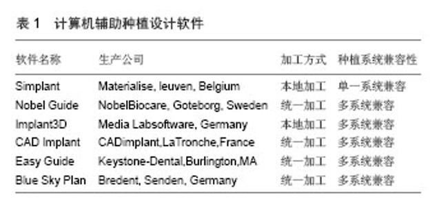
2.1 种植导板的分类及特点 种植导板是由导管与定位板组成,其中导管的位置和角度记录了术前设计种植体的位置、角度、深度信息,通过导管将这些信息转移到手术中,以便使种植体植入到准确位置。定位板起着定位作用,它与骨、牙齿或牙槽嵴表面相贴合[4,8]。为了保证种植体的精确性,往往还会制造不同内径的内管插入导管中以供不同直径的先锋钻或成型钻使用。 目前国内外学者对种植导板的分类各有不同,根据制作方法不同,可分为传统导板和CAD/CAM种植导板[9]。传统导板一般是在石膏模型上制作的,在一定程度上考虑了咬合和解剖情况,它制作简单,但精确性较差。CAD/CAM种植导板是结合数字影像、计算机技术、数字加工为一体的新型种植导板,它充分考虑了颌骨的三维解剖情况和咬合关系,精确性高,能简化手术和降低手术风险,但价格较昂贵[10]。 种植导板根据支持组织不同可分为黏膜支持式、骨支持式、牙支持式、混合支持式。①黏膜支持式多用于无牙颌的患者,导板直接覆盖在牙槽嵴上,手术过程中不用翻开黏骨膜瓣,但黏膜有一定的弹性,黏膜支持式的导板固位效果较差,往往需要增加固位钉固定。②骨支持式导板多适用于无牙颌及缺牙较多的患者,手术过程中需要翻开黏骨膜瓣。③牙支持式导板多用于缺牙数目较少的患者,缺牙区有邻牙,手术中根据情况既可翻瓣也可不翻瓣。 根据种植窝限制程度不同又可分为不限制式、部分限制式、完全限制式[11]。①不限制式导板只限制了植入位点,而没有限制种植体的深度、方向和直径。它的特点是制作简单,价格低廉。这类导板对牙槽骨条件差或邻近有重要解剖结构的情况有一定的局限性。②部分限制式导板只限制了植入的方向,而对植入深度、先锋钻和成型钻的直径没有限制,因此其精确性小于完全限制式。③完全限制式种植导板限制了种植体植入的每个环节,包括方向、直径和深度,精确度较高,现已成为种植导板发展的主要方向。 2.2 种植导板的制作 2.2.1 传统导板的制作 传统导板一般是用热压膜技术制作的。首先给患者取模并翻制石膏模型,在模型的缺牙区根据咬合关系和牙槽嵴的解剖情况雕刻蜡型,再翻制石膏模型,将有蜡型的石膏模型放置于抽真空压膜机上,用较厚的树脂薄膜压制导板,修剪导板,在缺牙区的相应位置即基台穿出的位置钻直径与导管外径等大的孔,插入导管,并将导管用透明树脂固定在导板上[4,12]。目前,还有一些研究报道了关于种植导板制作的改良方法。邓再喜等[13]介绍了一种制作种植体导板的新方法。首先,对需要进行种植体植入的缺损部位进行蜡牙形态恢复,然后使用手调式硅橡胶,制取印模,再使用透明注塑树脂进行灌注成形,最后进行打孔、磨光,完成种植体导板。George等[14]也报道了一种相似的方法,不同之处是他采用光敏树脂制作,且在制作过程中借助根尖片提供的信息,采用定位杆对拟植入种植体的位置进行校正。类似于这些种植导板的制作方法还有很多,但它们的一个共同特点是未能精确考虑颌骨的三维解剖结构。 2.2.2 CAD/CAM种植导板的制作 CAD/CAM种植导板制作原理:CAD/CAM导板的制作是基于CT数据基础上的。患者术前需要进行缺牙区颌骨的CT扫描,将扫描的数据以DICOM格式导入相应的软件,进行颌骨的三维重建,并且根据颌骨的三维解剖结构和咬合关系(用放射导板记录)设计种植体的最佳植入方案,包括种植体的位置、角度、数目、深度,进而根据其设计方案设计种植导板,最后将设计的导板进行加工制造[15-17]。 CAD/CAM导板制作中常用软件:现阶段导板设计的软件种类较多(表1)。每种设计软件所设计的种植导板的制作地点、加工工艺、种植系统兼容性各不相同,其中部分软件设计的导板需要国外制造,如瑞士Nobel Biocare 公司生产的Nobel Guide、美国生产的Easy Guide等;还有一部分软件设计的导板是可以本地加工制造的,如比利时Materialise 公司生产的Simplant 和Surgicase,德国生产的Implant 3D等。这些软件的使用程序大致为:根据导入的颌骨数据重建三维模型;测量缺牙区的牙槽嵴宽度、高度、离重要解剖结构的距离、骨密度等[18];根据缺牙区情况选择所需种植系统下不同型号的种植体,或者自行绘制不同型号的种植体进行模拟种植,要求种植体的位置能满足解剖结构和咬合的需求;根据所放置的种植体,轴向设计种植导板的导管结构,根据缺牙区周围邻牙和骨形态设计导板的固位结构,形成导板;把最后形成的导板以特定的格式(一般为STL格式)导入到制造设备中进行后续的制造。 CAD/CAM导板制作工艺:CAD/CAM种植导板后续的制作工艺主要有快速成型技术和数控技术。快速成型技术是将计算机设计的三维模型或者CT等断层扫描资料,转化为片层信息,应用快速成型设备依据这些片层信息,通过光固、烧结和黏结等工艺将材料逐层添加制作出实体来[19-20]。加工制造速度快是快速成型技术的特点之一,制作一个模型一般只需要几个小时,最长为十几个小时。由于快速成型技术是借助增加材料进行加工的方法,所以能够制造结构较复杂的模型,但其制造设备较昂贵,再加上受设备空间的限制,制造模型的尺寸有限,材料的选择范围也较窄[21-22]。数控技术是数字化控制技术的简称,它的成型原理是依据CAD三维模型的驱动,在整块材料上挖掘而成的技术方法,实质是去除材料加工,即减材加工。数控技术一般采用三维数控铣床或者雕刻机就可以进行生产,其加工制作过程速度较快、选择材料广泛、制作精度较高,由于数控设备加工范围大,所以加工尺寸也较大,但由于它是通过在整块材料上切割而成型的技术,这就决定了其在一些结构较复杂产品上的局限性[23-24]。 2.3 种植导板精确性的研究 种植外科导板作为最终的信息载体,是将术前设计与手术操作连接在一起的桥梁,对于一些多牙缺失、牙槽骨较差的情况,模拟的植入位置向实际植入位置转移是导板的关键,因此实际植入的精确度成为种植导板的关键[25]。 2.3.1 种植导板精确性的系统性评价 种植导板精确性的评价是通过把种植后的三维影像与术前模拟种植的三维影像进行配准,测量实际种植体与模拟种植体的偏差值(颈部、尖端、角度)来进行的。目前国内外对种植导板精确性评价的研究较多,但精确性有所差异。Tahmaseb等[26]在24例计算机辅助设计和制作的种植导板精确性研究的系统分析中(包含体内和体外研究共 1 532颗种植体),指出种植体颈部平均偏离为1.12 mm,其中最大值为4.5 mm,种植体尖端平均偏离值为 1.39 mm,最大值为7.1 mm。Ozan等[27]指出在临床研究中种植导板在下颌骨区角度偏离值小于上颌骨,分别为(3.32±1.90)°、(4.58±2.40)°。而在风险评估中,种植体头部的偏差极限值尤为重要,当水平偏差达1.86 mm或垂直偏差达2.7 mm都可能造成解剖结构的损害[28]。一些研究指示种植导板的精确性在体外研究中比体内研究中偏高,种植体顶端的偏离值大于颈部[29-30]。 2.3.2 不同类别种植导板的精确性 种植导板的种类较多,精确性也各有不同。Sarment等[31]比较传统种植导板与CAD/CAM种植导板的精确性,结果显示传统种植导板辅助下种植体颈部和尖端的偏离值分别为(1.50±0.70),(2.10±0.97) mm,在CAD/CAM种植导板辅助下种植体颈部与尖端的偏离值减小到(0.90±0.50),(1.00±0.60) mm。Turbush等[32]和Ozan等[27]分别在体内和体外进行了骨支持式、牙支持式、黏膜支持式导板精确性的比较,结果提示牙支持式种植导板在种植体角度、颈部、顶端的精确性都大于骨支持式和黏膜支持式;而在体外研究中,黏膜支持式种植导板在种植体颈部和顶端的精确性略差于骨支持式和牙支持式导板。两者均提示黏膜支持式导板精确性相对较差,而在牙、骨支持式导板精确性问题上存在分歧,可能是体外研究是在一个理想的环境下,排除了患者体位、唾液、血液特别是翻瓣后骨支持式导板固位不良的影响。 2.3.3 误差原因的分析 由于导板的制作方法、实验对象不同,研究者对导板精确度的评价也有所不同[33-34]。现分析如下:①CT扫描:颌骨的CT扫描为导板的制作提供三维影像数据,如选择的CT设备不合适或拍摄过程中患者不合作,都将影响数据的质量。目前口腔领域使用最多的为CBCT,因其拍摄时间短、放射剂量小、扫描层薄,所以能更好地提取口腔硬组织影像数据[35-36]。②三维重建:在颌骨或模型三维重建过程中,阈值的选择是至关重要的,若阈值选择不当,将影响组织的提取;如阈值过大,组织提取不完整,在此基础上制造的导板就位不良;阈值过小,制作的导板稳定性较差[37]。③导板制作工艺:选择何种制作工艺,将直接影响种植导板的精确性。④配准过程:由于种植导板精确性的评价是通过把种植后的三维影像与术前模拟种植的三维影像进行配准,测量实际种植体与模拟种植体的偏差值来进行的,所以配准与否也会影响精确度的评价。⑤手术过程:在手术过程中,操作不当导致导板就位不良、稳定性差都会影响精确度。如前牙区导板就位不良会加大种植体近远中偏差。 2.4 种植导板使用的临床效果 2.4.1 种植体的存活率 Tahmaseb等[26]在最近的1篇系统分析中报道,在种植导板辅助下种植体1年存活率为97.3%,而在无种植导板辅助下种植体的1年存活率为96%[38]。在种植导板辅助下,当上部修复结构为单冠时,种植体的5年存活率为96.8%;当上部修复结构为固定桥时,种植体的5年存活率为95.4%[38]。无种植导板辅助下,当上部修复结构为单冠时,5年存活率为94.5%[39];当上部修复结构固定桥时,5年存活率为91.5%[40]。由此可见,在种植导板辅助下的种植体有较高的存活率。 2.4.2 术中、术后并发症 由于适应证把握不当,种植导板使用方法不规范,手术操作失误等都会影响术后效果,严重情况下将会导致一系列的并发症。 术中并发症:①患者张口受限,导板就位困难。②术前未能精确的评价骨量与骨质,造成手术过程中需要计划外的骨增量。③导板使用不当或导板制造材料强度不足,导致术中导板破裂。④导板使用过程中牙槽嵴骨裂。⑤导板固位不良。 术后并发症:①导板固位钉口感染。②急性上颌窦炎。③ 附着龈不足。④龈缘瘘管。⑤种植体周围骨吸收等。 2.5 种植导板的优缺点 2.5.1 优点 ①术前能根据颌骨情况和咬合关系确定种植体的大小、位置和方向等,避免了手术的盲目性和术后因力传导不良而引起的并发症。②能精确测量牙槽骨的宽度和高度,以及下牙槽神经管、上颌窦、邻牙牙根等重要解剖结构至植牙区的距离,降低了术中风险。③多牙缺失或骨量缺损的情况往往增加了手术难度,需要临床经验丰富的医生来完成,而种植导板的使用将降低手术难度。④手术可不用翻开黏骨膜瓣,而直接用环切方式进行微创手术,减少了创伤和手术时间。 2.5.2 缺点 ①在手术过程中降低了术区的可视性和术者的触觉灵敏性。②对张口度不足的患者有一定的局限性。③种植导板的阻挡影响了种植窝洞的散热。④增加了患者经济负担。 "

| [1]Widmann G,Bale RJ.Accuracy in computer-aided implant surgery--a review. Int J Oral Maxillofac Implants.2006; 21(2): 305-313.
[2]Novellino MM,Sesma N,Lagana DC,et al.Linear and angular deviations of implants placed in experimental casts with stereolithographic drill guides fixed by o'ring ortho implant devices.Braz Dent J.2013;24(4):391-396.
[3]Dandekeri SS,Sowmya MK,Bhandary S.Stereolithographic surgical template: a review.J Clin Diagn Res.2013;7(9): 2093-2095.
[4]白石柱,刘宝林,陈小文,等.种植导板的制作及CAD-CAM技术的应用[J].实用口腔医学杂志,2011,27(1):138-142.
[5]Hultin M,Svensson KG,Trulsson M.Clinical advantages of computer-guided implant placement: a systematic review.Clin Oral Implants Res.2012;23 Suppl 6:124-135.
[6]Sohmura T,Kusumoto N,Otani T,et al.CAD/CAM fabrication and clinical application of surgical template and bone model in oral implant surgery. Clin Oral Implants Res.2009;20(1): 87-93.
[7]Lazic Z,Golubovic M,Markovic A,et al.Immunohistochemical analysis of blood vessels in peri-implant mucosa: a comparison between mini-incision flapless and flap surgeries in domestic pigs.Clin Oral Implants Res.2014.doi: 10.1111/clr.12337. [Epub ahead of print]
[8]Pattanaik S,Pattanaik BK.Fabrication of a surgical guide with help of a milling machine by ridge mapping method.J Indian Prosthodont Soc.2013;13(1):61-65.
[9]刘丽娜,杨敏,哈斯•巴根,等.牙种植定位导向模板的计算机辅助设计和制作[J].中国组织工程研究,2012,16(4):665-668.
[10]Liu H,Liu DX,Wang G,et al.Accuracy of surgical positioning of orthodontic miniscrews with a computer-aided design and manufacturing template. Am J Orthod Dentofacial Orthop.2010;137(6):721-728,728-729.
[11]D'Souza KM,Aras MA.Types of implant surgical guides in dentistry: a review. J Oral Implantol.2012;38(5):643-652.
[12]Stumpel LR.Cast-based guided implant placement: a novel technique. J Prosthet Dent.2008;100(1):61-69.
[13]邓再喜,张春宝,孙翔,等.使用手调式硅橡胶制作种植体导板[J].口腔医学,2009,29(9):501-502.
[14]George FM,Chan HL,Razzoog ME,et al.Fabrication of a cast-based implant surgical guide using guide sleeves.J Prosthet Dent.2011;106(6):409-412.
[15]Zhao XZ,Xu WH,Tang ZH,et al.Accuracy of computer-guided implant surgery by a CAD/CAM and laser scanning technique. Chin J Dent Res.2014;17(1):31-36.
[16]Moslehifard E,Nokar S.Designing a custom made gauge device for application in the access hole correction in the dental implant surgical guide.J Indian Prosthodont Soc.2012; 12(2):123-129.
[17]刘丽娜.牙种植模板的计算机辅助设计和制作的应用研究[D].天津医科大学,2012.
[18]Lee JH,Kim MJ,Kim SM,et al.The 3D CT superimposition method using image fusion based on the maximum mutual information algorithm for the assessment of oral and maxillofacial surgery treatment results.Oral Surg Oral Med Oral Pathol Oral Radiol.2012;114(2):167-174.
[19]安芬菊,梁永回.快速成型技术与数控技术在手版制作中的应用比较[J].机械制造,2012,50(2):54-56.
[20]Bickel B,Alexa M.Computational aspects of fabrication modeling, design, and 3D printing.IEEE Comput Graph Appl. 2013;33(6):24-25.
[21]Hochman JB,Kraut J,Kazmerik K,et al.Generation of a 3D printed temporal bone model with internal fidelity and validation of the mechanical construct. Otolaryngol Head Neck Surg.2014;150(3):448-454.
[22]Peterson GI,Larsen MB,Ganter MA,et al.3D-Printed Mechanochromic Materials. ACS Appl Mater Interfaces,2015; 7(1):577-583.
[23]王太勇,乔志峰,韩志国,等.高档数控装备的发展趋势[J].中国机械工程,2011,22(10):1247-1252.
[24]杨兆军,陈传海,陈菲,等.数控机床可靠性技术的研究进展[J].机械工程学报,2013,49(20):130-139.
[25]刘思玉,李宏卫,汤春波.种植体计算机辅助设计和制作导板的研究进展[J].口腔医学,2013,33(5):345-347.
[26]Tahmaseb A,Wismeijer D,Coucke W,et al.Computer technology applications in surgical implant dentistry: a systematic review.Int J Oral Maxillofac Implants.2014;29 Suppl:25-42.
[27]Ozan O,Turkyilmaz I,Ersoy AE,et al.Clinical accuracy of 3 different types of computed tomography-derived stereolithographic surgical guides in implant placement.J Oral Maxillofac Surg.2009;67(2):394-401.
[28]D'Haese J,Van De Velde T,Komiyama A,et al.Accuracy and complications using computer-designed stereolithographic surgical guides for oral rehabilitation by means of dental implants: a review of the literature.Clin Implant Dent Relat Res. 2012;14(3):321-335.
[29]Vieira DM,Sotto-Maior BS,Barros CA,et al.Clinical accuracy of flapless computer-guided surgery for implant placement in edentulous arches.Int J Oral Maxillofac Implants.2013;28(5): 1347-1351.
[30]Valente F,Schiroli G,Sbrenna A.Accuracy of computer-aided oral implant surgery: a clinical and radiographic study.Int J Oral Maxillofac Implants.2009;24(2):234-242.
[31]Sarment DP,Sukovic P,Clinthorne N.Accuracy of implant placement with a stereolithographic surgical guide.Int J Oral Maxillofac Implants.2003;18(4):571-577.
[32]Turbush SK,Turkyilmaz I.Accuracy of three different types of stereolithographic surgical guide in implant placement: an in vitro study.J Prosthet Dent.2012;108(3):181-188.
[33]刘洪,刘东旭,王克涛,等.种植体计算机辅助设计和制造导板精度的评价[J].华西口腔医学杂志,2010,28(5):517-521.
[34]郭树琴,王立军.数字化种植外科导板研究新进展[J].中国口腔种植学杂志,2013,18(4):205-209.
[35]Gonzalez-Garcia R,Monje F.The reliability of cone-beam computed tomography to assess bone density at dental implant recipient sites: a histomorphometric analysis by micro-CT.Clin Oral Implants Res.2013;24(8):871-879.
[36]Shah N,Bansal N,Logani A.Recent advances in imaging technologies in dentistry. World J Radiol.2014;6(10): 794-807.
[37]Yao W,Bekmezian S,Hardy D,et al.Cone-Beam Computed Tomographic Comparison of Surgically Assisted Rapid Palatal Expansion and Multipiece Le Fort I Osteotomy. J Oral Maxillofac Surg.2014.
[38]Jung RE,Schneider D,Ganeles J,et al.Computer technology applications in surgical implant dentistry: a systematic review.Int J Oral Maxillofac Implants.2009;24 Suppl:92-109.
[39]Jung RE,Pjetursson BE,Glauser R,et al. A systematic review of the 5-year survival and complication rates of implant-supported single crowns.Clin Oral Implants Res.2008; 19(2):119-130.
[40]Pjetursson BE,Thoma D,Jung R,et al.A systematic review of the survival and complication rates of implant-supported fixed dental prostheses (FDPs) after a mean observation period of at least 5 years.Clin Oral Implants Res.2012;23 Suppl 6:22-38. |
| [1] | Huo Hua, Cheng Yuting, Zhou Qian, Qi Yuhan, Wu Chao, Shi Qianhui, Yang Tongjing, Liao Jian, Hong Wei. Effects of drug coating on implant surface on the osseointegration [J]. Chinese Journal of Tissue Engineering Research, 2021, 25(22): 3558-3564. |
| [2] |
Zhang Xuan, Li Yunpeng, Zhang Xuejian, Yin Chuanrong, Deng Yue.
Guided bone regeneration using preformed titanium mesh combined with bioabsorbable membranes in aesthetic area [J]. Chinese Journal of Tissue Engineering Research, 2020, 24(26): 4112-4117. |
| [3] | Liu Dan, Min Changqin, Lu Shuai, Chen Yue, Sun Yong. Osseointegration induced by beta-tricalcium phosphate loaded with advanced platelet-rich fibrin [J]. Chinese Journal of Tissue Engineering Research, 2019, 23(6): 888-893. |
| [4] | Li Fang, Cheng Yuting, Huang Xiaolin, Zhou Qian, Wu Chao, Shi Qianhui, Wang Yong, Liao Jian. Maxillary sinus floor augmentation: with or without bone grafting [J]. Chinese Journal of Tissue Engineering Research, 2019, 23(6): 971-977. |
| [5] | Zou Yingnan, Wang Yibo, Ding Chao, Pan Xinyu, Shi Jiuhui . Three-dimensional finite element analysis of dental implant combined with residual tooth after hemisection under dynamic loads [J]. Chinese Journal of Tissue Engineering Research, 2019, 23(2): 178-183. |
| [6] | Wang Liping, Chen Weihong, Zha Jun, Chen Xili, Su Yucheng, Fang Ying, Dong Yu, Guo Xueqi, Ge Linhu. Short-term efficacy evaluation of Mis Seven implant system repairing dentition loss [J]. Chinese Journal of Tissue Engineering Research, 2019, 23(14): 2208-2214. |
| [7] | Li Ying, You Yapeng, Li Baoe, Song Yunjia, Ma Aobo, Chen Bo, Han Wen, Li Changyi. Type I collagen combined titanium dioxide nanotube composite coating modified titanium surface improves osteoblast adhesion and osseointegration [J]. Chinese Journal of Tissue Engineering Research, 2019, 23(14): 2169-2176. |
| [8] | Wei Wei, Shu Jing-ai, Wang Jing, Zheng Li-xia, Li Lu, Wang Qing-shan. Application of hydroxyapatite functional graded biomaterials in human hard tissue replacement [J]. Chinese Journal of Tissue Engineering Research, 2018, 22(6): 971-978. |
| [9] | Liu Ou, Gao Zhu, Li Tao, Zhou Bo. Minocycline hydrochloride ointment is available for the treatment of peri-implantitis [J]. Chinese Journal of Tissue Engineering Research, 2018, 22(6): 852-857. |
| [10] | Chang Lijun, Tang Tian, Zhang Xiaoge. Periodontal tissue changes in a Beagle dog model of peri-implantitis under orthodontic force [J]. Chinese Journal of Tissue Engineering Research, 2018, 22(34): 5464-5468. |
| [11] | Wang Zhong-da, Wang Yi-bo, Ding Chao, Shi Jiu-hui. Three-dimensional finite element analysis of implant denture for anterior tooth missing with different arch shapes [J]. Chinese Journal of Tissue Engineering Research, 2018, 22(30): 4818-4823. |
| [12] | Ma Yan, Zhang Hua, Shang Jian-ping, Jiang Qi. Acid etching and anodic oxidation of titanium surface of nanotubes promotes the proliferation and osteogenesis of bone marrow mesenchymal stem cells [J]. Chinese Journal of Tissue Engineering Research, 2018, 22(29): 4614-4619. |
| [13] | Huang Ya-xiang, Zhao Zi-ping, Wang Zhi-guo, Liao Qi-ming, Xie Yin-hao, Zhang Ping. Digital design of night-time scoliosis brace [J]. Chinese Journal of Tissue Engineering Research, 2018, 22(27): 4327-4331. |
| [14] | Lin Yi, Tao Xian-fa, Wang Yi-bo, Ding Chao, Xing Jian-yu, Xu Ran, Shi Jiu-hui. Three-dimensional finite element analysis of splinted crown restoration of molars with different implant platform positions [J]. Chinese Journal of Tissue Engineering Research, 2018, 22(26): 4123-4127. |
| [15] | Yu Jia-li, Nie Er-min, Jiang Rui, Zhang Chun-yuan, Huang Zhe-xun, Zhang Yu-hang, Lu Dun-lang. Digital technology in prosthodontics: impression, shade selection, material design and processing [J]. Chinese Journal of Tissue Engineering Research, 2018, 22(22): 3602-3608. |
| Viewed | ||||||
|
Full text |
|
|||||
|
Abstract |
|
|||||