Chinese Journal of Tissue Engineering Research ›› 2014, Vol. 18 ›› Issue (31): 5017-5023.doi: 10.3969/j.issn.2095-4344.2014.31.017
Previous Articles Next Articles
Establishment of digital template of cannulated screw fixation for femoral neck fractures
Cao Zhen-hua, Yin He-ping, Li Shu-wen
- Department of Minimally Invasive Spinal Surgery, Second Affiliated Hospital, Inner Mongolia Medical University, Hohhot 010010, Inner Mongolia Autonomous Region, China
-
Received:2014-06-17Online:2014-07-23Published:2014-07-23 -
Contact:Yin He-ping, Master, Chief physician, Professor, Department of Minimally Invasive Spinal Surgery, Second Affiliated Hospital, Inner Mongolia Medical University, Hohhot 010010, Inner Mongolia Autonomous Region, China -
About author:Cao Zhen-hua, Studying for doctorate, Attending physician, Department of Minimally Invasive Spinal Surgery, Second Affiliated Hospital, Inner Mongolia Medical University, Hohhot 010010, Inner Mongolia Autonomous Region, China
CLC Number:
Cite this article
Cao Zhen-hua, Yin He-ping, Li Shu-wen. Establishment of digital template of cannulated screw fixation for femoral neck fractures[J]. Chinese Journal of Tissue Engineering Research, 2014, 18(31): 5017-5023.
share this article
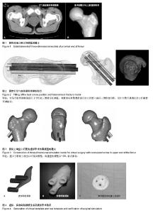
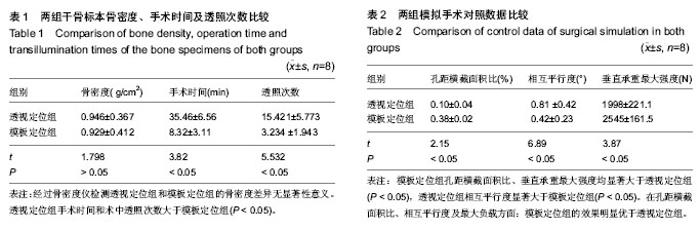
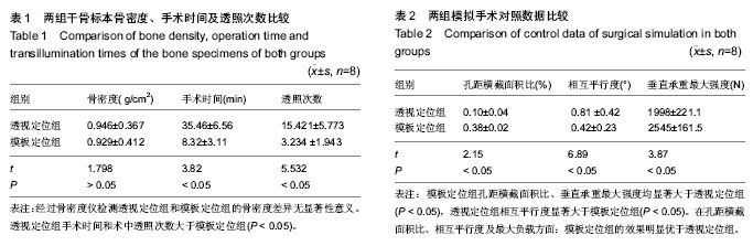
2.1 造模情况 对18例志愿者的股骨上端行连续薄层扫描,将得到的CT数据导入三维重建软件Mimics 10.0,成功建立了36个股骨颈三维模型(图2)。模型可三维旋转,模拟分析股骨上端的解剖结构,为设计数字通道及空心钉最佳穿刺途径,通过3D生物打印技术制作导航模板提供了模型基础。 2.2 设计数字通道及空心钉最佳穿刺途径 个性化地对每个股骨颈模型进行了三维数字化测量,根据指标参数数据进行空心钉置入途径三维形态分析,设计出了数字通道及空心钉最佳穿刺途径,见图2。 2.3 建立了股骨颈空心钉三维立体模型 激光扫描机扫描空心钉,对扫描盲点在Pro-E软件上构建,将得到的空心钉各种数据输入UG Imageware 12.1,建立了股骨上端空心钉复合模型,然后将重建的模型以*.STL格式保存,见图3。 2.4 虚拟导航模板生成 通过提取股骨颈置钉点区域的解剖形态,在UG Imageware 12.1中建立与髋部外侧部解剖结构互补的三维反向贴附模板,将模板与前面建立的股骨颈穿刺途径拟合为带有3个倒三角定位导向管的空心钉导航模板,可以三维任意旋转,见图4A。"

"
| [1] 王满宜,杨庆铭,曾炳芳,等.骨折治疗的AO原则[M].北京:华夏出版社,2005:445-449. [2] Thorngren KG, Hommel A, Norrman PO , et al. Epidemiology of femoral neck fractures. Injury. 2002;33 Suppl 3:C1-7. [3] 胥少汀,葛宝丰,徐印坎.实用骨科学[M].北京:人民军医出版社, 2010:415-447. [4] 马文辉,张英泽.股骨颈骨折:问题及对策[J].中国组织工程研究, 2014,18(9):1426-1433. [5] Zielinski SM, Bouwmans CA, Heetveld MJ, et al. FAITH trial investigators.The societal costs of femoral neck fracture patients treated with internal fixation. Osteoporos Int. 2014; 25(3):875-885. [6] 刘月驹.股骨颈骨折的基础与临床研究[D].河北医科大学,2012. [7] 王立江.不同钉位布局空心螺钉固定股骨颈骨折的生物力学研究[D].河北医科大学,2008. [8] Van Audekercke R, Martens M, Mulier JC, et al. Experimental study on internal fixation of femoral neck fractures. Clin Orthop Relat Res. 1979;(141):203-212. [9] Rubin R, Trent P, Arnold W, et al. Knowles pining of experimental femoral neck fracture:A biomechanical study. Trauma. 1981;21(12):1036-1039. [10] McCutchen JW, Carnesale PG.Comparison of fixation in the treatment of femoral neck fractures. Clin Orthop Relat Res. 1982;11:44-50. [11] 杨立. 三种典型固定钉固定不稳定股骨转子间骨折生物力学特征的三维有限元分析[D].四川大学,2005. [12] 沈惠良,王强,雍宜民.多枚7.0mm空心螺丝钉治疗股骨颈骨折23例[J].中华创伤杂志,2000, 16(3):20-22. [13] El’Sheikh HF, MacDonald BJ, Hashmi MS. Finite element simulation of the hip joint during stumbling: A comparison between static and dynamic loading. J Mater Process Technol. 2003;143/144(1): 249-255. [14] 高昂,王伟,杨琳.应用中空加压螺钉治疗股骨颈骨折手术失误原因分析[J].中国矫形外科杂志,2006,4(2):310. [15] Lu S, Xu YQ, Lu WW, et al. A novel patient-specific navigational template for cervical pedicle screw placement. Spine (Phila Pa 1976). 2009;34(26):E959-966. [16] Zhang YZ, Lu S, Yang YW, et al. Design and primary application of computer-assisted, patient-specific navigational templates in metal-on-metal hip resurfacing arthroplasty. Arthroplasty. 2011;26(7):1083-1087. [17] 罗永祥, Akkineni AR, Anja Lode W, et al. 3-D打印:一种个性化制备复杂支架和组织工程植入物的多功能快速成型技术(英文)[J]. 中国修复重建外科杂志,2014,28(3):279-285. [18] 张颖. 数字化导航模板对全髋置换术旋转中心定位的实验研究及临床应用[D].昆明医科大学,2013. [19] 梁金龙.个体化髋关节导航模板的设计及其在人工髋关节置换术中的实验研究[D].昆明医科大学,2013. [20] 张建雷.个体化导航模板辅助全膝关节置换的基础研究[D].昆明医科大学,2013. [21] 付茂庆. 辅助颈椎前路经椎弓根螺钉置入的生物安全的个性化导航模板研究[D].南方医科大学,2013. [22] 宋柯,付茂庆,孔祥雪,等. 生物安全性的下颈椎后路经椎弓根固定的个性化导航模板研究[J]. 中国临床解剖学杂志,2012,30(5): 509-512. [23] 曹振华,闫金玉,银和平,等.数字及逆向工程技术在复杂骨科应用进展[J].中国组织工程研究,2012,16(39):7370-7374. [24] 黄若昆,谢鸣,余嘉,等.应用动力髋螺钉治疗股骨转子下骨折导航模板与传统方法的效果比较[J].中国矫形外科杂志,2013,11(4): 327-331. [25] 张元智,陈斌,赵建民,等.快速成形导航模板在髋臼发育不良全髋关节置换中的应用[J].内蒙古医科大学学报,2013,35(1):47-51. [26] 陈鸿奋. 髋臼后柱顺行拉力螺钉置钉导航系统的研制与应用[D].南方医科大学,2012. [27] 宋柯.下颈椎后路经椎弓根固定的个性化导航模板研发及其置钉准确性分析[D].南方医科大学,2012. [28] 陈玉兵,陆声,徐永清,等.个体化导航模板辅助腰椎椎弓根螺钉置钉准确性实验研究[J].中国脊柱脊髓杂志,2009,19(8):623-626. [29] 陈玉兵,陆声,徐永清.个体化导航模板在胸椎椎弓根螺钉置入中的初步临床应用[J].中国脊柱脊髓杂志,2011,21(8):669-674. [30] 刘瑞,张元智,李志军,等.个体化导航模板辅助儿童下颈椎椎弓根螺钉置钉准确性实验研究[J].生物骨科材料与临床研究,2012, 9(1):13-16. [31] 刘瑞,张元智,李志军,等.个体化导航模板辅助儿童胸椎椎弓根螺钉置钉准确性实验研究[J].内蒙古医学院学报,2012,34(2): 99-103. [32] 张余,马立敏,尹庆水,等. 数字技术在骨肿瘤外科的应用临床数字骨科(三)[J]. 中国骨科临床与基础研究杂志,2012,4(1):68-72. [33] 李相伟.计算机导航模板辅助Oxford单髁置换术的实验研究及临床应用[D].昆明医科大学,2012. [34] 付军,郭征,王臻,等.多种3-D打印手术导板在骨肿瘤切除重建手术中的应用[J].中国修复重建外科杂志,2014,28(3):304-308. [35] 夏胜利,王秀会,王子平,等.新型空心钉导针定位器的研制及其在股骨颈骨折模型中的应用[J].中国骨与关节损伤杂志,2013, 28(8):707-709. [36] 张铁良,王沛,马信龙.临床骨科学[M].上海:上海世纪出版股份有限公司,2013:514-534. [37] 陆小庆,袁峰.老年髋部骨折手术疗效及失败原因分析[J].中国骨与关节损伤杂志,2012,27(4):334-335. [38] Samuel R,Sarker A.Placement of guidewire for fixation of intertrochanteric femoral neck fractures. Ann R Coll Surg Engl. 2005;87(3):211. [39] Levi N. Fracture of the femoral neck:optimal screw position and bone density determined by computer tomography. Injury. 1996;27(4): 287-289. [40] Hamelinck HK, Haagmans M, Snoeren MM, et al. Safety of Computer-assisted Surgery for Cannulated Hip Screws. Clin Orthop Relat Res. 2007;455:241-245. [41] 王立江,魏海强,郭连江,等.应用皮质支撑原理固定股骨颈骨折的生物力学研究[J].中华创伤杂志,2009,25(7):614-617. [42] Gurusamy K, Parker MJ, Rowlands TK.The complications of displaced intracapsular fractures of the hip:The effect of screw positioning and angulation on fracture healing.Bone Joint Surg Br. 2005;87: 632-634 [43] Zdero R, Keast-Butler O, Schemitsch EH.A biomechanical comparison of two triple-screw methods for femoral neck fracture fixation in a synthetic bone model.Trauma. 2010; 69(6):1537-1544. [44] Van Valin SE, Wenger DR. Value of the false-profile view to identify screw-tip position during treatment of slipped capital femoral epiphysis. A case report. J Bone Joint Surg Am. 2007; 89:643-648. [45] Magarelli N, Milano G, Baudi P, et al. Comparison between 2D and 3D computed tomography evaluation of glenoid bone defect in unilateral anterior gleno-humeral instability.Radiol Med.2012;117(1):102-111. [46] Xie A,Fang C,Huang Y,et al. Application of three-dimensionalreconstruction and visible simulation technique in reoperation of hepatolithiasis. Gastroenterol Hepatol. 2013;28(2):248-254. [47] Wu ZX,Huang LY,Sang HX, et al. Accuracy and safety assessment of pedicle screw placement using the rapid prototypingtechnique in severe congenital scoliosis. Spinal Disord Tech. 2011;24(7):444-450. [48] 陆声,徐永清,张元智,等.计算机辅助导航模板在下颈椎椎弓根定位中的临床应用[J].中华骨科杂志,2008,28(12):1002-1007. [49] Bose S, Vahabzadeh S, Bandyopadhyay A. Bone tissue engineering using 3D printing. Mater Today. 2013;16(12): 496-504. [50] Derby B. Printing and prototyping of tissues and scaffolds. Science. 2012;338(6109): 921-926. [51] Lantada AD, Morgado PL. Rapid prototyping for biomedical engineering: current capabilities and challenges.Annu Rev Biomed Eng. 2012;14: 73-96 |
| [1] | Fang Yi, Zhao Wenzhi, Pan Deyue, Han Xin, Zhang Lu, He Hongtao, Shi Feng, Tian Tingxiao. Acromioclavicular joint dislocation: how to achieve anatomical reduction, sustained stability and micro-motion [J]. Chinese Journal of Tissue Engineering Research, 2020, 24(5): 796-802. |
| [2] | Fu Jiaxin, Xiao Lianping, Wang Shusen, Li Xiaodong, Han Liqiang, Wang Tonghao. Therapeutic effects of paraspinal approach combined with internal fixation through pedicle of fractured vertebra versus traditional AF screw-rod system for thoracolumbar fractures [J]. Chinese Journal of Tissue Engineering Research, 2019, 23(8): 1177-1181. |
| [3] | Qiu Zhongpeng, Li Ke, Li Gang, Liu Keyu, Du Xinhui, Meng Defeng, Shi Chenhui, Wang Weishan. Different treatments for two-part and three-part proximal humeral fractures by Neer classification: follow-up results analyzed using clinical economics [J]. Chinese Journal of Tissue Engineering Research, 2019, 23(8): 1188-1195. |
| [4] | Ke Wei, Li Ke, Wang Sibo, Du Xinhui, Qiu Zhongpeng, Kang Zhilin, Wang Weishan, Li Gang . Open reduction and plate fixation versus closed reduction and external fixation for distal radius fractures: scores and linear regression analysis [J]. Chinese Journal of Tissue Engineering Research, 2019, 23(8): 1196-1202. |
| [5] | Fan Zhirong, Peng Jiajie, Zhong Degui, Zhou Lin, Su Haitao, Huang Yongquan, Wu Jianglin, Liang Yihao. Suture anchor combined with open reduction and internal fixation versus open reduction and internal fixation for ankle fracture combined with deltoid ligament injury: a meta-analysis [J]. Chinese Journal of Tissue Engineering Research, 2019, 23(8): 1307-1312. |
| [6] | Zhou Yu, Liu Yuehong, Liu Shuping, Chen Xi, Qin Wei, Li Qifeng. Spinal stability of intervertebral grafting reinforced by five or six augmenting screws versus transvertebral grafting reinforced by four augmenting screws for thoracolumbar vertebral fractures [J]. Chinese Journal of Tissue Engineering Research, 2019, 23(4): 505-511. |
| [7] | Li Xianzhou, Wang Qian, Zhang Cunxin . Lumbar spondylolisthesis: status and prospects of implant treatment [J]. Chinese Journal of Tissue Engineering Research, 2019, 23(4): 621-627. |
| [8] | Yin Hao, Zhou Enchang, Pan Zhengjun, Chen Guang, Jiang Hua. Finite element analysis of the four and three cannulated screws combined with buttress plate fixation for the treatment of Pauwels III femoral neck fractures [J]. Chinese Journal of Tissue Engineering Research, 2019, 23(32): 5133-5137. |
| [9] |
Wang Lei, Li Zilong, Yuan Binbin, Wu Qingwei, Tang Fengming.
Clinical effect of locking plate versus anterograde intramedullary nail in the treatment of adult humeral shaft fractures: a meta-analysis
[J]. Chinese Journal of Tissue Engineering Research, 2019, 23(24): 3924-3930.
|
| [10] | Hao Liang, Zhang Zhonglin, Wang Baodong, Bi Zhenggang. Intertrochanteric fracture of the femur: improvement of internal fixation device, surgical changes and related disputes [J]. Chinese Journal of Tissue Engineering Research, 2019, 23(18): 2927-2935. |
| [11] | Yao Liquan, Ling Qinjie, Li Jiaying, Zhong Letian, Zhou Xingping, Liu Jintao, He Erxing, Yin Zhixun. Antibiotic artificial bone implantation for treating pyogenic spondylodiscitis [J]. Chinese Journal of Tissue Engineering Research, 2019, 23(14): 2133-2139. |
| [12] | Gong Zhibing, Wu Zhaoke, Zhang Huantang, Xu Zhiqing, Zhuang Zhikun, Zhang Qianjin. Allogeneic cortical bone plate combined with locking plate for Vancouver type B1 and C osteoporotic periprosthetic femoral fractures after hip arthroplasty in older adults [J]. Chinese Journal of Tissue Engineering Research, 2019, 23(12): 1812-1817. |
| [13] | Li Xiaofeng, Xie Furong, Zhan Long, Yang Yuan. Design of locking compression plate through transoral approach [J]. Chinese Journal of Tissue Engineering Research, 2019, 23(12): 1824-1828. |
| [14] | Tian Youyong, Wang Zhiyong. Short-term follow-up of dynamic hip screw versus proximal femoral nail anti-rotation for type AO/OTA A1 intertrochanteric femoral fracture in older adults [J]. Chinese Journal of Tissue Engineering Research, 2019, 23(12): 1834-1839. |
| [15] | Huang Weiyan, Peng Jiewei, Wan Ming, Zheng Xiaoming, Wang Kangzhen. Four internal fixation methods of proximal femoral nail, locking compression plate, dynamic hip screw, and Gamma nail for treating senile intertrochanteric fractures [J]. Chinese Journal of Tissue Engineering Research, 2019, 23(12): 1846-1852. |
| Viewed | ||||||
|
Full text |
|
|||||
|
Abstract |
|
|||||