Chinese Journal of Tissue Engineering Research ›› 2014, Vol. 18 ›› Issue (24): 3773-3778.doi: 10.3969/j.issn.2095-4344.2014.24.001
Micro CT study on trabecular microstructure of tibial plateau in ovariectomized goats
Wang De-zhi, Chen Shi-chang, Liang Zheng-yang, Wang You
- Department of Orthopedics, Ninth People’s Hospital of Shanghai Jiao Tong University, Shanghai 200011, China
-
Revised:2014-04-26Online:2014-06-11Published:2014-06-11 -
Contact:Wang You, M.D., Professor, Chief physician, Doctoral supervisor, Department of Orthopedics, Ninth People’s Hospital of Shanghai Jiao Tong University, Shanghai 200011, China -
About author:Wang De-zhi, Studying for master’s degree, Department of Orthopedics, Ninth People’s Hospital of Shanghai Jiao Tong University, Shanghai 200011, China -
Supported by:the National Science & Technology Pillar Program of China, No. 2012BAI18B07
CLC Number:
Cite this article
Wang De-zhi, Chen Shi-chang, Liang Zheng-yang, Wang You. Micro CT study on trabecular microstructure of tibial plateau in ovariectomized goats[J]. Chinese Journal of Tissue Engineering Research, 2014, 18(24): 3773-3778.
share this article

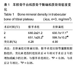
2.1 实验动物数量分析 纳入山羊12只,实验过程中无死亡和感染,最终全部计入结果分析。 2.2 胫骨平台松质骨骨密度 术后2,4年,去卵巢组胫骨平台松质骨骨密度均显著低于假手术组(P < 0.05);随术后时间的延长,去卵巢组与假手术组胫骨平台松质骨骨密度均发生显著性下降(P < 0.05)。见表1。"

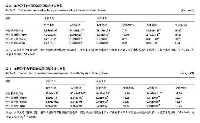
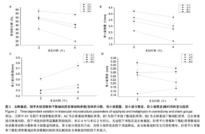
2.3 胫骨平台骨骺与干骺端松质骨微结构参数变化 术后2和4年,与假手术组相比,去卵巢组胫骨平台骨骺和干骺松质骨微观结构参数——骨体积分数、骨小梁数量和骨小梁厚度均降低(P < 0.05),骨小梁分离度均升高(P < 0.05),基本呈时间依赖性变化。仅在术后4年,去卵巢组骨骺松质骨微观结构参数骨小梁厚度与假手术组相比差异无显著性意义(P > 0.05),其骨小梁分离度、骨小梁厚度与去卵巢组术后2年相比差异无显著性意义(P > 0.05)。无论术后2或4年,与假手术组相比,去卵巢组干骺端松质骨微结构参数的改变均比骨骺松质骨明显。见表2,3,图2。"

"
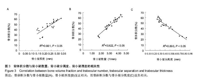
2.4 骨体积分数与骨小梁数量、骨小梁厚度和骨小梁分离度的相关性 见图3。对各标本胫骨平台骨骺与干骺端松质骨测得的骨体积分数与对应的骨小梁数量、骨小梁分离度和骨小梁厚度进行Pearson相关分析,发现骨体积分数与骨小梁数量和骨小梁厚度呈正相关关系(P < 0.05),相关系数分别为0.922,0.661,而骨体积分数与骨小梁分离度呈负相关关系(P < 0.05),相关系数为0.893。"

| [1] 李杨,冯世庆,杨宁,等.局部注射辛伐他汀对骨质疏松大鼠股骨髁骨小梁的改建效应[J].中国组织工程研究,2013,17(46): 7994-7999. [2] 王新祥,张允岭,吴坚.葛根对睾丸切除骨质疏松模型小鼠骨密度和骨构造的作用[J].中国组织工程研究与临床康复,2010,14(7): 1262-1266. [3] 韩亚军,帖小佳,伊力哈木•托合提.中国中老年人骨质疏松症患病率的Meta分析[J].中国组织工程研究,2014,18(7):1129-1134. [4] 赵玺,赵文,孙璟,等.骨代谢指标与骨关节炎及绝经后骨质疏松症的关系[J].中国组织工程研究,2014,18(2):245-250. [5] Raafat BM, Hassan NS, Aziz SW, et al. Bone mineral density (BMD) and osteoporosis risk factor in Egyptian male and female battery manufacturing workers. Toxicol Ind Health. 2012;28(3):245-52. [6] Blake GM, Griffith JF, Yeung DK, et al. Effect of increasing vertebral marrow fat content on BMD measurement, T-Score status and fracture risk prediction by DXA. Bone. 2009;44(3): 495-501. [7] McNamara LM. Perspective on post-menopausal osteoporosis: establishing an interdisciplinary understanding of the sequence of events from the molecular level to whole bone fractures. J R Soc Interface. 2010;7(44):353-372. [8] 张钧,江莉婷,王晋申,等.1型糖尿病小鼠下颌骨三维结构及组织形态[J].中国组织工程研究,2013,17(28):5101-5107. [9] 余文超,刘岩,袁文.脊髓损伤早期及制动大鼠股骨干骺端的显微CT观察[J].中国组织工程研究与临床康复,2010,15(30): 5596-5599. [10] Effendy NM, Khamis MF, Shuid AN. Micro-CT assessments of potential anti-osteoporotic agents. Curr Drug Targets. 2013; 14(13):1542-1551. [11] 王亮,张志敏,甄相周,等.去势雌性山羊骨质疏松模型的特点[J].中国组织工程研究,2012,16(7):1303-1306. [12] 李素萍.骨质疏松动物模型的研究现状[J].中国组织工程研究与临床康复,2011,15(20):3767-3770. [13] Newton B, Cooper RC, Gilbert JA, et al. The ovariectomized sheep as a model for human bone loss. J Comp Pathol. 2004; 130(4):323. [14] Oheim R, Amling M, Ignatius A, et al. Large animal model for osteoporosis in humans: the ewe. Eur Cell Mater. 2012;24: 372-385. [15] The Ministry of Science and Technology of the People’s Republic of China. Guidance Suggestions for the Care and Use of Laboratory Animals. 2006-09-30. [16] 丁文鸽.卵巢切除骨质疏松小鼠骨折愈合不同时期骨微结构及力学性能变化[J].中国组织工程研究与临床康复, 2008,12(42): 8247-8250. [17] Davison KS, Kendler DL, Ammann P, et al. Assessing fracture risk and effects of osteoporosis drugs:bone mineral density and beyond. Am J Med. 2009;122(11):992-997. [18] Rubin CD. Emerging concepts in osteoporosis and bone strength. Curr Med Res Opin. 2005;21(7):1049-1056. [19] 李冠武,汤光宇.Micro-CT及1H-MRS在骨质疏松骨质量研究中的应用[J].国际医学放射学杂志,2010,33(6):525-528. [20] 粱敏,张劼,粱杏欢,等.雌孕激素联合对去卵巢大鼠骨密度和骨形态计量学的影响[J].中国组织工程研究与临床康复, 2010, 14(24): 4380-4384. [21] Kim JE, Shin JM, Oh SO, et al. The three-dimensional microstructure of trabecular bone:Analysis of site-specific variation in the human jaw bone. Imaging Sci Den. 2013; 43(4):227-233. [22] Chen H, Wu M, Kubo KY. Combined treatment with a traditional Chinese medicine, Hachimi-jio-gan (Ba-Wei-Di-Huang-Wan) and alendronate improves bone microstructure in ovariectomized rats. J Ethnopharmacol. 2012;142(1):80-85. [23] Xu Y, Li D, Chen Q, et al. Full supervised learning for osteoporosis diagnosis using micro-CT images. Microsc Res Tech. 2013;76(4):333-341. [24] 吴子祥,雷伟,胡蕴玉,等.骨质疏松绵羊模型松质骨及皮质骨的微观结构及力学性能变化的研究[J].中国骨质疏松杂志, 2007, 13(8):537-541. [25] Yang J, Pham SM, Crabbe DL. High-resolution Micro-CT evaluation of mid- to long-term effects of estrogen deficiency on rat trabecular bone. Acad Radiol. 2003;10(10):1153-1158. [26] Jiang Y, Zhao J, Liao EY, et al. Application of micro-CT assessment of 3-D bone microstructure in preclinical and clinical studies. J Bone Miner Metab. 2005;23:122-131. [27] Frost HM. A 2003 update of bone physiology and Wolff's Law for clinicians. Angle Orthod. 2004;74(1):3-15. [28] Chen H, Washimi Y, Kubo KY, et al. Gender-related changes in three-dimensional microstructure of trabecular bone at the human proximal tibia with aging. Histol Histopathol. 2011; 26(5):563-570. [29] Thomsen JS, Niklassen AS, Ebbesen EN, et al. Age-related changes of vertical and horizontal lumbar vertebral trabecular 3D bone microstructure is different in women and men. Bone. 2013;57(1):47-55. [30] Milovanovic P, Djonic D, Marshall RP, et al. Micro-structural basis for particular vulnerability of the superolateral neck trabecular bone in the postmenopausal women with hip fractures. Bone. 2012;50(1):63-68. [31] Bevill G, Eswaran SK, Gupta A, et al. Influence of bone volume fraction and architecture on computed large-deformation failure mechanisms in human trabecular bone. Bone. 2006;39(6):218-225. [32] Arlot ME, Burt-Pichat B, Roux JP, et al. Microarchitecture influences microdamage accumulation in human vertebral trabecular bone. J Bone Miner Res. 2008;23(10):1613-1618. [33] Brouwers JE, van Rietbergen B, Huiskes R, et al. Effects of PTH treatment on tibial bone of ovariectomized rats assessed by in vivo micro-CT. Osteoporos Int. 2009;20(11):1823-1835. [34] 李展春,程光齐,张自明,等.骨质疏松与骨关节炎患者松质骨的显微结构[J].国际病理科学与临床杂志,2011,31(1):17-20. |
| [1] | Tang Hui, Yao Zhihao, Luo Daowen, Peng Shuanglin, Yang Shuanglin, Wang Lang, Xiao Jingang. High fat and high sugar diet combined with streptozotocin to establish a rat model of type 2 diabetic osteoporosis [J]. Chinese Journal of Tissue Engineering Research, 2021, 25(8): 1207-1211. |
| [2] | Li Zhongfeng, Chen Minghai, Fan Yinuo, Wei Qiushi, He Wei, Chen Zhenqiu. Mechanism of Yougui Yin for steroid-induced femoral head necrosis based on network pharmacology [J]. Chinese Journal of Tissue Engineering Research, 2021, 25(8): 1256-1263. |
| [3] | Hou Guangyuan, Zhang Jixue, Zhang Zhijun, Meng Xianghui, Duan Wen, Gao Weilu. Bone cement pedicle screw fixation and fusion in the treatment of degenerative spinal disease with osteoporosis: one-year follow-up [J]. Chinese Journal of Tissue Engineering Research, 2021, 25(6): 878-883. |
| [4] | Li Shibin, Lai Yu, Zhou Yi, Liao Jianzhao, Zhang Xiaoyun, Zhang Xuan. Pathogenesis of hormonal osteonecrosis of the femoral head and the target effect of related signaling pathways [J]. Chinese Journal of Tissue Engineering Research, 2021, 25(6): 935-941. |
| [5] | Kong Lingbao, Lü Xin. Effect of implant selection and approach on support in the operation of posterolateral tibial plateau fractures [J]. Chinese Journal of Tissue Engineering Research, 2021, 25(6): 942-947. |
| [6] | Xiao Fangjun, Chen Shudong, Luan Jiyao, Hou Yu, He Kun, Lin Dingkun. An insight into the mechanism of Salvia miltiorrhiza intervention on osteoporosis based on network pharmacology [J]. Chinese Journal of Tissue Engineering Research, 2021, 25(5): 772-778. |
| [7] | Liu Bo, Chen Xianghe, Yang Kang, Yu Huilin, Lu Pengcheng. Mechanism of DNA methylation in exercise intervention for osteoporosis [J]. Chinese Journal of Tissue Engineering Research, 2021, 25(5): 791-797. |
| [8] | Nie Shaobo, Li Jiantao, Sun Jien, Zhao Zhe, Zhao Yanpeng, Zhang Licheng, Tang Peifu. Mechanical stability of medial support nail in treatment of severe osteoporotic intertrochanteric fracture [J]. Chinese Journal of Tissue Engineering Research, 2021, 25(3): 329-333. |
| [9] | Zhong Yuanming, Wan Tong, Zhong Xifeng, Wu Zhuotan, He Bingkun, Wu Sixian. Meta-analysis of the efficacy and safety of percutaneous curved vertebroplasty and unilateral pedicle approach percutaneous vertebroplasty in the treatment of osteoporotic vertebral compression fracture [J]. Chinese Journal of Tissue Engineering Research, 2021, 25(3): 456-462. |
| [10] | Xie Chengxin, Wang Wei, Wang Chenglong, Li Qinglong, Yin Dong. Systematic review and meta-analysis of bone morphogenetic protein for the treatment of acute tibial fracture [J]. Chinese Journal of Tissue Engineering Research, 2021, 25(3): 486-492. |
| [11] | Lin Wang, Wang Yingying, Guo Weizhong, Yuan Cuihua, Xu Shenggui, Zhang Shenshen, Lin Chengshou. Adopting expanded lateral approach to enhance the mechanical stability and knee function for treating posterolateral column fracture of tibial plateau [J]. Chinese Journal of Tissue Engineering Research, 2021, 25(24): 3826-3827. |
| [12] | Zhu Yun, Chen Yu, Qiu Hao, Liu Dun, Jin Guorong, Chen Shimou, Weng Zheng. Finite element analysis for treatment of osteoporotic femoral fracture with far cortical locking screw [J]. Chinese Journal of Tissue Engineering Research, 2021, 25(24): 3832-3837. |
| [13] | Jie Ke, Deng Peng, Zeng Yirong. Application and comparison of four commonly used methods for patellar height measurement [J]. Chinese Journal of Tissue Engineering Research, 2021, 25(24): 3875-3881. |
| [14] | Feng Guancheng, Fang Jianming, Lü Haoran, Zhang Dongsheng, Wei Jiadong, Yu Bingbing. How does bone cement dispersion affect the early outcome of percutaneous vertebroplasty [J]. Chinese Journal of Tissue Engineering Research, 2021, 25(22): 3450-3457. |
| [15] | Liu Chang, Li Datong, Liu Yuan, Kong Lingbo, Guo Rui, Yang Lixue, Hao Dingjun, He Baorong. Poor efficacy after vertebral augmentation surgery of acute symptomatic thoracolumbar osteoporotic compression fracture: relationship with bone cement, bone mineral density, and adjacent fractures [J]. Chinese Journal of Tissue Engineering Research, 2021, 25(22): 3510-3516. |
| Viewed | ||||||
|
Full text |
|
|||||
|
Abstract |
|
|||||