| [1] Chan CW, Peng P. Failed back surgery syndrome. Pain Med. 2011;12(4):577-606.
[2] 董健,李超.腰椎手术失败综合征[J].中华骨科杂志,2012,32(10): 984-989.
[3] 徐文斌,范顺武,赵兴.腰椎手术失败综合征的再手术现状[J].中华骨科杂志,2012,32(10):979-981.
[4] Watkins MB. Posterolateral bonegrafting for fusion of the lumbar and lumbosacral spine. J Bone Joint Surg Am. 1959; 41-A(3):388-396.
[5] Wiltse LL, Bateman JG, Hutchinson RH, et al. The paraspinal sacrospinalis-splitting approach to the lumbar spine. J Bone Joint Surg Am. 1968;50(5):919-926.
[6] Wiltse LL, Spencer CW. New uses and refinements of the paraspinal approach to the lumbar spine. Spine (Phila Pa 1976). 1988;13(6):696-706.
[7] Vialle R, Court C, Khouri N, et al. Anatomical study of the paraspinal approach to the lumbar spine. Eur Spine J. 2005; 14(4):366-371.
[8] Vialle R, Wicart P, Drain O, et al. The Wiltse paraspinal approach to the lumbar spine revisited: an anatomic study. Clin Orthop Relat Res. 2006;445:175-180.
[9] Olivier E, Beldame J, Ould Slimane M, et al. Comparison between one midline cutaneous incision and two lateral incisions in the lumbar paraspinal approach by Wiltse: a cadaver study. Surg Radiol Anat. 2006;28(5):494-497.
[10] Palmer DK, Allen JL, Williams PA, et al. Multilevel magnetic resonance imaging analysis of multifidus-longissimus cleavage planes in the lumbar spine and potential clinical applications to Wiltse's paraspinal approach. Spine (Phila Pa 1976). 2011;36(16):1263-1267.
[11] Waschke A, Hartmann C, Walter J, et al. Denervation and atrophy of paraspinal muscles after open lumbar interbody fusion is associated with clinical outcome--electromyographic and CT-volumetric investigation of 30 patients. Acta Neurochir (Wien). 2014;156(2):235-244.
[12] Bresnahan LE, Smith JS, Ogden AT, et al. Assessment of Paraspinal Muscle Cross-Sectional Area Following Lumbar Decompression: Minimally Invasive versus Open Approaches. J Spinal Disord Tech. 2013 Oct 13.
[13] Mukai Y, Takenaka S, Hosono N, et al. Intramuscular pressure of the multifidus muscle and low-back pain after posterior lumbar interbody fusion: comparison of mini-open and conventional approaches. J Neurosurg Spine. 2013; 19(6):651-657.
[14] 蒋宋怡,胡志军,范顺武,等.两种入路在腰椎椎体间融合术中对多裂肌损伤的病例对照研究[J].中国骨伤,2013,26(9):735-740.
[15] 邵诗泽,张恩忠,付松,等. 腰骶段多裂肌的形态特点及功能意义[J].中国临床解剖学杂志,2010,28(1):17-19.
[16] Hu ZJ, Fang XQ, Zhou ZJ, et al. Effect and possible mechanism of muscle-splitting approach on multifidus muscle injury and atrophy after posterior lumbar spine surgery. J Bone Joint Surg Am. 2013;95(24):e192(1-9).
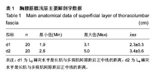
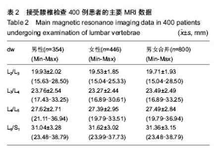
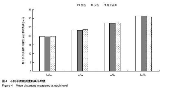
[17] 王洋,武汉,张子言,等.腰椎椎旁肌间隙至棘突正中线距离的MRI测量及意义[J].中国脊柱脊髓杂志,2013,23(4):316-319.
[18] 赵斌,赵轶波,马迅,等.经椎旁肌间隙入路在胸腰椎骨折治疗中的应用[J].中华骨科杂志,2011,31(10):1147-1151.
[19] 刘中浩,彭国栋,林勇,等.肌间隙入路并伤椎植骨内固定治疗胸腰椎骨折[J].中华创伤杂志,2013,29(6):503-506.
[20] 任忠明,吴宏飞,张远,等.肌间隙入路与传统入路椎弓根螺钉内固定治疗胸腰椎骨折[J].中华创伤杂志,2013,29(9):845-848.
[21] 蔡福金,朱建平,骆宇春,等.经椎旁肌间隙入路椎弓根钉棒系统置入内固定治疗胸腰椎爆裂骨折:与传统方法比较[J].中国组织工程研究,2012,16(30):5676-5680.
[22] 戴胡明,方诗元,夏睿,等.传统入路与椎旁肌间隙入路置入椎弓根螺钉治疗胸腰段骨折[J].中国组织工程研究,2013,17(13):2339- 2345.
[23] 郑燕平,刘新宇,原所茂. Wiltse入路经椎间孔腰椎椎体间融合术治疗单节段腰椎峡部裂性滑脱[J].中华骨科杂志,2011,31(9): 921-926.
[24] 张铭华,董靖,卢旻鹏,等. Wiltse手术入路和后正中入路经椎间孔腰椎椎体间融合术治疗退变性腰椎滑脱症[J].中华创伤杂志, 2012,28(7):624-628.
[25] 任忠明,吴宏飞,张远,等.双侧小切口椎旁肌间隙入路在下腰椎融合术中应用[J].中国矫形外科杂志,2012,20(15):1356-1359.
[26] 陈宣煌,许卫红,胡建伟,等.小切口椎旁肌间隙入路和传统开放式入路腰椎后路融合术的比较[J].脊柱外科杂志,2012,10(2): 101-104.
[27] 吴剑飞,谢德胜,龚进红,等.椎旁肌间隙入路治疗椎间孔外型腰椎间盘突出症15例[J].颈腰痛杂志, 2012,33(6):435-437.
[28] 高先政,杨滨,邹德威,等.经椎旁肌间隙入路与传统后正中入路腰椎融合术治疗L5S1腰椎间盘突出症对比研究[J].中国矫形外科杂志,2013,21(9):874-878.
[29] 黎庆初,尹刚辉,张忠民,等. 微创Wiltse入路与传统后正中入路手术治疗双节段腰椎管狭窄症的疗效比较[J].中国脊柱脊髓杂志,2012,22(9):812-817.
[30] 林定坤,陈树东,苏国义,等.经肌间隙入路经椎间孔腰椎椎体间融合术并椎管潜行减压治疗腰椎管狭窄症[J].广东医学,2013, 34 (15):2336-2339.
[31] 陆军海,贝抗胜,熊英辉.椎旁肌间隙入路在胸腰椎手术中的应用进展[J].中华损伤与修复杂志(电子版),2013,8(1):62-65. |