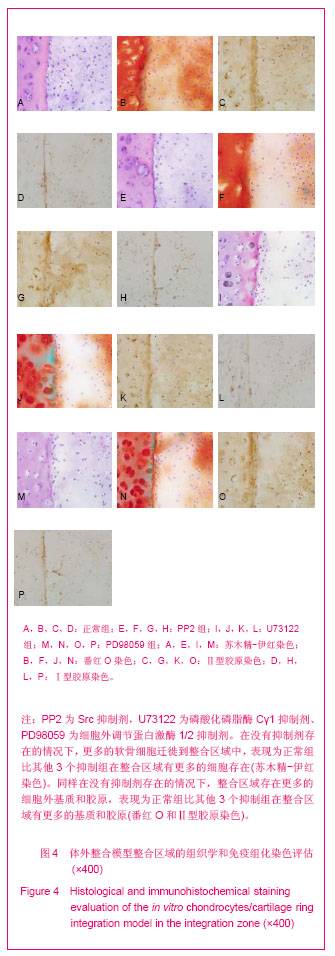
| [1]Khan IM, Gilbert SJ, Singhrao SK, et al. Cartilage integration: evaluation of the reasons for failure of integration during cartilage repair. A review. Eur Cell Mater. 2008;16:26-39.
[2]Zhang Z, McCaffery JM, Spencer RG, et al. Growth and integration of neocartilage with native cartilage in vitro. J Orthop Res. 2005;23(2):433-439.
[3]Pabbruwe MB, Esfandiari E, Kafienah W, et al. Induction of cartilage integration by a chondrocyte/collagen-scaffold implant.Biomaterials. 2009;30(26):4277-4286.
[4]Theodoropoulos JS, De Croos JN, Park SS, et al. Integration of tissue-engineered cartilage with host cartilage: an in vitro model. Clin Orthop Relat Res. 2011;469(10):2785-2795.
[5]Beier F, Loeser RF. Biology and pathology of Rho GTPase, PI-3 kinase-Akt, and MAP kinase signaling pathways in chondrocytes. J Cell Biochem. 2010;110(3):573-580.
[6]Whitney NP, Lamb AC, Louw TM, et al. Integrin-mediated mechanotransduction pathway of low-intensity continuous ultrasound in human chondrocytes. Ultrasound Med Biol. 2012;38(10):1734-1743.
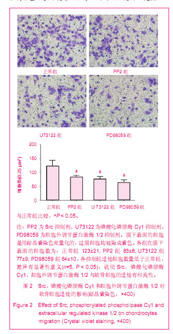
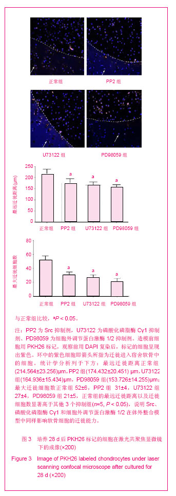
[7]Nong L, Yin G, Ren K, et al. Periodic mechanical stress enhances rat chondrocyte area expansion and migration through Src-PLCgamma1-ERK1/2 signaling. Eur J Cell Biol. 2010;89(9):705-711.
[8]Enders JT, Otto TJ, Peters HC, et al. A model for studying human articular cartilage integration in vitro. J Biomed Mater Res A. 2010;94(2):509-514.
[9]Bos PK, DeGroot J, Budde M, et al.Specific enzymatic treatment of bovine and human articular cartilage: implications for integrative cartilage repair. Arthritis Rheum. 2002;46(4):976-985.
[10]Ahsan T, Sah R. Biomechanics of integrative cartilage repair. Osteoarthritis Cartilage. 1999;7:29-40.
[11]Ruggiero F, Petit B, Ronziere M, et al. Composition and organization of the collagen network produced by fetal bovine chondrocytes cultured at high density. J Histochem Cytochem. 1993;41:867-875.
[12]Seol D, McCabe DJ, Choe H, et al. Chondrogenic progenitor cells respond to cartilage injury. Arthritis Rheum. 2012;64(11): 3626-3637.
[13]Raggatt LJ, Jefcoat SC Jr, Choudhury I, et al. Matrix metalloproteinase-13 influences ERK signalling in articular rabbit chondrocytes. Osteoarthritis Cartilage. 2006;14(7): 680-689.
[14]Kudirka JC, Panupinthu N, Tesseyman MA, et al. P2Y nucleotide receptor signaling through MAPK/ERK is regulated by extracellular matrix: involvement of beta3 integrins. J Cell Physiol. 2007;213(1):54-64.
[15]Bradley EW, Drissi MH. Wnt5b regulates mesenchymal cell aggregation and chondrocyte differentiation through the planar cell polarity pathway. J Cell Physiol. 2011;226(6): 1683-1693.
[16]Bursell L, Woods A, James CG, et al. Src kinase inhibition promotes the chondrocyte phenotype. Arthritis Res Ther. 2007;9(5):R105.
[17]Ryan JA, Eisner EA, DuRaine G, et al. Mechanical compression of articular cartilage induces chondrocyte proliferation and inhibits proteoglycan synthesis by activation of the ERK pathway: implications for tissue engineering and regenerative medicine. J Tissue Eng Regen Med. 2009;3(2): 107-116.
[18]Yoon YM, Kim SJ, Oh CD, et al. Maintenance of differentiated phenotype of articular chondrocytes by protein kinase C and extracellular signal-regulated protein kinase. J Biol Chem. 2002; 277(10):8412-8420.
[19]DiMicco MA, Waters SN, Akeson WH, et al. Integrative articular cartilage repair: dependence on developmental stage and collagen metabolism. Osteoarthritis Cartilage. 2002;10(3): 218-225. |