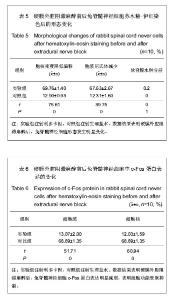
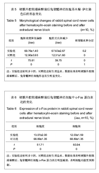
| [1] |
Zhu Xuefen, Huang Cheng, Ding Jian, Dai Yongping, Liu Yuanbing, Le Lixiang, Wang Liangliang, Yang Jiandong.
Mechanism of bone marrow mesenchymal stem cells differentiation into functional neurons induced by glial cell line derived neurotrophic factor
[J]. Chinese Journal of Tissue Engineering Research, 2021, 25(7): 1019-1025.
|
| [2] |
Wang Feng, Zhou Liyu, Saijilafu, Qi Shibin, Ma Yanxia, Wei Shanwen.
CaMKII-Smad1 promotes axonal regeneration of peripheral nerves
[J]. Chinese Journal of Tissue Engineering Research, 2021, 25(7): 1064-1068.
|
| [3] |
Deng Zhibo, Li Zhi, Wu Yahong, Mu Yuan, Mu Yuexi, Yin Liangjun.
Local infiltration anesthesia versus femoral nerve block for pain control and safety after total knee arthroplasty: a meta-analysis
[J]. Chinese Journal of Tissue Engineering Research, 2021, 25(21): 3401-3408.
|
| [4] |
Chen Deng, Zhang Yaxin, Dai Jihang, Chen Duoyun, Sun Yu.
Analysis on relative factors affecting pyrexia following total hip replacement
[J]. Chinese Journal of Tissue Engineering Research, 2021, 25(18): 2846-2850.
|
| [5] |
Wang Shao, Yuan Dajiang, Li Yanyan, Li Xiaoya.
Dexmedetomidine combined with local anesthetic for brachial plexus block: a systematic review and meta-analysis
[J]. Chinese Journal of Tissue Engineering Research, 2021, 25(12): 1951-1958.
|
| [6] |
Xiong Guobing, Liu Aibo, Wang Shize, Wang Yu, Qiu Mingxing.
Urine turbulent shear stress system of bionic human bladder based on bacterial biofilm reactor: in vitro construction#br#
[J]. Chinese Journal of Tissue Engineering Research, 2021, 25(10): 1560-1565.
|
| [7] |
Wang Guoyu, Cheng Zhijian, Yang Baohui, Li Haopeng, He Xijing.
Olfactory ensheathing cell transplantation promotes the
ultrastructure repair at the lesion site of rat models of spinal cord injury
[J]. Chinese Journal of Tissue Engineering Research, 2020, 24(5): 699-703.
|
| [8] |
Wang Jing, Lu Changfeng, Peng Jiang, Zhu Chen, Xu Wenjing, Cheng Xiaoqing, Fang Jie, Zhu Yaqiong, Zhao Yanxu, Jiang Wen, Xu Hongguang, Wang Yu.
Establishment and evaluation of traumatic neuroma model
[J]. Chinese Journal of Tissue Engineering Research, 2020, 24(5): 716-719.
|
| [9] |
Liu Yongpan, Gong Xiaofang.
Effects of dexmedetomidine on perioperative brain protection in patients undergoing craniocerebral surgery under inhalation anesthesia with sevoflurane: a randomized controlled study
[J]. Chinese Journal of Tissue Engineering Research, 2020, 24(35): 5688-5694.
|
| [10] |
Du Xiaowen, Lin Dapeng, Tu Guanjun.
S100A4 promotes differentiation of neural stem cells through
up-regulation of brain-derived neurotrophic factor
[J]. Chinese Journal of Tissue Engineering Research, 2020, 24(19): 3029-3034.
|
| [11] |
Liu Jianhang, Liu Hao, Chen Daoyun, Xu Zhiwei, Xie Guixin, Li Jinwei.
Advantages and values of magnetic resonance diffusion tensor imaging in clinical diagnosis and treatment
[J]. Chinese Journal of Tissue Engineering Research, 2019, 23(8): 1241-1247.
|
| [12] |
Peng Xianjiang1, Liu Bozhi2, Duan Haifeng1, Yang Qianzi1, Hu Sheng1 .
Anesthesia management for aortic valve plus mitral valve replacement in Small-Tailed Han sheep
[J]. Chinese Journal of Tissue Engineering Research, 2019, 23(7): 1052-1056.
|
| [13] |
Xia Ling, Wang Pan, Wu Chunfang, Zhang Zhaobo.
Application value of surface electromyography in the repair of peripheral nerve injury
[J]. Chinese Journal of Tissue Engineering Research, 2019, 23(7): 1142-1148.
|
| [14] |
Xie Yuchen, Chen Wendong, Ma Li .
Anatomical differences in three-dimensional finite element model of difficult airway patients
[J]. Chinese Journal of Tissue Engineering Research, 2019, 23(4): 562-566.
|
| [15] |
Gong Jiao, Liu Ming.
Human induced pluripotent stem cell transplantation for hypoxic-ischemic encephalopathy in neonatal mice
[J]. Chinese Journal of Tissue Engineering Research, 2019, 23(33): 5322-5327.
|