Chinese Journal of Tissue Engineering Research ›› 2013, Vol. 17 ›› Issue (36): 6423-6429.doi: 10.3969/j.issn.2095-4344.2013.36.007
Previous Articles Next Articles
In vitro osteogenic differentiation of bone marrow mesenchymal stem cells from ovariectomied osteoporotic rats
Wang Yun, Bao Xiao-ming, Hou Yong-xin, Li Jun, Zhang Min
- Department of Orthopedics, the Second Hospital of Shanxi Medical University, Taiyuan 030001, Shanxi Province, China
-
Received:2013-01-07Revised:2013-02-19Online:2013-09-03Published:2013-09-03 -
Contact:Zhang Min, M.D., Associate professor, Department of Orthopedics, the Second Hospital of Shanxi Medical University, Taiyuan 030001, Shanxi Province, China zhangminty@yahoo.com.cn -
About author:Wang Yun★, Studying for master’s degree, Department of Orthopedics, the Second Hospital of Shanxi Medical University, Taiyuan 030001, Shanxi Province, China wangyunff@163.com -
Supported by:Key Science and Technology Project of Shanxi Province, No. 20100311098-2*
CLC Number:
Cite this article
Wang Yun, Bao Xiao-ming, Hou Yong-xin, Li Jun, Zhang Min. In vitro osteogenic differentiation of bone marrow mesenchymal stem cells from ovariectomied osteoporotic rats[J]. Chinese Journal of Tissue Engineering Research, 2013, 17(36): 6423-6429.
share this article
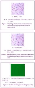
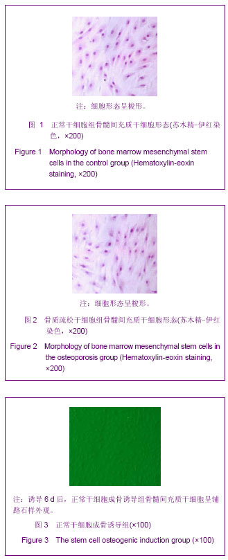
2.1 实验动物数量分析 实验选用大鼠40只,分为4组,实验过程无脱失,全部进入结果分析。 2.2 骨髓间充质干细胞(成骨诱导后)形态学观察 正常干细胞组和骨质疏松干细胞组的骨髓间充质干细胞经培养、传代后,细胞状态逐渐稳定,两组的细胞形态均呈梭形,无明显差异,见图1,2。正常干细胞成骨诱导组和骨质疏松干细胞成骨诱导组的骨髓间充质干细胞在加入成骨诱导液后形态开始改变,逐渐由梭形转变为圆形、立方形。其中,正常干细胞成骨诱导组骨髓间充质干细胞的细胞形态在诱导后6 d明显变为铺路石样外观;而骨质疏松干细胞成骨诱导组骨髓间充质干细胞在诱导6 d后,除立方形细胞外,仍存有部分梭形细胞,见图3,4。诱导8 d后,骨质疏松干细胞成骨诱导组细胞形态明显变为立方型、呈铺路石样外观,与正常干细胞成骨诱导组无明显差别。"
2.3 正常及骨质疏松大鼠骨髓间充质干细胞增殖指数变化 正常干细胞组和骨质疏松干细胞组的细胞周期分布、增殖指数见表1。"
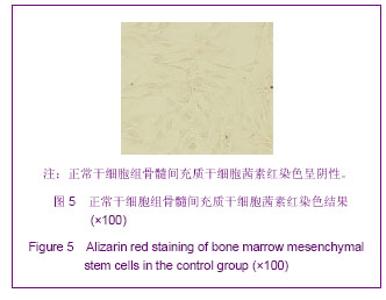
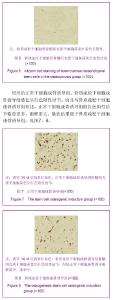
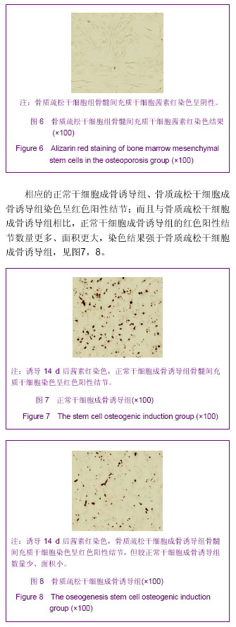
由表1可见,正常干细胞组的骨髓间充质干细胞增殖指数值明显高于骨质疏松干细胞组,差异有显著性意义 (P < 0.05),表明骨质疏松干细胞组的细胞增殖活性降低。 2.4 骨髓间充质干细胞成骨诱导后茜素红染色 成骨诱导14 d后,茜素红对正常干细胞组、骨质疏松干细胞组及相应的正常干细胞成骨诱导组、骨质疏松干细胞成骨诱导组细胞进行染色。其中正常干细胞组、骨质疏松干细胞组的染色结果呈阴性,见图5,6。"
"
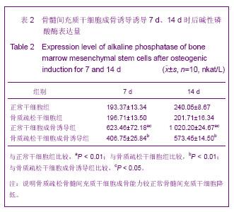
2.5 各组骨髓间充质干细胞的碱性磷酸酶染色表达量变化 培养(或成骨诱导)后各组骨髓间充质干细胞的碱性磷酸酶表达量见表2。在培养的第7天(或成骨诱导的第7天),正常干细胞组和骨质疏松干细胞组的碱性磷酸酶表达量差异无显著性意义(P > 0.05)。正常干细胞成骨诱导组和骨质疏松干细胞成骨诱导组碱性磷酸酶的表达量分别是相应正常干细胞组的3.22倍(P < 0.01)、 骨质疏松干细胞组的2.07倍(P < 0.01)。正常干细胞成骨诱导组碱性磷酸酶表达量是骨质疏松干细胞成骨诱导组的1.53倍(P < 0.05)。 在培养的第14天(成骨诱导的第14天),正常干细胞组和骨质疏松干细胞组的碱性磷酸酶表达量差异无显著性意义(P > 0.05)。正常干细胞成骨诱导组和骨质疏松干细胞成骨诱导组碱性磷酸酶的表达量分别是相应正常干细胞组的4.25倍(P < 0.01)、骨质疏松干细胞组的2.84倍(P < 0.01)。正常干细胞成骨诱导组碱性磷酸酶表达量是骨质疏松干细胞成骨诱导组的1.78倍 (P < 0.05)。"

| [1] Adams JE. Advances in bone imaging for osteoporosis. Nat Rev Endocrinol. 2012; 9(1): 28-42.
[2] Coipeau P,Rosset P,Langonne A,et al.Impaired differentiation potential of human trabecular bone mesenchymal stromal cells from elderly patients.Cytotherapy.2009; 11: 584-594.
[3] Wagner W, Wein F, Seckinger A, et al. Comparative characteristics of mesenchymal stem cells from human bone marrow, adipose tissue, and umbilical cord blood. Exp Hematol. 2005;33(11): 1402-1416.
[4] Yoshimura H, Muneta T, Nimura A, et al. Comparison of rat mesenchymal stem cells derived from bone marrow, synovium, periosteum, adipose tissue, and muscle. Cell Tissue Res.2007;327 (3): 449- 462.
[5] Srouji S,Livne E. Bone marrow stem cells and biological scaffold for bone repair in aging and disease. Mech Ageing Dev.2005;126( 2): 281-287.
[6] Zhu X, Du J, Liu G. The comparison of multilineage differentiation of bone marrow and adipose-derived mesenchymal stem cells. Clin Lab. 2012;58: 897-903.
[7] 中华人民共和国科学技术部.关于善待实验动物的指导性意见. 2006-09-30.
[8] 李素萍. 骨质疏松动物模型的研究现状[J].中国组织工程研究与临床康复, 2011, 15(20): 3767-3770.
[9] Chachra D, Lee JM, Kasram, et al. Differential effects of ovariectomy on the mechanical properties of cortical and cancellous bones in rat femora and vertebrae. Biomed Sci Instrum.2000;36: 123-128.
[10] Liu Y, Wang L, Fatahi R, et al. Isolation of murine bone marrow derived mesenchymal stem cells using Twist2 Cre transgenic mice. Bone. 2010;47: 916-925.
[11] Javazon EH, Beggs K, Flake AW. Mesenchymal stem cells: paradoxes of passaging. Exp Hematol.2004;32(5): 414-425.
[12] Delmas P. Clinical usefulness of biochemical markers of bone remodeling in osteoporosis. Rev Prat. 1995;45(9):1102-1105.
[13] Bala Y, Farlay D, Chapurlat RD, et al.Modifications of bone material properties in postmenopausal osteoporotic women long-term treated with alendronate. Eur J Endocrinol. 2011; 165(4): 647-655.
[14] Pinzon R. The clinical profile and risk factors of postmenopausal lumbar osteoporosis. International Journal of Rheumatic Diseases, 2010,13:171.
[15] 中华医学会骨质疏松和骨矿盐疾病分会.原发性骨质疏松症诊治指南(2011年).中华骨质疏松和骨矿盐疾病杂志, 2011, 4: 2-17.
[16] Shil AB, Greer JR. Screening strategies for treatment of osteoporosis in long-term care residents. Am Geriatr Soc. 2012;60(12): 2383-2384.
[17] 王强,赵兵,李超,等.干细胞移植治疗骨质疏松[J].中华骨质疏松和骨矿盐疾病杂志,2012, 5(3): 225-229.
[18] Kumar S, Chanda D, Ponnazhagan S. Therapeutic potential of genetically modified mesenchymal stem cells. Gene Ther. 2008;15(10):711-715.
[19] Rosen CJ, Bouxsein ML.Mechanism of disease: Is osteoporosis the obesity of bone? Nat Clin Pract Rheumatol. 2006;2(1):35-43.
[20] Astudillo P,Rios S,Pastenes L,et al.Increased adipogenesis of osteoporotic human-mesenchymal stem cells ( MSCs) characterizes by impaired leptin action.Cell Biochem.2008; 103: 1054-1065.
[21] Kretlow JD, Jin YQ, Liu W, et al. Donor age and cell passage affects differentiation potential of murine bone marrowderived stem cells. BMC Cell Biol. 2008;9: 60.
[22] Zhou S, Greenberger JS, Epperly MW, et al. Age-related intrinsic changes in human bone-marrow-derived mesenchymal stem cells and their to osteoblasts. Aging Cell.2008 ;7: 335-343.
[23] Rodriguez JP, Montecinos L, Rios R, et al. Mesenchymal stem cells from osteoporotic patients produce a type I collagen- deficient extracellular matrix favoring adipogenic differentiation. Cell Biochem.2000;79: 557-565.
[24] Peng S, Xia R, Fang H, et al. Effect of epimedium-derived phytoestrogen on bone turnover and bone microarchitecture in OVX-induced osteoporotic rats. J Huazhong Univ Sci Technolog Med Sci. 2008;28(2): 167-170.
[25] Tahompson D,Simmons HA,Pirie CM,et al.FDA guidelines and animal models for osteopomsis.Bone.1995;17(2): 125-133.
[26] 郑良朴,李远志,胡海霞,等.不同月龄大鼠去卵巢骨质疏松模型的比较[J].福建中医药,2009,40(2):50-51.
[27] 蔡鹏,朱绍兴,苏一鸣,等.全骨髓贴壁法分离培养大鼠骨髓间充质干细胞及其诱导分化[J].中国组织工程研究与临床康复, 2009, 13(36):7073-7077.
[28] Schonau E, Rauch F. Markers of bone and collagen metabolism-problems and perspectives in paediatrics. Horm Res.1997;48: 50-59.
[29] See EY, Toh SL, Goh JC. Multilineage potential of bone-marrow-derived mesenchymal stem cell cell sheets: implications for tissue engineering. Tissue Eng Part A. 2010; 16(4): 1421-1431.
[30] 张英,袁月,孙富丽,等.成骨细胞胞内胞外碱性磷酸酶含量比较[J]. 中国医科大学学报, 2011, 40(10): 874-884.
[31] Imene B, Suganya S, William TW. Autologous bone marrow derived rat mesenchymal stem cells promote PDX-1 and insulin expression in the islets,alter T cell cytokine pattern and preserve regulatory T cells in the periphery and induce sustained normoglycemia. Autoimmun.2009;32(1)33-42.
[32] Farley JR, Stilt-Coflfing B. Apoptosis may determine the release of skeletal alkaline phosphatase activity from human osteoblast-line cell. Calcif Tissue Int. 2001;6868(1): 43-50.
[33] Ovesen O, Riegels P, Lindequist S, et al. The diagnostic value of digital substractiotn, arthrography and vadionuclide bone scan in revision hip arthroplasty. Jarthrolasty.2003;18 (6): 735- 740.
[34] Versluis RG, Papapoulos SE, Zwinderman AH, et al.Clinical risk factors as predictors of postmenopausal osteoporosis in general practice. Br J Gen Pract. 2001;51: 806-810.
[35] Tondreau T, Dejeneffe M, Meuleman N, et al. Gene expression pattern of functional neuronal cells derived from human bone marrow mesenchymal stromal cells. BMC Genomics.2008;11(9): 166-171.
[36] Yamaza T, Miura Y, Bi Y, et al. Pharmacologic stem cell based intervention as a new approach to osteoporosis treatment in rodents. Plos One.2008;3(7): 2615-2619. |
| [1] | Ren Rong, Li Ling-wei, Guo Qi-fa. Feasibility of implantation of a cemented femoral stem in the treatment of osteoporotic femoral neck fracture in elderly patients: study protocol of a randomized controlled trial [J]. Chinese Journal of Tissue Engineering Research, 2016, 20(48): 7261-7266. |
| [2] | Wang Jian-ji, Yang Long, Li Jing, Sun Qi, Zuo Wei-min, Ren Qi-feng, Sun Yu, Wu Zhan-yu, Zou Qiang, Ma Min-xian, Ye Chuan. Development and application of special-purpose grafter by femoral head decompression combined with bone marrow mesenchymal stem cells transplantation based on three-dimensional printing technology [J]. Chinese Journal of Tissue Engineering Research, 2016, 20(44): 6636-6642. |
| [3] | Zhang Yan, Yang Qiu-ping, Zhao Yan, Zhao Yu-mei, Tan Hong, Du Si-cheng. Establishing a rat model of type 2 diabetes: its bone metabolism level [J]. Chinese Journal of Tissue Engineering Research, 2016, 20(40): 6041-6047. |
| [4] | Wei Jun-qiang, Liu Li-rui, Wang Xin-yu, Yan Shi, Jin Yu, Feng Zhen. Proximal femoral nail antirotation fixation for osteoporotic intertrochanteric fracture in the elderly: characteristics of deep venous thrombosis of lower extremity [J]. Chinese Journal of Tissue Engineering Research, 2016, 20(35): 5224-5230. |
| [5] | Zhang Yi-long, Ren Lei, Sun Zhi-jie, Wang Ya-hui, Sun He. New vertebral compression fractures after vertebroplasty: association with osteoporosis and spinal sagittal imbalances [J]. Chinese Journal of Tissue Engineering Research, 2016, 20(35): 5263-5269. |
| [6] | Wu Jian-jun. Biocompatibility of Sextant minimally invasive pedicle screw fixation for osteoporotic vertebral fractures in the elderly [J]. Chinese Journal of Tissue Engineering Research, 2016, 20(31): 4603-1609. |
| [7] | Ning Xu, Zhuang Yong, Liu Miao, Zhang Hao, Huang Ming-zhi. Biomechanical properties of lower anterior vertebral pedicle screw system and its effects on osteoporotic vertebral stability [J]. Chinese Journal of Tissue Engineering Research, 2016, 20(31): 4665-4670. |
| [8] | Liu Yang, Liu Dan, Xiao Yun-xiang, Chen Hai-dan, Zhao Hong-wei. Biomechanical properties of a novel pourable cement pedicle screw and its application to osteoporotic lumbar degeneration [J]. Chinese Journal of Tissue Engineering Research, 2016, 20(31): 4671-4676. |
| [9] | Yu Hai-ming, Li Yi-zhong, Yao Xue-dong, Lin Jin-kuang, Pan Yuan-cheng, Zhuang Hua-feng,Wang Pei-wen. Percutaneous vertebroplasty or percutaneous kyphoplasty for Kummell’s disease with vertebral posterior wall collapse: how to treat individually? [J]. Chinese Journal of Tissue Engineering Research, 2016, 20(26): 3856-3862. |
| [10] | Ren Zhi-shuai, Cheng Zhao-jun, Sun He-jun, Sun Zhen-hui, Cui Zi-jian, Zhang Li-long, Lin Yong-zhi, Zhang Ren-zan, Peng Bing, Zhang Xue-li. Osteoporosis-related factors in patients with knee osteoarthritis before total knee arthroplasty [J]. Chinese Journal of Tissue Engineering Research, 2016, 20(22): 3212-3218. |
| [11] | Zhou Chang-yan, Zhou Qing-huan, Bian Jing, Chen Ke, Chen Wen. Bone marrow mesenchymal stem cells combined with calcium phosphate cement to repair articular cartilage defects in rabbits [J]. Chinese Journal of Tissue Engineering Research, 2015, 19(8): 1195-1199. |
| [12] | Du Hui, Fu Qin. Allogenic bone transplantation versus autologous bone grafting for repair of limb comminuted fractures: bony union and bone activity [J]. Chinese Journal of Tissue Engineering Research, 2015, 19(8): 1206-1210. |
| [13] | Xu Xiang, Yin He-ping. Platelet-rich plasma accelerates the proliferation of bone marrow mesenchymal stem cells [J]. Chinese Journal of Tissue Engineering Research, 2015, 19(14): 2144-2148. |
| [14] | Ma Fa-ku, Wang Huan, Liu Bin, Yang Yan-li, Su Qin-jun, Qian Zhen, Dong Liang. Expression of CD44+/C-myc+ cancer stem cells and its relationship with the prognosis of patients in colorectal tumors [J]. Chinese Journal of Tissue Engineering Research, 2015, 19(14): 2161-2166. |
| [15] |
Chen Ping, Wang Jian.
Enrichment of lung cancer stem cells and expression of related markers
[J]. Chinese Journal of Tissue Engineering Research, 2015, 19(14): 2167-2171.
|
| Viewed | ||||||
|
Full text |
|
|||||
|
Abstract |
|
|||||