Chinese Journal of Tissue Engineering Research
Previous Articles Next Articles
Achilles tendon graft matches with bone tunnel of different sizes for anterior cruciate ligament reconstruction
Yang Xiao, Wang Yue, Lü Bo
- Sichuan Provincial People’s Hospital, Chengdu 610041, Sichuan Province, China
-
Received:2013-03-05Revised:2013-04-09Online:2013-07-30Published:2013-07-30 -
Contact:Yang Xiao, Master, Attending physician, Sichuan Provincial People’s Hospital, Chengdu 610041, Sichuan Province, China yangmed@126.com -
About author:Yang Xiao★, Master, Attending physician, Sichuan Provincial People’s Hospital, Chengdu 610041, Sichuan Province, China yangmed@126.com
CLC Number:
Cite this article
Yang Xiao, Wang Yue, Lü Bo. Achilles tendon graft matches with bone tunnel of different sizes for anterior cruciate ligament reconstruction[J]. Chinese Journal of Tissue Engineering Research, doi: 10.3969/j.issn.2095-4344.2013.31.007.
share this article
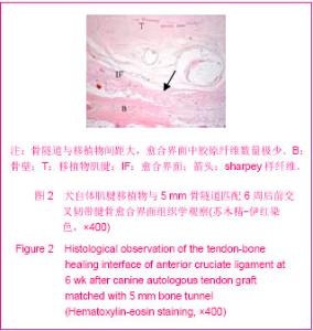
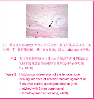
2.1 参与者数量分析 术后1只犬麻醉未能苏醒后死亡,予以补充。其余动物在观察期间存活好。观察6周,最后均进入结果分析。 2.2 大体观察 各组移植后6周时,关节腔内前交叉韧带均有明显滑膜包裹;各组直径均较正常前交叉韧带增粗;关节液清亮。5 mm骨隧道组1例6周的标本,股骨外侧髁近内侧及相应胫骨平台部分有软骨损伤表现,表面失去正常光泽,部分崩裂,可见软骨下骨。其余各组标本未见有半月板和软骨损伤表现。 2.3 组织学情况 正常前交叉韧带与骨的连接点具有典型的4层结构。第1层是韧带本身,第2层是未矿化的软骨区,第3层是钙化的软骨区,在这个区,纤维软骨矿化并且插入至第4层软骨下骨板。其中在纤维软骨和钙化软骨之间有“潮线”相隔。 前交叉韧带重建6周时,移植物与骨隧道之间的愈合界面由含有新生毛细血管的肉芽组织和胶原纤维形成。5,4.5 mm骨隧道组骨隧道与移植物间距大,愈合界面中胶原纤维数量少。4 mm骨隧道组骨隧道与移植物接触较好,愈合界面sharpey样纤维连接增多。3.5 mm骨隧道组移植物与骨隧道结合紧密,编织骨和移植物间的愈合界面其他组更加致密,可以见到更多的sharpey样纤维连接,但排列还不规则,见图2。"
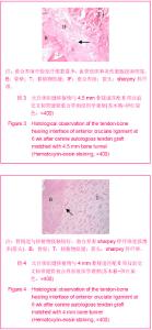
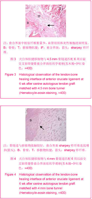
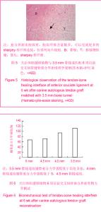
5,4.5 mm骨隧道组腱骨愈合界面中还存在一些血管组织,胶原纤维排列无序,类软骨样细胞数量较少,分布零乱。4 mm骨隧道组腱骨愈合界面的胶原纤维数 量含量增加,排列也更加有序。3.5 mm骨隧道组腱骨之间的sharpey样纤维连接致密有序,大片编织骨转化成板层骨,基质中的类软骨样细胞有排列成行的趋势,但未观察到分区和“潮线”形成。见图3-5。"
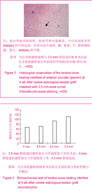
2.4 生物力学情况 移植物重建后 6周时,每组标本的移植物均从骨隧道中拖出,腱骨愈合界面发生撕脱。6周时,各组标本生物力学测试数据分别为5 mm骨隧道组(67.16±8.2) N,4.5 mm骨隧道组(71.82±12.06) N, 4 mm骨隧道组(93.94±14.26) N,3.5 mm骨隧道组(113.62±15.58) N,在移植物重建后6周时,3.5 mm骨隧道组腱骨愈合力学强度优于其他3组,4 mm骨隧道组腱骨愈合力学强度优于5,4.5 mm骨隧道组。除了5,4.5 mm骨隧道组之间差异无显著性意义外,其余各组间比较,均有显著性意义。见图6。"
| [1] 刘东波,沙鹏.运动性膝关节交叉韧带损伤:重建生物材料的选择[J].中国组织工程研究,2012,16(38):7169-7176.[2] Fanelli GC, Edson CJ. Surgical treatment of combined PCL-ACL medial and lateral side injuries (global laxity): surgical technique and 2- to 18-year results. J Knee Surg. 2012;25(4):307-316. [3] Scanlan SF, Lai J, Donahue JP, et al. Variations in the three-dimensional location and orientation of the ACL in healthy subjects relative to patients after transtibial ACL reconstruction. J Orthop Res. 2012;30(6):910-918. [4] Zantop T, Ferretti M, Bell KM, et al. Effect of tunnel-graft length on the biomechanics of anterior cruciate ligament-reconstructed knees: intra-articular study in a goat model. Am J Sports Med. 2008;36(11):2158-2166.[5] Chudik S, Beasley L, Potter H, et al. The influence of femoral technique for graft placement on anterior cruciate ligament reconstruction using a skeletally immature canine model with a rapidly growing physis. Arthroscopy. 2007;23(12):1309-1319. [6] 李敏,王丽霞,张瑞存.异体肌腱及跟腱移植后移植物的免疫学特点[J].中国组织工程研究与临床康复,2012,16(5):915-918.[7] 刘心,冯华,张辉.同种异体跟腱移植物重建内侧副韧带浅层结构治疗膝关节外翻不稳定的临床研究[J].中国运动医学杂志,2012, 31(11):949-956. [8] Sun L, Hou C, Wu B, et al. Effect of muscle preserved on tendon graft on intra-articular healing in anterior cruciate ligament reconstruction. Knee Surg Sports Traumatol Arthrosc. 2012 Aug 29. [9] Kondo E, Yasuda K, Katsura T,et al. Biomechanical and histological evaluations of the doubled semitendinosus tendon autograft after anterior cruciate ligament reconstruction in sheep. Am J Sports Med. 2012;40(2):315-324.[10] Ahn JH, Jeong HJ, Ko CS, et al. Three-Dimensional Reconstruction Computed Tomography Evaluation of Tunnel Location during Single-Bundle Anterior Cruciate Ligament Reconstruction: A Comparison of Transtibial and 2-Incision Tibial Tunnel-Independent Techniques. Clin Orthop Surg. 2013;5(1):26-35. [11] Beynnon BD, Johnson RJ, Fleming BC. The science of anterior cruciate ligament rehabilitation. Clin Orthop Relat Res. 2002;(402):9-20. [12] Cabaud HE, Feagin JA, Rodkey WG. Acute anterior cruciate ligament injury and augmented repair. Experimental studies. Am J Sports Med. 1980;8(6):395-401.[13] Petersen W, Tillmann B. Anatomy and function of the anterior cruciate ligament. Orthopade. 2002;31(8):710-718.[14] Mainil-Varlet P, Schiavinato A, Ganster MM. Efficacy Evaluation of a New Hyaluronan Derivative HYADD® 4-G to Maintain Cartilage Integrity in a Rabbit Model of Osteoarthritis. Cartilage. 2013;4(1):28-41.[15] Carey T, Oliver D, Pniewski J, et al. Anterior cruciate ligament augmentation for rotational instability following primary reconstruction with an accelerated physical therapy protocol.J Surg Orthop Adv. 2013;22(1):59-65.[16] Sutton KM, Bullock JM. Anterior cruciate ligament rupture: differences between males and females.J Am Acad Orthop Surg. 2013;21(1):41-50. [17] Kurosaka M, Yoshiya S, Andrish J. A biomechanical comparison of different surgical techniques of grart fixation in anterior cruciate ligament reconstruction. A J Sports Med. 1987; 15(3):225-229 .[18] Lambert K. Vascularized patellar tendon graft with rigid internal fixation for anterior cruciate ligament insufficiency. Clin Orthop.1983;172:85-89.[19] Hoher J, Moller HD, Fu FH. Bone tunnel enlargement after anterior cruciate ligament reconsruction: fact or fiction? Knee Surg Sports Traumatol Arthrosc. 1998;6(4):231-240.[20] Messner K. Postnatal development of the cruciate ligament insertions in the rat knee: morphological evalution and immunohistochemical study of collagens types Ⅰ and Ⅱ. Acta Anat (Basel). 1997;160(4):261-268.[21] Ye S, Dongyang C, Zhihong X, et al.The incidence of deep venous thrombosis after arthroscopically assisted anterior cruciate ligament reconstruction. Arthroscopy.2013;29(4): 742-747.[22] Dwyer T, Whelan D. Anatomical considerations in multiligament knee injury and surgery.J Knee Surg. 2012;25(4):263-274. [23] Gao J, Rasanen T, Persliden J, et al. The morphology of ligament insertions after failure at low strain velocity. An evaluation of ligament enthuses in the rabbit knee. J Anat. 1996;189(Pt 1):127-133.[24] Winiarski S, Czamara A. Evaluation of gait kinematics and symmetry during the first two stages of physiotherapy after anterior cruciate ligament reconstruction.Acta Bioeng Biomech. 2012;14(2):91-100.[25] Carpenter JE, Hankenson KD. Animal models of tendon and ligament injuries for tissue engineering applications. Biomaterials. 2004;25(9):1715-1722.[26] Ntoulia A, Papadopoulou F, Ristanis S,et al.Revascularization process of the bone--patellar tendon--bone autograft evaluated by contrast-enhanced magnetic resonance imaging 6 and 12 months after anterior cruciate ligament reconstruction.Am J Sports Med. 2011;39(7):1478-1486. [27] Vogrin M, Rupreht M, Dinevski D, et al. Effects of a platelet gel on early graft revascularization after anterior cruciate ligament reconstruction: a prospective, randomized, double-blind, clinical trial. Eur Surg Res. 2010;45(2):77-85.[28] Weiler A, Hoffmann RFG, Bail HJ, et al. Tendon healing in a bone tunnel. Part II: histologic Analysis after biodegradable interference fit fixation in a model of anterior cruciate ligament reconstruction in sheep. Arthroscopy. 2002;18(2): 124-135.[29] Muller B, Bowman KF Jr, Bedi A. CL graft healing and biologics.Clin Sports Med. 2013;32(1):93-109. [30] Kondo E, Yasuda K, Katsura T, et al. Biomechanical and histological evaluations of the doubled semitendinosus tendon autograft after anterior cruciate ligament reconstruction in sheep.Am J Sports Med. 2012;40(2): 315-324. [31] Buelow JU, Siebold R, Ellermann A, et al. A prospective evaluation of tunnel enlargement in anterior cruciate ligament reconstruction with hamstrings: extracortical versus anatomical fixation. Knee Surg Sports Traumatol Arthrose. 2002;10(2):80-85.[32] Parker MG.Biomechanical and histological concepts in the rehabilitation of patients with anterior cruciate ligament reconstructions. J Orthop Sports Phys Ther. 1994;20(1): 44-50.[33] Jorgensen U, Thomsen HS. Behavior of the graft within the bone tunnels following anterior cruciate ligament reconstruction,studied by cinematic magnetic resonance imaging. Knee Surg Sports Traumatol Arthrose.2000;8(1): 32-35.[34] Segawa H, Omori G, Tomita S,et al.Bone tunnel enlargement after anterior cruciate ligament reconstruction using hamstring tendons. Knee Surg Sports Traumatol Arthrose.2001;9(4): 206-210.[35] Yamakado K, Kitaoka K, Yamada H, et al. Periosteal augmentation of a tendon graft improves tendon healing in the bone tunnel. Clin Orthop. 2004;419:223-231.[36] Rodeo SA, Arnoczky SP, Torzilli PA, et al. Tendon-healing in a bone tunnel: a biomechanical and histological study in the dog. J Bone J Surg(Am).1993;75(2):1795-1803.[37] Darabos N, Hundric-Haspl Z, Haspl M,et al. Correlation between synovial fluid and serum IL-1beta levels after ACL surgery-preliminary report.Int Orthop. 2009;33(2):413-4388. [38] Roy S, Fernhout M, Stanley R, et al.Tibial interference screw fixation in anterior cruciate ligament reconstruction with and without autograft bone augmentation.Arthroscopy. 2010; 26(7):949-956. [39] Tien YC, Chih HW, Cheng YM, et al. The influence of the gap size on the interfacial union between the bone and the tendon. Kaohsiung J Med Sci. 1999;15(10):581-588 |
| [1] | Zhang Tongtong, Wang Zhonghua, Wen Jie, Song Yuxin, Liu Lin. Application of three-dimensional printing model in surgical resection and reconstruction of cervical tumor [J]. Chinese Journal of Tissue Engineering Research, 2021, 25(9): 1335-1339. |
| [2] | Liang Yan, Zhao Yongfei, Xu Shuai, Zhu Zhenqi, Wang Kaifeng, Liu Haiying, Mao Keya. Imaging evaluation of short-segment fixation and fusion for degenerative lumbar scoliosis assisted by highly selective nerve root block [J]. Chinese Journal of Tissue Engineering Research, 2021, 25(9): 1423-1427. |
| [3] | Liu Shaohua, Zhou Guanming, Chen Xicong, Xiao Keming, Cai Jian, Liu Xiaofang. Influence of anterior cruciate ligament defect on the mid-term outcome of fixed-bearing unicompartmental knee arthroplasty [J]. Chinese Journal of Tissue Engineering Research, 2021, 25(6): 860-865. |
| [4] | Liu Lihua, Sun Wei, Wang Yunting, Gao Fuqiang, Cheng Liming, Li Zirong, Wang Jiangning. Type L1 steroid-induced osteonecrosis of the femoral head through femoral head and neck junction decompression by fenestration: a single-center prospective clinical study [J]. Chinese Journal of Tissue Engineering Research, 2021, 25(6): 906-911. |
| [5] | Yang Xin, Jin Zhe, Feng Xu, Lu Bing. The current situation of knowledge and attitudes towards organ, eye tissue, body donation of residents in Shenyang [J]. Chinese Journal of Tissue Engineering Research, 2021, 25(5): 779-784. |
| [6] | Li Chenjie, Lü Linwei, Song Yang, Liu Jingna, Zhang Chunqiu. Measurement and statistical analysis of trabecular morphological parameters of titanium alloy peri-prosthesis under preload [J]. Chinese Journal of Tissue Engineering Research, 2021, 25(4): 516-520. |
| [7] | Ma Ziyue, Ju Xiaochen, Zhang Lei, Sun Rongxin. Tendon-bone healing in anterior cruciate ligament reconstruction with and without remnant preservation [J]. Chinese Journal of Tissue Engineering Research, 2021, 25(4): 582-587. |
| [8] | Zhang Yu, Feng Shuo, Yang Zhi, Zhang Ye, Sun Jianning, An Lun, Chen Xiangyang. Three-dimensional gait of patients with developmental dysplasia of hip undergoing total hip arthroplasty with high hip center [J]. Chinese Journal of Tissue Engineering Research, 2021, 25(3): 350-355. |
| [9] | He Jie, Chang Qi. Biological reconstruction of large bone defects after resection of malignant tumor of extremities [J]. Chinese Journal of Tissue Engineering Research, 2021, 25(3): 420-425. |
| [10] | Xing Hao, Zhang Yonghong, Wang Dong. Advantages and disadvantages of repairing large-segment bone defect [J]. Chinese Journal of Tissue Engineering Research, 2021, 25(3): 426-430. |
| [11] | Zhou Jihui, Li Xinzhi, Zhou You, Huang Wei, Chen Wenyao. Comparison of the advantages and disadvantages of multiple implants in treatment of traumatic dislocation of sternoclavicular joint [J]. Chinese Journal of Tissue Engineering Research, 2021, 25(3): 443-448. |
| [12] | Xie Jingshu, Zhang Xianglin, Liu Jinlei, Wen Jing. Application of High Resolution reconstruction algorithm in precision CT scans of the middle and inner ears [J]. Chinese Journal of Tissue Engineering Research, 2021, 25(23): 3614-3618. |
| [13] | Wang Shengjun, Yin Fei, Jiang Maoyu. Application of titanium ossicular prosthesis in one-stage open tympanoplasty for middle ear cholesteatoma [J]. Chinese Journal of Tissue Engineering Research, 2021, 25(22): 3505-3509. |
| [14] | Chen Jie, Liao Chengcheng, Zhao Hongbo, Zhao Wei, Chen Zhiwei, Wang Yan. Application of tissue engineering urethral stent and its preparation technology in urethral reconstruction [J]. Chinese Journal of Tissue Engineering Research, 2021, 25(22): 3591-3596. |
| [15] | Xiong Xiaolong, Wang Guangji, Fang Yehan, Du Xiufan, Hang Hui, Ye Zhifang. Meta-analysis of comparison of the effect of tibial fixation using absorbable screws and metal screws in anterior cruciate ligament reconstruction with autologous hamstrings [J]. Chinese Journal of Tissue Engineering Research, 2021, 25(21): 3438-3444. |
| Viewed | ||||||
|
Full text |
|
|||||
|
Abstract |
|
|||||