Chinese Journal of Tissue Engineering Research ›› 2013, Vol. 17 ›› Issue (23): 4240-4247.doi: 10.3969/j.issn.2095-4344.2013.23.009
Previous Articles Next Articles
Monitoring transplanted stem cells in rat Achilles tendon by in vivobioluminescent imaging
Huang De-qing1, Gary Balian2
- 1 Department of Orthopedics, Tianjin No. 3 Municipal Hospital, Tianjin 200250, China
2 Department of Orthopedic Surgery, University of Virginia Health System, Charlottesville, Virginia 22908, USA
-
Online:2013-06-04Published:2013-06-04 -
About author:Huang De-qing☆, M.D., Chief physician, Department of Orthopedics, Tianjin No. 3 Municipal Hospital, Tianjin 300250, China deqinghuang@yahoo.com.cn -
Supported by:Tianjin Research Program of Application Foundation and Advanced Technology in 2008, No. 08JCYBJC27100*;
Scientific Foundation of Tianjin Municipal Health Bureau (General Program), No. 07KR01*
CLC Number:
Cite this article
Huang De-qing, Gary Balian. Monitoring transplanted stem cells in rat Achilles tendon by in vivobioluminescent imaging[J]. Chinese Journal of Tissue Engineering Research, 2013, 17(23): 4240-4247.
share this article
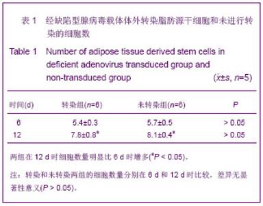
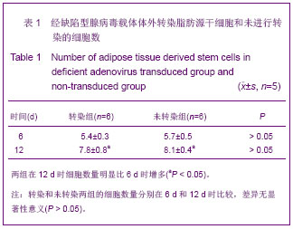
2.1 实验动物数量分析 体外实验纳入8只大鼠在脱颈处死前均无死亡,设自身对照,双侧后肢手术及分组情况如前述,术后动物无死亡,伤口无感染,早期有跟腱无力步态,1周后步态逐渐恢复正常。 2.2 脂肪源性干细胞的分离、培养和鉴定 倒置显微镜下观察,原代细胞接种4-6 h后开始贴壁,初为小的圆形细胞,24 h后细胞逐渐伸展成纺锤形,细胞密度大约为50%,72 h后细胞数量明显增加,密度为80%,细胞呈短梭形或多角形,随着培养时间的延长,梭形细胞逐渐占据优势。经1∶2传代后的细胞生长速度明显快,72 h后细胞即已长满培养瓶底,融合成片状,细胞排列更加整齐有序,杂质细胞基本消失,多次传代后细胞分布更加均匀有序,经鉴定为脂肪源性间充质干细胞[12] 。 2.3 缺陷型腺病毒载体转染后脂肪源干细胞的形态变化及增殖情况 转染与未转染组细胞数量见表1。缺陷型腺病毒载体转染脂肪源干细胞后可见细胞贴壁生长,形态正常,细胞核清晰,并且成倍增殖,未见到胞浆毒性作用。在12 d时细胞数量明显比6 d时增多(P < 0.05),并且转染和未转染两者的细胞数量分别在6 d和12 d时相比较差异无显著性意义(P > 0.05)。说明缺陷型腺病毒载体的转染对脂肪源干细胞的生长状态和增殖没有明显影响。"
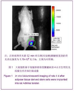
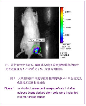
2.4 活体生物发光成像结果 用活体成像软件分析追踪植入跟腱脂肪源干细胞荧光素酶的表达。结果显示建模后24 h右侧(实验侧)细胞植入的部位检测到荧光素酶的表达,生物发光信号最大强度平均为(1.22±0.43)×106光子/s。第4天的荧光表达增强,为(1.81±0.76)×106光子/s,7 d时荧光表达达到峰值,为(1.88±0.69)×106 光子/s,14 d仍可检测的荧光表达,但明显减弱,为(0.89±0.26)×105光子/s。荧光表达的衰减可能表明细胞死亡、迁徙、基因丢失或与再生组织以及鼠毛的厚度增加有关。在跟腱以外的区域未发现荧光表达。而左侧(对照侧)跟腱修复区域始终未检测到绿色荧光的表达,见图1。"

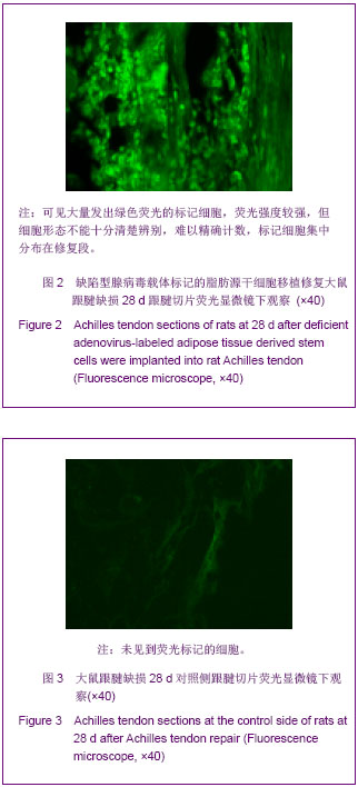
2.5 跟腱切片荧光显微镜观察 大鼠跟腱修复段标本冰冻切片在荧光显微镜下观察,缺陷型腺病毒载体标记的脂肪源干细胞移植侧可以找到大量发出绿色荧光的标记细胞,荧光强度较强,但细胞形态不能十分清楚辨别,难以精确计数,标记细胞集中分布在修复段,修复段以外区域未找到标记细胞,见图2;而对照侧,冰冻切片中未能找到绿色荧光细胞,见图3。 2.6 不良反应 大鼠跟腱修复区域未见明显炎症反应和异物反应,此次实验未作组织切片的苏木精伊红染色,还有待于将进一步研究。"
| [1]Lee SK. Modern tendon repair techniques. Hand Clin. 2012; 28(4):565-570.[2]Karabekmez FE, Zhao C. Surface treatment of flexor tendon autograft and allograft decreases adhesion without an effect of graft cellularity: a pilot study. Clin Orthop Relat Res. 2012; 470(9):2522-2527.[3]Dy CJ, Daluiski A, Do HT, et al. The epidemiology of reoperation after flexor tendon repair. J Hand Surg Am. 2012; 37(5):919-924.[4]Griffin M, Hindocha S, Jordan D, et al. An overview of the management of flexor tendon injuries. Open Orthop J. 2012;6:28-35. [5]Butler DL, Awad HA. Perspectives on cell and collagen composites for tendon repair. Clin Orthop Relat Res. 1999; (367 Suppl):S324-332.[6]Zuk PA, Zhu M, Mizuno H,et al.Multilineage cells from human adipose tissue: implications for cell-based therapies. Tissue Eng. 2001;7(2):211-228.[7]Shen JF, Sugawara A, Yamashita J, et al. Dedifferentiated fat cells:an alternative source of adult multipotent cells from the adipose tissues. Int J Oral Sci. 2011;3(3):117-124.[8]Levi B, Longaker MT. Concise review:adipose-derived stromal cells for skeletal regenerative medicine. Stem Cells. 2011;29(4):576-582.[9]Hou K, Li M, Li JR, et al. Zhongguo Xiandai Yisheng Zazhi. 2011;21(8):929-933.侯凯,李梅,李金茹,等.体外诱导大鼠脂肪源间充质干细胞成肌腱潜能的研究[J].中国现代医学杂志,2011,21(8):929-933.[10]Leo BM, Li X, Balian G, et al. In vivo bioluminescent imaging of virus-mediated gene transfer and transduced cell transplantation in the intervertebral disc. Spine. 2004; 29(8): 838-844.[11]Murphy KD, Mushkudiani IA, Kao D, et al. Successful incorporation of tissue-engineered porcine small-intestinal submucosa as substitute flexor tendon graft is mediated by elevated TGF-beta1 expression in the rabbit. J Hand Surg Am. 2008;33(7):1168-1178.[12]Hou K, Li M, Yang YK, et al. Zhongguo Zuzhi Gongcheng Yanjiu yu Linchuang Kangfu. 2011;15(1):29-32.侯凯,李梅,杨逸昆,等. SD大鼠脂肪源间充质干细胞分离培养特性及影响因素[J].中国组织工程研究与临床康复,2011,15(1): 29-32.[13]Galle J, Bader A, Hepp P, et al. Mesenchyal stem cells in cartilage repair: state of the art and methods to monitor cell growth, differentiation and cartilage regeneration. Curr Med Chem. 2010;17(21):2274-2291.[14]Togel F, Yang Y, Zhang P, et al. Bioluminescent imaging to mornitor the in vivo distribution of mesenchyal stem cells in acute kidney injury. Am J Physiol Renal Physiol. 2008;295 (1):315-321. [15]Hwang do W, Jang SJ, Kim YH, et al. Real-time in vivo monitoring of viable stem cells implanted on biocompatible scaffolds. Eur J Nucl Med Mol Imaging. 2008;35(10): 1887-1898. [16]Li HX, Zhou YF, Jiang B, et al. Yixue Yanjiusheng Xuebao. 2013;26(3):228-231.李红霞,周亚峰,蒋彬,等.大鼠骨髓间充质干细胞转染GATA-4的表达及鉴定[J].医学研究生学报,2013,26(3):228-231.[17]Wang Q, Chen GX, Guo L, et al. Zhonghua Shiyan Waike Zazhi. 2013;30(3):599-601.王庆,陈光兴,郭林,等. 腺病毒介导的骨形态发生蛋白-2和骨形态发生蛋白-7基因联合转染大鼠脂肪来源干细胞对成骨分化的影响[J]. 中华实验外科杂志,2013,30(3):599-601.[18]Li Y, Tian Y, Wu ZC, et al. Zhongguo Zuzhi Gongcheng Yanjiu. 2012;16(41):7607-7611.李洋,田瑜,武志超,等.稳定转染增强型绿色荧光蛋白大鼠骨髓间充质干细胞系的建立[J].中国组织工程研究, 2012,16(41): 7607-7611.[19]Duan L, Song ZG, Gong DJ, et al. Jiepouxue Zazhi. 2012; 35(4):431-435.段炼,宋智钢,龚德军,等.腺病毒介导HCN2基因转染对人脂肪干细胞生物学特性的影响[J].解剖学杂志, 2012,35(4):431- 435.[20]Luker KE, Luker GD. Applications of bioluminescence imaging to antiviral research and therapy: multiple luciferase enzymes and quantitation. Antiviral Res. 2008;78(3):179-187. [21]Prinz A, Reither G, Diskar M, et al. Fluorescence and bioluminescence procedures for functional proteomics. Proteomics. 2008;8(6):1179-1196.[22]Feng G, Wan Y, Balian G, et al.Adenovirus-mediated expression of growth and differentiation factor-5 promotes chondrogenesis of adipose stem cells. Growth Factors. 2008;26(3):132-142.[23]Hou Y, Mao Z, Wei X, et al. Effects of transforming growth factor-beta1 and vascular endothelial growth factor 165 gene transfer on Achilles tendon healing. Matrix Biol. 2009;28(6): 324-335. [24]Shao L, Wu WS. Gene-delivery systems for iPS cell generation. Expert Opin Biol Ther. 2010;10(2):231-42.[25]Kyritsis AP, Sioka C, Rao JS. Viruses, gene therapy and stem cells for thetreatment of human glioma. Cancer Gene Ther. 2009;16(10):741-752. [26]Chen J, Yuan W, Song DW, et al. Zhongguo Zuzhi Gongcheng Yanjiu. 2012;16(49):9139-9145.陈剑,袁文,宋滇文,等.骨形态发生蛋白2和7共转染骨髓间充质千细胞的成骨分化[J].中国组织工程研究,2012,16(49): 9139-9145.[27]Wang DL, Xing DG, Wu JJ, et al. Zhonghua Chuangshang Zazhi. 2012;28(11):1042-1045.王德亮,邢德国,吴建军,等. 骨形态发生蛋白-2基因转染骨髓间充质干细胞移植对糖尿病大鼠骨折愈合的影响[J]. 中华创伤杂志,2012,28(11):1042-1045.[28]Huang D, Balian G, Chhabra AB. Tendon tissue engineering and gene transfer: the future of surgical treatment. J Hand Surg Am. 2006;31(5):693-704.[29]Murray SJ, Santangelo KS, Bertone AL. Evaluation of early cellular influences of bone morphogenetic proteins 12 and 2 on equine superficial digital flexor tenocytes and bone marrow-derived mesenchymal stem cells in vitro. Am J Vet Res. 2010;71(1):103-114. [30]Hou Y, Mao Z, Wei X, et al. Effects of transforming growth factor-beta1 and vascular endothelial growth factor 165 gene transfer on Achilles tendon healing. Matrix Biol. 2009; 28(6): 324-335. [31]Thorfinn J, Saber S, Angelidis IK, et al. Flexor tendon tissue engineering: temporal distribution of donor tenocytes versus recipient cells.Plast Reconstr Surg. 2009;124(6):2019-2026.[32]Hogan MV, Bagayoko N, James R, et al. Tissue engineering solutions for tendon repair. J Am Acad Orthop Surg. 2011; 19(3):134-142. [33]Wang HG, Li HM, Liu DL, et al. Zhongguo Zuzhi Gongcheng Yanjiu. 2013;17(1):106-111.王和庚,黎洪棉,柳大烈,等.缺氧诱导因子1α基因转染脂肪干细胞与自体脂肪移植[J].中国组织工程研究,2013,17(1): 106-111.[34]Sun L, Tian XB, Hu RY, et al. Zhongguo Zuzhi Gongcheng Yanjiu. 2012;16(27):5017-5021. 孙立,田晓滨,胡如印,等. plRES-骨形态发生蛋白2质粒转染大鼠骨髓间充质干细胞后的持续表达[J].中国组织工程研究,2012, 16(27):5017-5021.[35]Guo WT, Wang H, Liu SJ, et al. Zhongguo Zuzhi Gongcheng Yanjiu. 2012;16(23):4273-4427.郭伟韬,王辉,刘思景,等.人骨形态发生蛋白2和人成纤维细胞生长因子2双基因共表达腺病毒载体转染人骨髓基质干细胞[J]. 中国组织工程研究,2012,16(23):4273-4427.[36]Han D, Xu H, Zheng YX, et al. Zhongguo Shiyan Zhenduanxue. 2012;16(5):766-769.韩冬,徐煌,郑永霞,等. Klotho基因转染促进脂肪间充质干细胞增殖的实验研究[J].中国实验诊断学,2012,16(5):766-769.[37]Dong F, Chen HH, Yang XH, et al. Zhongguo Jiaoxing Waike Zazhi. 2010,19:1631-1634.董飞,陈鸿辉,杨小红,等.骨髓间充质干细胞促进骨-肌腱结合部位早期愈合的动物生物力学实验研究[J].中国矫形外科杂志, 2010,25(19):1631-1634.[38]Butler DL, Gooch C, Kinneberg KR, et al. The use of mesenchymal stem cells in collagen-based scaffolds for tissue-engineered repair of tendons. Nat Protoc. 2010; 5(5):849-863.[39]Grange S. Current issues and regulations in tendon regeneration and musculoskeletal repair with mesenchymal stem cells. Curr Stem Cell Res Ther. 2012;7(2):110-114. [40]Yin Z, Chen X, Chen JL, et al. Stem cells for tendon tissue engineering and regeneration. Expert Opin Biol Ther. 2010; 10(5):689-700. [41]Park A, Hogan MV, Kesturu GS, et al. Adipose-tissue derived mesenchyal stem cells treated with growth differentiation factor-5 express tendon-specific markers. Tissue Eng Part A. 2010;16(9):2941-2951.[42]Uysal AC, Mizuno H. Tendon regeneration and repair with adipose derived stem cells. Curr Stem Cell Res Ther. 2010; 5(2):161-167. |
| [1] | Pu Rui, Chen Ziyang, Yuan Lingyan. Characteristics and effects of exosomes from different cell sources in cardioprotection [J]. Chinese Journal of Tissue Engineering Research, 2021, 25(在线): 1-. |
| [2] | Zhang Tongtong, Wang Zhonghua, Wen Jie, Song Yuxin, Liu Lin. Application of three-dimensional printing model in surgical resection and reconstruction of cervical tumor [J]. Chinese Journal of Tissue Engineering Research, 2021, 25(9): 1335-1339. |
| [3] | Shen Jinbo, Zhang Lin. Micro-injury of the Achilles tendon caused by acute exhaustive exercise in rats: ultrastructural changes and mechanism [J]. Chinese Journal of Tissue Engineering Research, 2021, 25(8): 1190-1195. |
| [4] | Zhang Xiumei, Zhai Yunkai, Zhao Jie, Zhao Meng. Research hotspots of organoid models in recent 10 years: a search in domestic and foreign databases [J]. Chinese Journal of Tissue Engineering Research, 2021, 25(8): 1249-1255. |
| [5] | Wang Zhengdong, Huang Na, Chen Jingxian, Zheng Zuobing, Hu Xinyu, Li Mei, Su Xiao, Su Xuesen, Yan Nan. Inhibitory effects of sodium butyrate on microglial activation and expression of inflammatory factors induced by fluorosis [J]. Chinese Journal of Tissue Engineering Research, 2021, 25(7): 1075-1080. |
| [6] | Wang Xianyao, Guan Yalin, Liu Zhongshan. Strategies for improving the therapeutic efficacy of mesenchymal stem cells in the treatment of nonhealing wounds [J]. Chinese Journal of Tissue Engineering Research, 2021, 25(7): 1081-1087. |
| [7] | Liao Chengcheng, An Jiaxing, Tan Zhangxue, Wang Qian, Liu Jianguo. Therapeutic target and application prospects of oral squamous cell carcinoma stem cells [J]. Chinese Journal of Tissue Engineering Research, 2021, 25(7): 1096-1103. |
| [8] | Xie Wenjia, Xia Tianjiao, Zhou Qingyun, Liu Yujia, Gu Xiaoping. Role of microglia-mediated neuronal injury in neurodegenerative diseases [J]. Chinese Journal of Tissue Engineering Research, 2021, 25(7): 1109-1115. |
| [9] | Li Shanshan, Guo Xiaoxiao, You Ran, Yang Xiufen, Zhao Lu, Chen Xi, Wang Yanling. Photoreceptor cell replacement therapy for retinal degeneration diseases [J]. Chinese Journal of Tissue Engineering Research, 2021, 25(7): 1116-1121. |
| [10] | Jiao Hui, Zhang Yining, Song Yuqing, Lin Yu, Wang Xiuli. Advances in research and application of breast cancer organoids [J]. Chinese Journal of Tissue Engineering Research, 2021, 25(7): 1122-1128. |
| [11] | Wang Shiqi, Zhang Jinsheng. Effects of Chinese medicine on proliferation, differentiation and aging of bone marrow mesenchymal stem cells regulating ischemia-hypoxia microenvironment [J]. Chinese Journal of Tissue Engineering Research, 2021, 25(7): 1129-1134. |
| [12] | Zeng Yanhua, Hao Yanlei. In vitro culture and purification of Schwann cells: a systematic review [J]. Chinese Journal of Tissue Engineering Research, 2021, 25(7): 1135-1141. |
| [13] | Kong Desheng, He Jingjing, Feng Baofeng, Guo Ruiyun, Asiamah Ernest Amponsah, Lü Fei, Zhang Shuhan, Zhang Xiaolin, Ma Jun, Cui Huixian. Efficacy of mesenchymal stem cells in the spinal cord injury of large animal models: a meta-analysis [J]. Chinese Journal of Tissue Engineering Research, 2021, 25(7): 1142-1148. |
| [14] | Hou Jingying, Yu Menglei, Guo Tianzhu, Long Huibao, Wu Hao. Hypoxia preconditioning promotes bone marrow mesenchymal stem cells survival and vascularization through the activation of HIF-1α/MALAT1/VEGFA pathway [J]. Chinese Journal of Tissue Engineering Research, 2021, 25(7): 985-990. |
| [15] | Shi Yangyang, Qin Yingfei, Wu Fuling, He Xiao, Zhang Xuejing. Pretreatment of placental mesenchymal stem cells to prevent bronchiolitis in mice [J]. Chinese Journal of Tissue Engineering Research, 2021, 25(7): 991-995. |
| Viewed | ||||||
|
Full text |
|
|||||
|
Abstract |
|
|||||