Chinese Journal of Tissue Engineering Research ›› 2013, Vol. 17 ›› Issue (20): 3635-3642.doi: 10.3969/j.issn.2095-4344.2013.20.004
Previous Articles Next Articles
The subchondral bone has no mechanical changes after acute injury for 2 weeks
Guo Xin-yu, Zhang Yuan, Wang Zhi-bing, Qin Chuan, Hao Yong, Zhang Yu-mei, Zhang Xia
- Department of Orthopedics, Xinqiao Hospital, Third Military Medical University of PLA, Chongqing 400037, China
-
Received:2013-02-14Revised:2013-03-15Online:2013-05-14Published:2013-05-14 -
Contact:Zhang Xia, M.D., Associate professor, Department of Orthopedics, Xinqiao Hospital, Third Military Medical University of PLA, Chongqing 400037, China zhangsw199254330@163.com -
About author:Guo Xin-yu, Department of Orthopedics, Xinqiao Hospital, Third Military Medical University of PLA, Chongqing 400037, China 03011935@163.com -
Supported by:the National Natural Science Foundation of China, No. 81171720
CLC Number:
Cite this article
Guo Xin-yu, Zhang Yuan, Wang Zhi-bing, Qin Chuan, Hao Yong, Zhang Yu-mei, Zhang Xia. The subchondral bone has no mechanical changes after acute injury for 2 weeks[J]. Chinese Journal of Tissue Engineering Research, 2013, 17(20): 3635-3642.
share this article
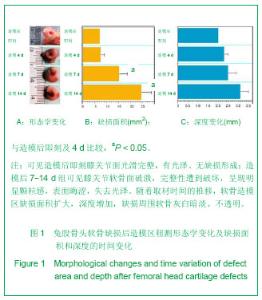
2.1 实验动物数量分析 实验选用大白兔24只,分为4组,无脱失,均进入结果分析。 2.2 兔软骨缺损大体观察结果 见图1。"
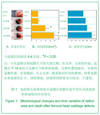
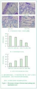
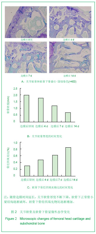
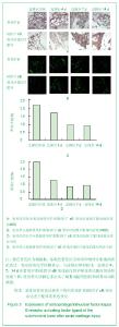
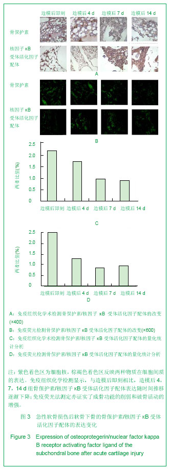
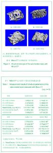
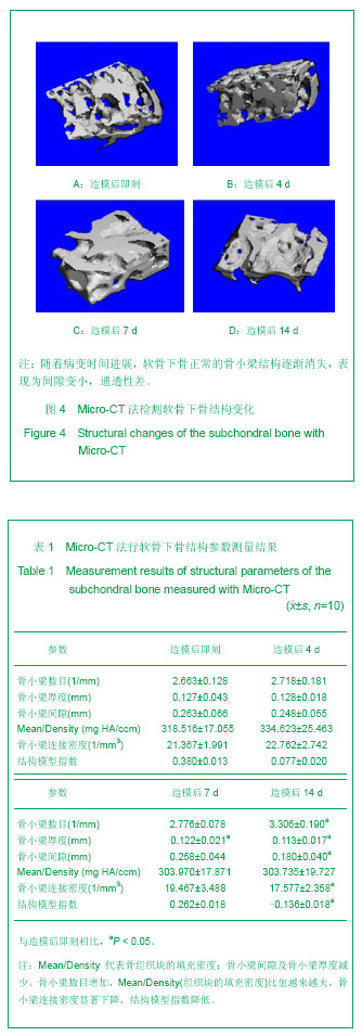
直视状态下对软骨缺损造模后进行观察,可见造模后即刻组膝关节面光滑完整,有光泽、无缺损形成;造模后4,7,14 d组可见膝关节软骨面破溃,完整性遭到破坏,呈现明显颗粒感,表面晦涩,失去光泽。随着取材时间的推移,软骨造模区缺损面积扩大,深度增加,缺损周围软骨灰白暗淡、不透明。利用MIG2000软件测定其缺损面积及深度,发现造模后7-14 d缺损面积和深度比较差异有显著性意义(P < 0.05)。 2.3 显微形态学分析结果 采用番红-固绿染色法观察软骨及软骨下骨的组织学变化,由图2可见关节软骨和软骨下骨分别被标记为红色和绿色。镜下观察、采样结合MIG2000软件定量可对关节软骨和软骨下骨进行量化评价,见图2。可见软骨缺损模型后,关节软骨厚度不断下降,至造模后4,7,14 d分别为造模后即刻组的85.2%,63.7%,38.5%(P < 0.05)。而软骨下正常骨小梁结构逐渐破坏,表现为软骨下骨组织填充物比逐渐增加,统计结果显示分别增加0.5,1.1,1.8倍,差异无显著性意义(P > 0.05)。 2.4 骨转换分析结果 骨保护素和核因子κB受体活化因子配体分别为成骨-破骨活动标志物,两者在不同时期软骨下骨中的表达,可以作为指标来反映软骨下骨内骨吸收/骨重建的动态变化。 实验采用免疫组织化学和免疫荧光法分别评价骨保护素/核因子κB受体活化因子配体,见图3A。紫色着色区为细胞核,棕褐色着色区反映两种物质在细胞间质的表达。利用IPP软件对阳性区域进行量化测定,发现软骨下骨中骨转化标志物的表达发生早期改变。与造模后即刻相比,造模后4,7,14 d组骨保护素/核因子κB受体活化因子配体表达随时间推移逐渐下降,造模后4,7,14 d的表达量分别为造模后即刻的78.1%,43.2%,40%。与之对应,免疫荧光法测定亦证实了成骨功能的削弱和破骨活动的增强,造模后4,7,14 d的骨保护素/核因子κB受体活化因子配体分别下调了48.7%,71.2%,61.7%。 2.5 软骨下骨微观形态学分析结果 随着病变时间进展,软骨下骨正常的骨小梁结构逐渐消失,表现为间隙变小,通透性差。采集数据发现:骨小梁间隙及骨小梁厚度减少、骨小梁数目增加,Mean/Density(组织块的填充密度)比值越来越大,骨小梁连接密度显著下降,结构模型指数降低,造模后7-14 d变化最大,差异有显著性意义(P < 0.05)。见图4,表1。"
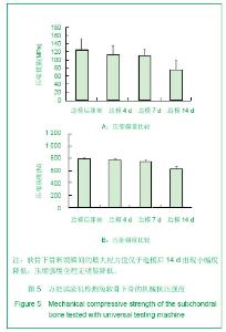
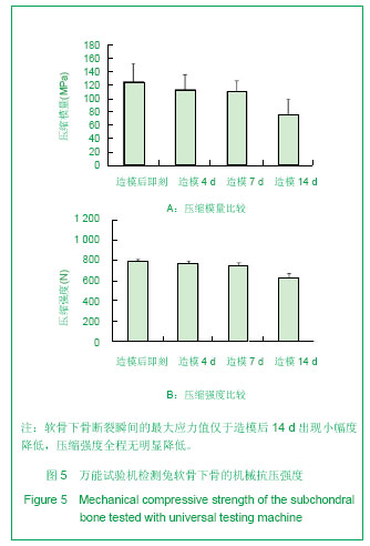
2.6 兔软骨下骨生物力学分析结果 通过抗压应力实验采集数据,建立应力/应变曲线,发现软骨下骨断裂瞬间的最大应力值在造模后差异无显著性意义(P > 0.3),仅于造模后14 d出现小幅度降低,而最大斜率反映的压缩模量全程差异均无显著性意义。见图5。"
"
"
| [1] Noyes FR, Bassett RW, Grood ES,et al. Arthroscopy in acute traumatic hemarthrosis of the knee. Incidence of anterior cruciate tears and other injuries. J Bone Joint Surg Am 1980; 62(5): 687-695, 757.[2] Curl WW, Krome J, Gordon ES, et al. Cartilage injuries: a review of 31,516 knee arthroscopies. Arthroscopy 1997; 13(4): 456-460.[3] Orth P, Cucchiarini M, Kaul G, et al. Temporal and spatial migration pattern of the subchondral bone plate in a rabbit osteochondral defect model. Osteoarthritis and Cartilage. 2012(20):1161-1169.[4] Hunziker EB. Articular cartilage repair: basic science and clinical progress. A review of the current status and prospects. Osteoarthritis Cartilage. 2002;10:432-463.[5] Pallante AL, Görtz S, Chen AC,et al. Treatment of articular cartilage defects in the goat with frozen versus fresh osteochondral allografts: effects on cartilage stiffness, zonal composition, and structure at six months. J Bone Joint Surg Am. 2012;94(21):1984-1995.[6] Pan J, Wang B, Li W, et al. Elevated cross-talk between subchondral bone and cartilage in osteoarthritic joints.Bone 2012;(51):212-217.[7] Botter SM,Van OG,Waarsing JH,et al. Quantification of subehondral bone changes in a murine osteoarthritis model using micro-CT. Biorheology.2006;43(3-4):379-388. [8] Hayami T, Pickarski M,Zhuo Y,et al.Charaeterization of articular cartilage and subchondral bone changes in the rat anterior crueiate ligament transection and men iscectomized models of osteoarthritis.Bone.2006;38 (2):234-243. [9] Ahern BJ, Parvizi J, Boston R,et al.Preclinical animal models in single site cartilage defect testing:a systematic review. Osteoarthritis and Cartilage.2009; 17: 705-713.[10] Che JH, Zhang ZR, Li GZ, et al.Application of tissue-engineered cartilage with BMP-7 gene to repair knee joint cartilage injury in rabbits. Knee Surg Sports Traumatol Arthrosc. 2010(18):496-503.[11] Mc Kinley TO, Bay BK.Trabecular bone strain changes associated with subchondral stiffening of the proximal tibia.Journal of Biomechanics.2003;36(2):155-163.[12] Thambyah A, Broom N. On new bone formation in the preosteoarthritic joint. Osteoarthritis Cartilage.2009;17(4): 456-463.[13] Lajeunesse D, Reboul P. Subchondral bone in osteoarthritis:a biologic link with articular cartilage leading to abnormalremodeling. Curr Opin Rheumatol 2003;15(5): 628-633.[14] Henriksen K, Neutzsky-Wulff AV, Bonewald LF,et al.Local communication on and within bone controls bone remodeling. Bone. 2009;44(6):1026-1033.[15] Borges P, Midha N, Marenzana M. Automated microstructural analysis of tibial subchondral bone in a surgical model of mouse osteoarthritis.Osteoarthritis and Cartilage.2012;(20): S54-S296.[16] Burguera EF, Vela-Anero A, Blanco FJ.Modulation of gene expression in human subchondral bone cells co-cultured with human articular chondrocytes. Osteoarthritis and Cartilage. 2012; (20) S113-S114.[17] Irie K ,Uchiyama E.Intra articular inflammatory cytokines in acute anterior cruciate ligament injured knee.knee.2003;10(1):93-96.[18] Walsh NC ,Crotti TN,Goldring SR,Rheumatic diseases :the effects of inflammation on bone .Immunol Rev.2005;208; 228-251[19] Boyce BF, Xing L.Functions of RANK/RANKL/OPG in bone modeling and remodeling. Arch Biochem Biophys.2008;473: 139-146.[20] Kearns AE, Khosla S, Kostenuik PJ.Receptor activator of nuclear factor kB ligand and osteoprotegerin regulation of bone remodeling in health and disease. Endocr Rev.2008;29: 155-192.[21] Boyle WJ, Simonet WS, Lacey DL. Osteoclast differentiation and activation. Nature 2003;423:337-342.[22] Hofbauer LC. Pathophysiology of RANK ligand (RANKL) and osteoprotegerin (OPG). Ann Endocrinol.2006;67(2):139-141.[23] Huebner JL Hanes MA Beekman B,et al.A comparative analysis of bone and cartilage metabolism in two strains of guinea pig with varying degrees of naturally occurring osteoarthritis J.Osteoarthritis Cartilage.2002;10(10): 758-767.[24] Mansell JP Bailey AJ.Increased metabolism of bone collagen in post menopausal female osteoporotic femoral heads J.Int J Biochem Cell Biol.2003;35(4):522-529.[25] Bailey AJ,Mansell JP,Sims TJ,et al.Biochemical and mechanical properties of subchondral bone in osteoarthritis. Biorheology.2004;41(3):349-358.[26] Brittberg M,Nilsson A,Lindahl A,et al.Rabbit articular cartilage defects treated with autologous cultured chondrocytes.Clin Orthop Relat Res.1996;(326):270-283. |
| [1] | Xu Feng, Kang Hui, Wei Tanjun, Xi Jintao. Biomechanical analysis of different fixation methods of pedicle screws for thoracolumbar fracture [J]. Chinese Journal of Tissue Engineering Research, 2021, 25(9): 1313-1317. |
| [2] | Chen Xinmin, Li Wenbiao, Xiong Kaikai, Xiong Xiaoyan, Zheng Liqin, Li Musheng, Zheng Yongze, Lin Ziling. Type A3.3 femoral intertrochanteric fracture with augmented proximal femoral nail anti-rotation in the elderly: finite element analysis of the optimal amount of bone cement [J]. Chinese Journal of Tissue Engineering Research, 2021, 25(9): 1404-1409. |
| [3] | Zhou Jihui, Li Xinzhi, Zhou You, Huang Wei, Chen Wenyao. Multiple problems in the selection of implants for patellar fracture [J]. Chinese Journal of Tissue Engineering Research, 2021, 25(9): 1440-1445. |
| [4] | Liu Zhichao, Zhang Fan, Sun Qi, Kang Xiaole, Yuan Qiaomei, Liu Genzhe, Chen Jiang. Morphology and activity of human nucleus pulposus cells under different hydrostatic pressures [J]. Chinese Journal of Tissue Engineering Research, 2021, 25(8): 1172-1176. |
| [5] | Guan Qian, Luan Zuo, Ye Dou, Yang Yinxiang, Wang Zhaoyan, Wang Qian, Yao Ruiqin. Morphological changes in human oligodendrocyte progenitor cells during passage [J]. Chinese Journal of Tissue Engineering Research, 2021, 25(7): 1045-1049. |
| [6] | Song Chengjie, Chang Hengrui, Shi Mingxin, Meng Xianzhong. Research progress in biomechanical stability of lateral lumbar interbody fusion [J]. Chinese Journal of Tissue Engineering Research, 2021, 25(6): 923-928. |
| [7] | Xu Yulin, Shen Shi, Zhuo Naiqiang, Yang Huilin, Yang Chao, Li Yang, Zhao Heng, Zhao Lu. Biomechanical comparison of three different plate fixation methods for acetabular posterior column fractures in standing and sitting positions [J]. Chinese Journal of Tissue Engineering Research, 2021, 25(6): 826-830. |
| [8] | Cai Qunbin, Zou Xia, Hu Jiantao, Chen Xinmin, Zheng Liqin, Huang Peizhen, Lin Ziling, Jiang Ziwei. Relationship between tip-apex distance and stability of intertrochanteric femoral fractures with proximal femoral anti-rotation nail: a finite element analysis [J]. Chinese Journal of Tissue Engineering Research, 2021, 25(6): 831-836. |
| [9] | Xie Chongxin, Zhang Lei. Comparison of knee degeneration after anterior cruciate ligament reconstruction with or without remnant preservation [J]. Chinese Journal of Tissue Engineering Research, 2021, 25(5): 735-740. |
| [10] | Deng Zhenhan, Huang Yong, Xiao Lulu, Chen Yulin, Zhu Weimin, Lu Wei, Wang Daping. Role and application of bone morphogenetic proteins in articular cartilage regeneration [J]. Chinese Journal of Tissue Engineering Research, 2021, 25(5): 798-806. |
| [11] | Zhou Jihui, Li Xinzhi, Zhou You, Huang Wei, Chen Wenyao. Comparison of the advantages and disadvantages of multiple implants in treatment of traumatic dislocation of sternoclavicular joint [J]. Chinese Journal of Tissue Engineering Research, 2021, 25(3): 443-448. |
| [12] | Nie Shaobo, Li Jiantao, Sun Jien, Zhao Zhe, Zhao Yanpeng, Zhang Licheng, Tang Peifu. Mechanical stability of medial support nail in treatment of severe osteoporotic intertrochanteric fracture [J]. Chinese Journal of Tissue Engineering Research, 2021, 25(3): 329-333. |
| [13] | Tan Jiachang, Yuan Zhenchao, Wu Zhenjie, Liu Bin, Zhao Jinmin. Biomechanical analysis of elastic nail combined with end caps and wire fixation for long oblique femoral shaft fractures [J]. Chinese Journal of Tissue Engineering Research, 2021, 25(3): 334-338. |
| [14] | Chen Lu, Zhang Jianguang, Deng Changgong, Yan Caiping, Zhang Wei, Zhang Yuan. Finite element analysis of locking screw assisted acetabular cup fixation [J]. Chinese Journal of Tissue Engineering Research, 2021, 25(3): 356-361. |
| [15] | Ren Wenbo, Liao Yuanpeng. Visualization analysis of traumatic osteoarthritis research hotspots and content based on CiteSpace [J]. Chinese Journal of Tissue Engineering Research, 2021, 25(21): 3374-3381. |
| Viewed | ||||||
|
Full text |
|
|||||
|
Abstract |
|
|||||