Chinese Journal of Tissue Engineering Research ›› 2013, Vol. 17 ›› Issue (15): 2661-2668.doi: 10.3969/j.issn.2095-4344.2013.15.001
Dynamic observation of rat hippocampal neuronal death process by hopping probe ion conductance microscopy
Yang Guo-wei, Zhu Hui, Zhou Zi-wei, Li Ying, Zhang Yan-jun, Zhang Jian-ning
- Tianjin Medical University General Hospital, Tianjin Neurological Institute, Key Laboratory of Post-trauma Neuro-repair and Regeneration in Central Nervous System, Ministry of Education, Tianjin Key Laboratory of Injuries, Variations and Regeneration of Nervous System, Tianjin 300052, China
-
Received:2013-02-16Revised:2013-02-27Online:2013-04-09Published:2013-04-09 -
Contact:Zhang Jian-ning, Doctor, Professor, Doctoral supervisor, Tianjin Medical University General Hospital, Tianjin Neurological Institute, Key Laboratory of Post-trauma Neuro-repair and Regeneration in Central Nervous System, Ministry of Education, Tianjin Key Laboratory of Injuries, Variations and Regeneration of Nervous System, Tianjin 300052, China jianningzhang@hotmail.com -
About author:Yang Guo-wei★, Studying for master’s degree, Tianjin Medical University General Hospital, Tianjin Neurological Institute, Key Laboratory of Post-trauma Neuro-repair and Regeneration in Central Nervous System, Ministry of Education, Tianjin Key Laboratory of Injuries, Variations and Regeneration of Nervous System, Tianjin 300052, China yangguowei@tijmu.edu.cn
CLC Number:
Cite this article
Yang Guo-wei, Zhu Hui, Zhou Zi-wei, Li Ying, Zhang Yan-jun, Zhang Jian-ning. Dynamic observation of rat hippocampal neuronal death process by hopping probe ion conductance microscopy[J]. Chinese Journal of Tissue Engineering Research, 2013, 17(15): 2661-2668.
share this article
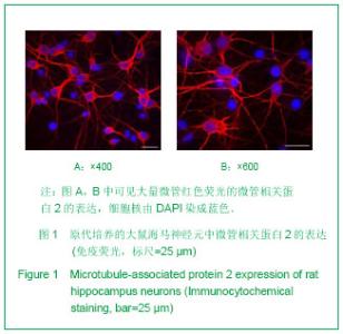
2.1 原代培养的大鼠海马神经元鉴定 新生大鼠海马神经元培养7 d后进行微管相关蛋白2荧光染色鉴定。微管相关蛋白2特异表达于神经元细胞骨架。图1可见400和600倍荧光显微镜下观察细胞染色情况。其中微管相关蛋白2呈红色荧光,细胞核由DAPI染成蓝色,原代培养大鼠海马神经元阳性率>99%。 2.2 高剂量过氧化氢对大鼠海马神经元坏死的影响 实验用较高浓度过氧化氢(1 mmol/L)致神经元坏死。为观察坏死效果,神经元经过氧化氢作用30 min后更换为普通neurobasal/B27培养基继续培养,3 h后行Annexin-V、PI荧光双标染色。Annexin-V可特异结合磷脂酰丝氨酸(PS),细胞发生凋亡或坏死时,PS可被Annexin-V检测到[9]。细胞坏死时细胞膜完整性破坏,PI可以通过破损细胞膜与细胞核特异结合。分别在100、200和400倍荧光显微镜下观察过氧化氢组与对照组Annexin-V(绿色)及PI(红色)的表达情况,可见大部分过氧化氢组神经元呈Annexin-V、PI双阳性,而对照组神经元Annexin-V和PI仅有少量阳性表达,见图2。"
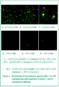
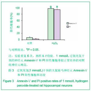
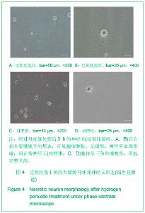
过氧化氢组 Annexin-V和PI阳性率大于对照组为(P < 0.01)。Annexin-V、PI染色结果证明大部分神经元经1 mmol/L 过氧化氢处理后发生坏死性改变。见图3。 2.3 相差显微镜下过氧化氢干预的大鼠海马坏死神经元形态 见图4。 经过氧化氢处理后的神经元3 h后在相差显微镜下的形态,可见胞体肿胀,呈球形,神经突串珠形成;而正常神经元胞体呈三角形或梭形,突起完整光滑,见图4。"
"
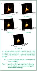
2.4 跳跃式离子电导显微镜扫描单个神经元坏死后形态 过氧化氢处理30 min后立即换成L15培养基,通过跳跃式离子电导显微镜观察神经元形态,连续扫描9 h。扫描期间可见神经元胞体从1 h后开始肿胀,形状从开始的三角形或梭形逐渐变成球形。同时在扫描1 h时,可见轴突和树突形成串珠样改变,随扫描时间延长轴突和树突逐渐断裂直至溶解消失,见图5。"
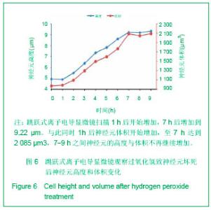
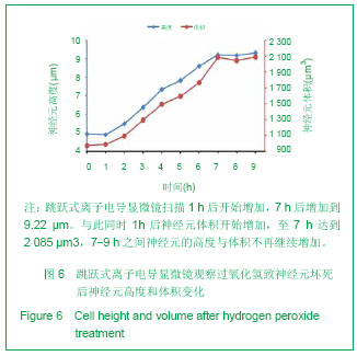
同时由扫描离子电导显微镜 Image Viewer生成的扫描细胞的高度和体积信息可见此神经元细胞起始高度为4.92 μm,于扫描1 h后开始增加,7 h后增加到 9.22 μm,约为原来的两倍。与此同时1 h后神经元体积开始增加,至7 h达到2 085 μm3约为初始体积(973 μm3)的2倍。7-9 h之间神经元的高度与体积不再继续增加,处于稳定水平,见图6。"
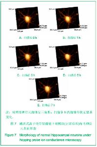
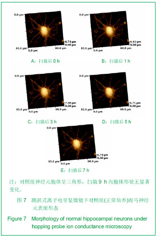
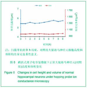
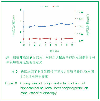
经跳跃式离子电导显微镜连续扫描9 h发现:对照组神经元胞体呈三角形,扫描9 h内胞体形状没有明显变化,轴突和树突等神经突起光滑完整,扫描9 h内没有断裂或减少,见图7。扫描开始时细胞高度为5.45 μm,扫描9 h结束时高度为5.77 μm,扫描开始时细胞体积为972 μm3,9 h体积为989 μm3,均变化不大,见图8。"
"
与对照组相比,过氧化氢组细胞高度比和体积比从扫描1 h后开始增加,并于2-7 h之间持续增加(P < 0.05),7-9 h之间维持于相对稳定水平。扫描7 h时达顶峰,并维持在初始高度和体积的2倍水平。见图9。"
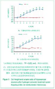
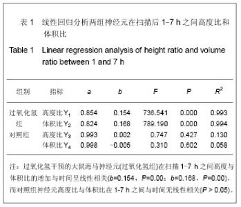
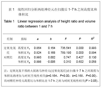
回归分析结果表明:过氧化氢组神经元在扫描1- 7 h之间高度与体积比的增加与时间呈线性相关(b=0.15,P=0.00;b=0.17,P=0.00),而对照组神经元高度比与体积比在1-7 h之间与时间无线性相关 (P > 0.05)。见表1。"

"
| [1] Saisho Y, Manesso E, Gurlo T, et al. Development of factors to convert frequency to rate for beta-cell replication and apoptosis quantified by time-lapse video microscopy and immunohistochemistry. Am J Physiol Endocrinol Metab. 2009; 296(1):E89-96.[2] Gilloteaux J, Jamison JM, Arnold D, et al. Autoschizis of human ovarian carcinoma cells: scanning electron and light microscopy of a new cell death induced by sodium ascorbate: menadione treatment. Scanning. 2003;25(3):137-149.[3] Korchev YE, Bashford CL, Milovanovic M, et al. Scanning ion conductance microscopy of living cells. Biophys J. 1997;73(2): 653-658.[4] Zhao XL, Liu X, Lu HJ, et al. Hopping probe ion conductance microscopy and its combined patch-clamping: a powerful tool for structural and functional studies in neural nanobiology. Mater Sci Forum. 2011;694:54-58.[5] Liu X, Yang X, Zhang B, et al. High-resolution morphological identification and characterization of living neuroblastoma sk-n-sh cells by hopping probe ion conductance microscopy. Brain Res. 2011;1386:35-40.[6] Yang X, Liu X, Zhang X, et al. Investigation of morphological and functional changes during neuronal differentiation of PC12 cells by combined hopping probe ion conductance microscopy and patch-clamp technique. Ultramicroscopy. 2011;111(8):1417-1422.[7] Chen X, Zhang Q, Cheng Q, et al. Protective effect of salidroside against H2O2-induced cell apoptosis in primary culture of rat hippocampal neurons. Mol Cell Biochem. 2009; 332(1-2):85-93.[8] Troyano A, Sancho P, Fernandez C, et al. The selection between apoptosis and necrosis is differentially regulated in hydrogen peroxide-treated and glutathione-depleted human promonocytic cells. Cell Death Differ. 2003;10(8):889-898.[9] Leventis PA, Grinstein S. The distribution and function of phosphatidylserine in cellular membranes. Annu Rev Biophys. 2010;39:407-427.[10] Kim KT, Chung KJ, Lee HS,et al. Neuroprotective effects of tadalafil on gerbil dopaminergic neurons following cerebral ischemia. Neural Regen Res.2013;8(8): 693-701.[11] Golstein P, Kroemer G. Cell death by necrosis: towards a molecular definition. Trends Biochem Sci. 2007;32(1):37-43.[12] Kroemer G, Galluzzi L, Vandenabeele P, et al. Classification of cell death: Recommendations of the nomenclature committee on cell death 2009. Cell Death Differ. 2009;16(1): 3-11.[13] Cellerino A, Galli-Resta L, Colombaioni L. The dynamics of neuronal death: A time-lapse study in the retina. J Neurosci. 2000;20(16):RC92.[14] Hansma PK, Drake B, Marti O, et al. The scanning ion-conductance microscope. Science. 1989;243(4891): 641-643.[15] Novak P, Li C, Shevchuk AI, et al. Nanoscale live-cell imaging using hopping probe ion conductance microscopy. Nat Methods. 2009;6(4):279-281.[16] Zhu H, Li Y, Liu X, et al. Shenwu Wuli Xuebao. 2012;28(8): 644-653.朱晖,李瑛,刘晓,等.跳跃探针式离子电导显微镜技术及其在生物学研究中的应用[J].生物物理学报,2012,28(8):644-653.[17] Zong WX, Thompson CB. Necrotic death as a cell fate. Genes Dev. 2006;20(1):1-15.[18] Simon F, Varela D, Riveros A, et al. Non-selective cation channels and oxidative stress-induced cell swelling. Biol Res. 2002;35(2):215-222.[19] Xiao AY, Wei L, Xia S, et al. Ionic mechanism of ouabain-induced concurrent apoptosis and necrosis in individual cultured cortical neurons. J Neurosci. 2002;22(4): 1350-1362.[20] Greenwood SM, Connolly CN. Dendritic and mitochondrial changes during glutamate excitotoxicity. Neuropharmacology. 2007;53(8):891-898.[21] Takeuchi H, Mizuno T, Zhang G, et al. Neuritic beading induced by activated microglia is an early feature of neuronal dysfunction toward neuronal death by inhibition of mitochondrial respiration and axonal transport. J Biol Chem. 2005;280(11):10444-10454.[22] Greenwood SM, Mizielinska SM, Frenguelli BG, et al. Mitochondrial dysfunction and dendritic beading during neuronal toxicity. J Biol Chem. 2007;282(36):26235-26244.[23] The Ministry of Science and Technology of the People’s Republic of China. Guidance Suggestions for the Care and Use of Laboratory Animals. 2006-09-30. |
| [1] | Liu Xiangxiang, Huang Yunmei, Chen Wenlie, Lin Ruhui, Lu Xiaodong, Li Zuanfang, Xu Yaye, Huang Meiya, Li Xihai. Ultrastructural changes of the white zone cells of the meniscus in a rat model of early osteoarthritis [J]. Chinese Journal of Tissue Engineering Research, 2021, 25(8): 1237-1242. |
| [2] | Li Zhongfeng, Chen Minghai, Fan Yinuo, Wei Qiushi, He Wei, Chen Zhenqiu. Mechanism of Yougui Yin for steroid-induced femoral head necrosis based on network pharmacology [J]. Chinese Journal of Tissue Engineering Research, 2021, 25(8): 1256-1263. |
| [3] | Lun Zhigang, Jin Jing, Wang Tianyan, Li Aimin. Effect of peroxiredoxin 6 on proliferation and differentiation of bone marrow mesenchymal stem cells into neural lineage in vitro [J]. Chinese Journal of Tissue Engineering Research, 2021, 25(7): 1014-1018. |
| [4] | Zhu Xuefen, Huang Cheng, Ding Jian, Dai Yongping, Liu Yuanbing, Le Lixiang, Wang Liangliang, Yang Jiandong. Mechanism of bone marrow mesenchymal stem cells differentiation into functional neurons induced by glial cell line derived neurotrophic factor [J]. Chinese Journal of Tissue Engineering Research, 2021, 25(7): 1019-1025. |
| [5] | Wang Feng, Zhou Liyu, Saijilafu, Qi Shibin, Ma Yanxia, Wei Shanwen. CaMKII-Smad1 promotes axonal regeneration of peripheral nerves [J]. Chinese Journal of Tissue Engineering Research, 2021, 25(7): 1064-1068. |
| [6] | Liu Lihua, Sun Wei, Wang Yunting, Gao Fuqiang, Cheng Liming, Li Zirong, Wang Jiangning. Type L1 steroid-induced osteonecrosis of the femoral head through femoral head and neck junction decompression by fenestration: a single-center prospective clinical study [J]. Chinese Journal of Tissue Engineering Research, 2021, 25(6): 906-911. |
| [7] | Liu Zhao, Xu Xilin, Shen Yiwei, Zhang Xiaofeng, Lü Hang, Zhao Jun, Wang Zhengchun, Liu Xuzhuo, Wang Haitao. Guiding role and prospect of staging and classification combined collapse prediction method for osteonecrosis of femoral head [J]. Chinese Journal of Tissue Engineering Research, 2021, 25(6): 929-934. |
| [8] | Li Shibin, Lai Yu, Zhou Yi, Liao Jianzhao, Zhang Xiaoyun, Zhang Xuan. Pathogenesis of hormonal osteonecrosis of the femoral head and the target effect of related signaling pathways [J]. Chinese Journal of Tissue Engineering Research, 2021, 25(6): 935-941. |
| [9] | Zheng Xiaolong, He Xiaoming, Gong Shuidi, Pang Fengxiang, Yang Fan, He Wei, Liu Shaojun, Wei Qiushi. Bone turnover characteristics in patients with alcohol-induced osteonecrosis of the femoral head [J]. Chinese Journal of Tissue Engineering Research, 2021, 25(5): 657-661. |
| [10] | Zhao Xiang, Wei Cuilan, Zhang Yeting. Neurogenesis and neuroinflammation under exercise: alteration and regulation [J]. Chinese Journal of Tissue Engineering Research, 2021, 25(5): 813-820. |
| [11] | Chen Junyi, Wang Ning, Peng Chengfei, Zhu Lunjing, Duan Jiangtao, Wang Ye, Bei Chaoyong. Decalcified bone matrix and lentivirus-mediated silencing of P75 neurotrophin receptor transfected bone marrow mesenchymal stem cells to construct tissue-engineered bone [J]. Chinese Journal of Tissue Engineering Research, 2021, 25(4): 510-515. |
| [12] | Zeng Xianghong, Liang Bowei. A new strategy for the treatment of osteonecrosis of the femoral head [J]. Chinese Journal of Tissue Engineering Research, 2021, 25(3): 431-437. |
| [13] | Jiang Shengyuan, Li Dan, Jiang Jianhao, Shang-you Yang, Yang Shuye. Biological response of Co2+ to preosteoblasts during aseptic loosening of the prosthesis [J]. Chinese Journal of Tissue Engineering Research, 2021, 25(21): 3292-3299. |
| [14] | Huang Zhusong, Lin Yu, Chen Xiang, Lan Jinfu, Guan Yong, Gao Xi. Alcohol extract of Morinda officinalis improves lipid metabolism and bone metabolism in ovariectomized obese rats [J]. Chinese Journal of Tissue Engineering Research, 2021, 25(2): 205-210. |
| [15] | Chen Yutong, Li Chenchen, Liu Yang, Zheng Yaqin, Yang Xihua, An Meiwen. Establishment of an acute radioactive skin injury model in Wistar rats [J]. Chinese Journal of Tissue Engineering Research, 2021, 25(2): 237-241. |
| Viewed | ||||||
|
Full text |
|
|||||
|
Abstract |
|
|||||