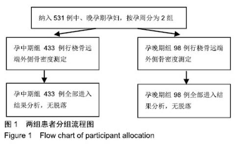
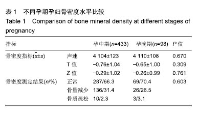
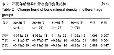
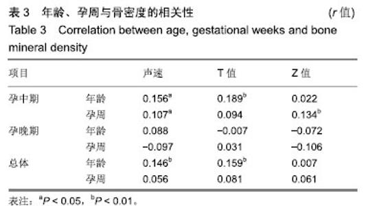
| [1]Yun KY, Han SE, Kim SC, et al. Pregnancy-related osteoporosis and spinal fractures. Obstet Gynecol Sci. 2017;60(1):133-137.[2]Kovacs CS. Bone development and mineral homeostasis in the fetus and neonate: roles of the calciotropic and phosphotropic hormones. Phys Rev. 2014;94(4):1143-1218.[3]Kovacs CS. Calcium, phosphorus and bone metabolism in the fetus and newborn. Early Human Dev. 2015;91(11):623-628.[4]Miyatake N, Muta H, Murota C, et al. Bone status assessment in Japanese subjects using speed of sound along the tibia. Chi Med J. 2002;115(2):254.[5]Karlsson MK, Ahlborg HG, Karlsson C. Maternity and bone mineral density. Acta Orthopaedica.2005;76(1):2-13.[6]杨晓红,熊琼英,左惠萍,等.妊娠期糖尿病患者骨密度检测及影响因素分析[J].中国骨质疏松杂志,2015,21(6):708-711.[7]谢乐瑜,宋兵文,邓如芝,等.产后妇女骨密度影响因素分析[J].实用预防医学,2011,18(7):1300-1301.[8]赵银花,刘春源,温勇,等.妊娠期妇女超声跟骨骨密度测定的临床观察[J]. 婚育与健康·实用诊疗,2014,22(7):45-46.[9]张欣.妊娠期骨密度检测对孕期饮食指导的意义[J].基层医学论坛,2013,17(18):2433-2434.[10]李晋娥,司丽芬,王海生.妊娠期妇女不同孕期骨量监测结果分析[J].中国实用医刊,2014,41(22):66-67.[11]郑彦,杨洋.妊娠期妇女骨密度状况监测分析[J].护理研究,2013, 27(17):1745-1746.[12]Della Martina M, Biasioli A, Vascotto L, et al. Bone ultrasonometry measurements during pregnancy. Arch Gynecol Obstet. 2010;281(3):401-407.[13]Moller UK, Viqstreym S, Mosekilde L, et al. Changes in bone mineral density and body composition during pregnancy and postpartum. A controlled cohort study. Osteoporos Int. 2012; 23(4):1213-1223.[14]胡丽萍.妊娠期骨密度检测对孕期的指导作用[J].中国卫生标准管理,2016,7(2):168-169.[15]杨凤英,海向军.妊娠期妇女骨密度的研究进展[J].中国骨质疏松杂志,2014,20(7):854-857.[16]武亮,祁桠楠,李丽,等.296例妊娠妇女超声骨密度检测分析[J]. 医学信息,2016,29(6):300-301.[17]江滢. 3320例孕妇骨密度测定结果分析[J].中国妇幼保健,2012, 27(28):4377-4378.[18]王士艳,蒋洁,蒋霞. 1056例产妇骨密度检测结果分析[J].实用临床医药杂志,2017,21(21):228-229.[19]徐艳红.不同年龄段孕产妇的骨密度表现比较[J].河北医药,2016, 38(1):108-109.[20]王凯.骨密度检测联合早期干预对孕妇钙丢失的影响[J].医疗装备, 2015,29(15):181-182.[21]凌静,戴新民,叶细锋,等. 19708例健康体检人员跟骨定量超声检测骨密度结果分析[J].中华保健医学杂志,2011,13(2):149-150.[22]张红,王芳芳,白云,等.不同年龄孕期女性骨密度的比较分析[J]. 中国药物与临床,2014,14(1):16-18.[23]李俊.武昌区孕妇骨密度及影响因素分析[J].医学信息,2013,26(22):80-80.[24]吴秀菊,林文雅,黎亮儿.不同孕期孕妇骨密度干预结果与对比分析[J].现代诊断与治疗,2015,26(5):1147-1148.[25]谢晓敏,梁慧芬.不同孕期超声桡骨远端外侧骨密度调查研究[J]. 临床和实验医学杂志,2015,14(10):809-811.[26]龚华虹,刘海静,蒋维,等.孕期钙营养指导及运动干预对孕妇骨密度的影响[J].中国妇幼保健,2013,28(11):1734-1735.[27]邓红.广州市30-49岁女教师骨质与营养状况的相关性分析[J].中国组织工程研究,2018,22(4):499-504.[28]Kyoko Y, Junko I. The effects of dietary calcium and protein intake on changes in bone mineral density during early and late stages of pregnancy. Nihon Koshu Eisei Zasshi. 2010; 57(10):871-880.[29]Kovacs CS. Calcium and bone metabolism disorders during pregnancy and lactation. Endocrinol Metab Clin North Am. 2011;40(4):795-826. |