Chinese Journal of Tissue Engineering Research ›› 2019, Vol. 23 ›› Issue (33): 5249-5255.doi: 10.3969/j.issn.2095-4344.1843
Potential mechanisms of bone marrow stem cells in the treatment of ischemic stroke based on bioinformatics
An Taijian1, Zhang Wei2, Yang Hong2, Wang Qingfeng3
- 1Department of Encephalopathy K2, 2Department of Encephalopathy K3, Affiliated Hospital of Liaoning University of Traditional Chinese Medicine, Shenyang 110032, Liaoning Province, China; 3Liaoning University of Traditional Chinese Medicine, Shenyang 110032, Liaoning Province, China
-
Revised:2019-07-02Online:2019-11-28Published:2019-11-28 -
Contact:Zhang Wei, MD, Associate chief physician, Department of Encephalopathy K3, Affiliated Hospital of Liaoning University of Traditional Chinese Medicine, Shenyang 110032, Liaoning Province, China; Yang Hong, Master, Attending physician, Department of Encephalopathy K3, Affiliated Hospital of Liaoning University of Traditional Chinese Medicine, Shenyang 110032, Liaoning Province, China -
About author:An Taijian, Master, Attending physician, Department of Encephalopathy K2, Affiliated Hospital of Liaoning University of Traditional Chinese Medicine, Shenyang 110032, Liaoning Province, China -
Supported by:the National Natural Science Foundation of China, No. 81703993 (to ZW)
CLC Number:
Cite this article
An Taijian, Zhang Wei, Yang Hong, Wang Qingfeng. Potential mechanisms of bone marrow stem cells in the treatment of ischemic stroke based on bioinformatics[J]. Chinese Journal of Tissue Engineering Research, 2019, 23(33): 5249-5255.
share this article
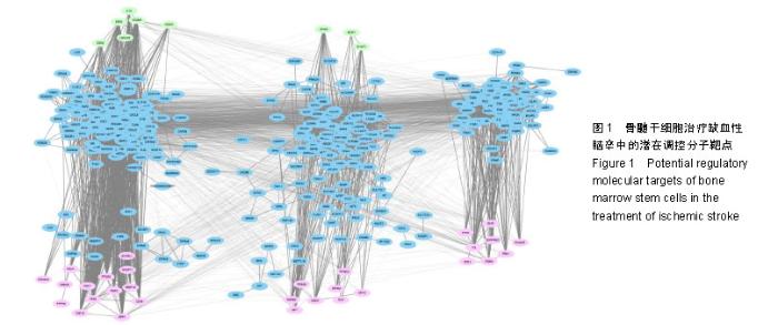
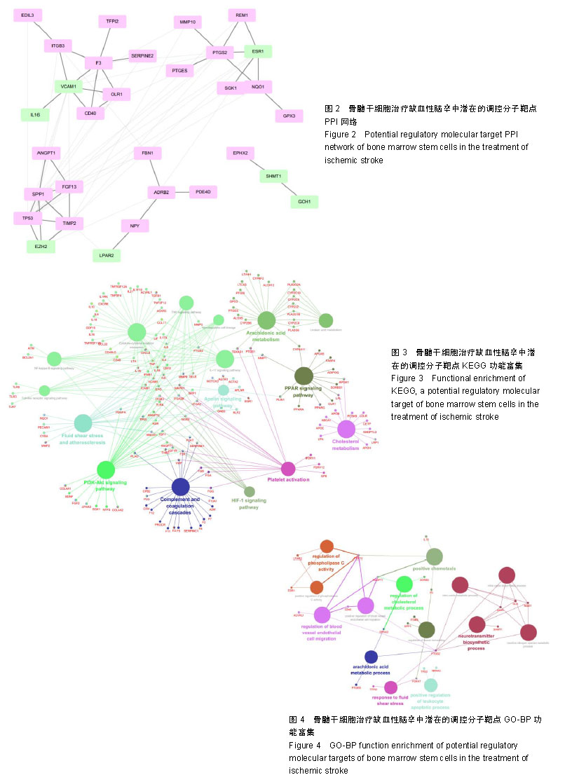
2.1 骨髓干细胞治疗缺血性脑卒中的潜在调控分子靶点 在DisGeNET数据库内,以ischemic cerebrovascular accident(NO.C0948008)为关键词,获得缺血性脑卒中的调控分子靶标共计393个(Summary模式),再以String构建上述分子靶标对应的网络数据库;同时根据GEO数据库GSE21393数据集的运算结果,获得的差异表达基因对上述网络进行打靶处理,最终获得的结果如图1所示。根据上述结果可知,其对应的下调靶点包括FBN1,NPY,MMP10,ITGB3,GLA,SEMA6A,GPX3,TIMP2,FGF13,PTGS2,F3,TFPI2,PTGES,OLR1,NQO1,PTPRG,SERPINE2,SGK1,CD40,PDE4D,PLA2G7,EDIL3,SPP1,ADRB2,ACVRL1,NR4A3,ANGPT1,TP53,REM1,EPHX2等30个分子靶点,其对应的上调靶点包括EZH2,ESR1,IL16,LGALS2,GCH1,SHMT1,VCAM1,P2RX7,LPAR2等9个靶点。 2.2 骨髓干细胞治疗缺血性脑卒中潜在分子靶点的互作关系 对上述靶点进行整理后,由String数据库获得其对应的互作关系,再由Cytoscape构建其PPI网络,见图2。剔除离散节点后,对该网络进行拓扑结构分析可知:该网络中的节点共计31个,对应的边共计66个,平均节点度为4.26,在全部29个节点中,共有10个节点大于平均节点度,分别为TP53,SPP1,VCAM1,PTGS2,ESR1,F3,TIMP2,CD40,EZH2,ITGB3,除了ESR1和EZH2是上调的基因外,其余均为下调的基因。可将上述10个分子靶点视作骨髓干细胞疗法治疗缺血性脑卒中过程中的重要潜在分子靶点。 2.3 骨髓干细胞治疗缺血性脑卒中潜在分子靶点功能分析 对已经获得的全部39个靶点进行相应的功能分析。由于需要分析上述靶点对缺血性脑卒中背景网络的全部调控通路,因此并未直接以上述靶点进行KEGG功能富集,而是进行了网络扩展后,再行分析:首先,在String数据库内,获得上述靶点的相邻靶点;其次,应用Cytoscape软件对获得的全部靶点进行PPI网络构建;最后,将该PPI网络放入Cytoscape的clueGO插件内进行分析(P ≤ 0.05),最终获得上述关键靶点所调控的KEGG通路图,见图3。 由图3可知,已经获得的靶点主要介入的功能有3类,第1类诸如血管剪切力调节及松弛素信号通路等直接作用于血管的通路;第2类为诸如IL-7,NFKB等炎症相关的信号通路,第3类为神经活性配体-受体,同种异体排斥反应相关的信号通路。由该结果可知,在应用骨髓干细胞治疗缺血性脑卒中过程中,模型小鼠症状好转受多个KEGG通路所调控。 为进一步获得上述39个靶点所对应的生物学功能,对上述靶点进行基因本体论的生物过程(GO-BP)功能分析,直接将上述靶点放入Cytoscape的clueGO插件内(P ≤ 0.05),所得的功能富集结果见图4。由图4可知,已经获得的靶点主要介入的功能有3类:第1类诸如直接介入血氧代谢的过程,如一氧化氮合成/代谢等;第2类为组织修复,如组织重塑、血管内皮细胞迁移等;第3类为代谢调控,如花生四烯酸代谢、血小板活化等功能。该结果表明,在应用骨髓干细胞治疗缺血性脑卒中的过程中,所获得的差异表达基因可通过调节多个生物学过程以实现对缺血性脑卒中的治疗。"
"
| [1]陈红霞,杨志敬,潘锐焕,等.中西医结合康复方案对脑卒中后偏瘫患者运动功能、日常生活活动能力和生活质量的影响[J].中国中西医结合杂志,2016,36(4):395-398.[2]中华医学会神经病学分会,中华医学会神经病学分会脑血管病学组.中国急性缺血性脑卒中诊治指南2018[J].中华神经科杂志,2018, 51(9):666-682.[3]柏晓玲,王天兰,石国凤,等. 临床护理路径在脑梗死患者健康教育中应用效果的Meta分析[J].中华医院管理杂志,2016, 32(2): 158-160.[4]张永明,姚奕,王金标,等.骨髓间充质干细胞动脉移植对缺血性脑损伤小鼠行为学的影响[J].中华行为医学与脑科学杂志,2016,25(9): 791-796.[5]郑臻,母得志.骨髓间充质干细胞治疗缺氧缺血性脑损伤的研究现状[J].中华妇幼临床医学杂志(电子版),2017,13(4):373-377.[6]Donega V, van Velthoven CT, Nijboer CH, et al. Intranasal mesenchymal stem cell treatment for neonatal brain damage: long-term cognitive and sensorimotor improvement. PLoS One. 2013;8(1):e51253.[7]van Velthoven CT, Kavelaars A, van Bel F, et al. Mesenchymal stem cell treatment after neonatal hypoxic-ischemic brain injury improves behavioral outcome and induces neuronal and oligodendrocyte regeneration. Brain Behav Immun. 2010;24(3): 387-393.[8]Jellema RK, Wolfs TG, Lima Passos V, et al. Mesenchymal stem cells induce T-cell tolerance and protect the preterm brain after global hypoxia-ischemia. PLoS One. 2013;8(8):e73031.[9]Donega V, Nijboer CH, van Tilborg G, et al. Intranasally administered mesenchymal stem cells promote a regenerative niche for repair of neonatal ischemic brain injury. Exp Neurol. 2014;261:53-64.[10]van Velthoven CT, Kavelaars A, van Bel F, et al. Mesenchymal stem cell transplantation changes the gene expression profile of the neonatal ischemic brain. Brain Behav Immun. 2011;25(7): 1342-1348.[11]Hu ZL, Li N, Wei X, et al. Neuroprotective effects of BDNF and GDNF in intravitreally transplanted mesenchymal stem cells after optic nerve crush in mice. Int J Ophthalmol. 2017;10(1):35-42.[12]Liu X, Wang X, Li A, et al. Effect of mesenchymal stem cell transplantation on brain-derived neurotrophic factor expression in rats with Tourette syndrome. Exp Ther Med. 2016;11(4):1211-1216.[13]Wang Q, Sun G, Gao C, et al. Bone marrow mesenchymal stem cells attenuate 2,5-hexanedione-induced neuronal apoptosis through a NGF/AKT-dependent pathway. Sci Rep. 2016;6:34715.[14]Sohni A, Verfaillie CM. Mesenchymal stem cells migration homing and tracking. Stem Cells Int. 2013;2013:130763.[15]Wei ZZ, Gu X, Ferdinand A, et al. Intranasal delivery of bone marrow mesenchymal stem cells improved neurovascular regeneration and rescued neuropsychiatric deficits after neonatal stroke in rats. Cell Transplant. 2015;24(3):391-402.[16]Cui C, Cui Y, Gao J, et al. Intraparenchymal treatment with bone marrow mesenchymal stem cell-conditioned medium exerts neuroprotection following intracerebral hemorrhage. Mol Med Rep. 2017;15(4):2374-2382.[17]Hasan A, Deeb G, Rahal R, et al. Mesenchymal Stem Cells in the Treatment of Traumatic Brain Injury. Front Neurol. 2017;8:28.[18]马志波. CTA与DSA在脑血管疾病诊断中的价值研究[J].中国医学创新,2013,10(5):110-111.[19]周平,谢伟杰,孙桂波,等.缺血性脑卒中发病机制及药物干预研究进展[J].世界最新医学信息文摘,2018,18(34):53-54,61.[20]孟文婷,李东翔,佟玲.缺血性脑卒中的治疗研究进展[J].中国新药杂志,2016,25(10):1114-1120.[21]付睿,戴威,席春江,等.不同剂量阿托伐他汀对缺血性脑卒中介入诊疗术后对比剂肾损害的预防作用研究[J].中国全科医学,2017,20(7): 812-816.[22]王伊龙.短暂性脑缺血发作的中国专家共识[J].中国实用乡村医生杂志,2013,20(2):883-885.[23]Compston A. Brain repair: an overview. J Neurol. 1994;242 (1 Suppl 1):S1-4.[24]易娟,李贵南.神经干细胞移植治疗新生儿脑损伤的前景展望[J].中国新生儿科杂志, 2017, 32(5):398-400.[25]Song M, Kim YJ, Kim YH, et al. Long-term effects of magnetically targeted ferumoxide-labeled human neural stem cells in focal cerebral ischemia. Cell Transplant. 2015;24(2): 183-190.[26]Chen L, Qiu R, Li L, et al. The role of exogenous neural stem cells transplantation in cerebral ischemic stroke. J Biomed Nanotechnol. 2014;10(11):3219-3230.[27]Wang J, Xia J, Zhang F, et al. Galectin-1-secreting neural stem cells elicit long-term neuroprotection against ischemic brain injury. Sci Rep. 2015;5:9621.[28]Tavares I. Human neural stem cell transplantation in spinal cord injury models: how far from clinical application. Stem Cell Res Ther. 2013;4(3):61.[29]林小娟,唐毅,陈建豪,等.脂肪干细胞移植对大鼠脑缺血后微血管生成及MT1-MMP表达的影响[J].中国实用神经疾病杂志, 2018, 21(14):1514-1520.[30]Yao Y, Zheng XR, Zhang SS, et al. Transplantation of vascular endothelial growth factor-modified neural stem/progenitor cells promotes the recovery of neurological function following hypoxic-ischemic brain damage. Neural Regen Res. 2016; 11(9):1456-1463.[31]Komatsu K, Honmou O, Suzuki J, et al. Therapeutic time window of mesenchymal stem cells derived from bone marrow after cerebral ischemia. Brain Res. 2010;1334:84-92.[32]吴中华.脑外源性神经因子基因修饰神经干细胞对脑卒中的神经保护机制[J].中国组织工程研究, 2015,19(45):7331-7336.[33]Li M, Zhou ZP, Sun M, et al. Reduced Nicotinamide Adenine Dinucleotide Phosphate, a Pentose Phosphate Pathway Product, Might Be a Novel Drug Candidate for Ischemic Stroke. Stroke. 2016;47(1):187-195.[34]Gudkov AV, Komarova EA. Pathologies associated with the p53 response. Cold Spring Harb Perspect Biol. 2010;2(7):a001180.[35]Vaseva AV, Marchenko ND, Ji K, et al. p53 opens the mitochondrial permeability transition pore to trigger necrosis. Cell. 2012;149(7):1536-1548.[36]Yan YP, Lang BT, Vemuganti R, et al. Osteopontin is a mediator of the lateral migration of neuroblasts from the subventricular zone after focal cerebral ischemia.Neurochem Int.2009;55(8):826-832.[37]Ellison JA, Velier JJ, Spera P, et al. Osteopontin and its integrin receptor alpha(v)beta3 are upregulated during formation of the glial scar after focal stroke. Stroke.1998;29(8):1698-1706.[38]李欣遥,张良荣,张硕,等.急性脑梗死患者血清骨桥蛋白水平的改变及其对预后的影响[J]. 中风与神经疾病杂志, 2016, 33(3): 211-214.[39]张文婷,侯华娟,赵昊,等. P2Y1和ITGB3基因多态性与中国汉族大动脉粥样硬化性卒中患者阿司匹林抵抗的相关性[J].国际脑血管病杂志, 2017,25(11):1018-1022.[40]Furie B, Furie BC. Thrombus formation in vivo. J Clin Invest. 2005; 115(12):3355-3362.[41]陈乔,朱金华,李征峰,等.红景天苷对新生大鼠缺氧缺血脑组织MMP-9、TIMP-1含量的影响[J].亚太传统医药,2017,13(13):6-8.[42]Montaner J, Alvarez-Sabín J, Molina C, et al. Matrix metalloproteinase expression after human cardioembolic stroke: temporal profile and relation to neurological impairment. Stroke. 2001;32(8):1759-1766.[43]Romanic AM, White RF, Arleth AJ, et al. Matrix metalloproteinase expression increases after cerebral focal ischemia in rats: inhibition of matrix metalloproteinase-9 reduces infarct size. Stroke. 1998;29(5):1020-1030.[44]Bendeck MP, Hou G, Chen J, et al. Matrix, Matrix Metalloproteinases and Smooth Muscle Cell Function in Atherosclerosis. International Congress.2004;1262(2): 486-489.[45]张毕奎. CD40/CD40L系统在脑梗塞疾病发生发展中的作用及机制研究[D].长沙:中南大学, 2013.[46]陈建昌,孙其中,郝跃伟.阿司匹林抵抗的动脉粥样硬化缺血性脑卒中患者COX-2基因启动子区-765G/C多态性观察[J].山东医药,2016, 56(35):91-93.[47]陈忠云,卫景沛,张洁,等.环氧化酶-2基因-765G/C位点多态性与缺血性脑卒中相关性的Meta分析[J].中国循证医学杂志, 2016,16(1): 48-53.[48]丁利静,李菲,石京山,等.淫羊藿苷对大脑中动脉阻塞诱导的缺血性脑卒中模型大鼠的保护作用[J].遵义医学院学报, 2015,38(2): 137-145.[49]李孝明.异补骨脂素通过雌激素α受体对脊髓的神经保护作用机制研究[D].上海:第二军医大学, 2017.[50]杨青梅,白松,雷又鸣,等. EZH2的功能及在肿瘤中的表达[J].中国老年保健医学, 2014,12(6):72-75. |
| [1] | Luo Lin, Song Naiqing, Huang Jin, Zou Xiaodong. Review and prospect of international research on preschool children’s movement development assessment: a CiteSpace-based visual analysis [J]. Chinese Journal of Tissue Engineering Research, 2021, 25(8): 1270-1276. |
| [2] | Hou Jingying, Yu Menglei, Guo Tianzhu, Long Huibao, Wu Hao. Hypoxia preconditioning promotes bone marrow mesenchymal stem cells survival and vascularization through the activation of HIF-1α/MALAT1/VEGFA pathway [J]. Chinese Journal of Tissue Engineering Research, 2021, 25(7): 985-990. |
| [3] | Liang Xueqi, Guo Lijiao, Chen Hejie, Wu Jie, Sun Yaqi, Xing Zhikun, Zou Hailiang, Chen Xueling, Wu Xiangwei. Alveolar echinococcosis protoscolices inhibits the differentiation of bone marrow mesenchymal stem cells into fibroblasts [J]. Chinese Journal of Tissue Engineering Research, 2021, 25(7): 996-1001. |
| [4] | Geng Yao, Yin Zhiliang, Li Xingping, Xiao Dongqin, Hou Weiguang. Role of hsa-miRNA-223-3p in regulating osteogenic differentiation of human bone marrow mesenchymal stem cells [J]. Chinese Journal of Tissue Engineering Research, 2021, 25(7): 1008-1013. |
| [5] | Lun Zhigang, Jin Jing, Wang Tianyan, Li Aimin. Effect of peroxiredoxin 6 on proliferation and differentiation of bone marrow mesenchymal stem cells into neural lineage in vitro [J]. Chinese Journal of Tissue Engineering Research, 2021, 25(7): 1014-1018. |
| [6] | Zhu Xuefen, Huang Cheng, Ding Jian, Dai Yongping, Liu Yuanbing, Le Lixiang, Wang Liangliang, Yang Jiandong. Mechanism of bone marrow mesenchymal stem cells differentiation into functional neurons induced by glial cell line derived neurotrophic factor [J]. Chinese Journal of Tissue Engineering Research, 2021, 25(7): 1019-1025. |
| [7] | Pei Lili, Sun Guicai, Wang Di. Salvianolic acid B inhibits oxidative damage of bone marrow mesenchymal stem cells and promotes differentiation into cardiomyocytes [J]. Chinese Journal of Tissue Engineering Research, 2021, 25(7): 1032-1036. |
| [8] | Wang Shiqi, Zhang Jinsheng. Effects of Chinese medicine on proliferation, differentiation and aging of bone marrow mesenchymal stem cells regulating ischemia-hypoxia microenvironment [J]. Chinese Journal of Tissue Engineering Research, 2021, 25(7): 1129-1134. |
| [9] | Pan Qile, Zhang Hong, Zhou Huikang, Cai Guang. Comparison of the Greulich-Pyle method, the CHN method and the China 05 method for assessing bone age in children and adolescents [J]. Chinese Journal of Tissue Engineering Research, 2021, 25(5): 662-667. |
| [10] | Chen Junyi, Wang Ning, Peng Chengfei, Zhu Lunjing, Duan Jiangtao, Wang Ye, Bei Chaoyong. Decalcified bone matrix and lentivirus-mediated silencing of P75 neurotrophin receptor transfected bone marrow mesenchymal stem cells to construct tissue-engineered bone [J]. Chinese Journal of Tissue Engineering Research, 2021, 25(4): 510-515. |
| [11] | Zhang Yu, Feng Shuo, Yang Zhi, Zhang Ye, Sun Jianning, An Lun, Chen Xiangyang. Three-dimensional gait of patients with developmental dysplasia of hip undergoing total hip arthroplasty with high hip center [J]. Chinese Journal of Tissue Engineering Research, 2021, 25(3): 350-355. |
| [12] | Jiang Tao, Ma Lei, Li Zhiqiang, Shou Xi, Duan Mingjun, Wu Shuo, Ma Chuang, Wei Qin. Platelet-derived growth factor BB induces bone marrow mesenchymal stem cells to differentiate into vascular endothelial cells [J]. Chinese Journal of Tissue Engineering Research, 2021, 25(25): 3937-3942. |
| [13] | Sun Jianwei, Yang Xinming, Zhang Ying. Effect of montelukast combined with bone marrow mesenchymal stem cell transplantation on spinal cord injury in rat models [J]. Chinese Journal of Tissue Engineering Research, 2021, 25(25): 3962-3969. |
| [14] | Zhang Lishu, Liu Anqi, He Xiaoning, Jin Yan, Li Bei, Jin Fang. Alpl gene affects the therapeutic effect of bone marrow mesenchymal stem cells on ulcerative colitis [J]. Chinese Journal of Tissue Engineering Research, 2021, 25(25): 3970-3975. |
| [15] | Hao Xiaona, Zhang Yingjie, Li Yuyun, Xu Tao. Bone marrow mesenchymal stem cells overexpressing prolyl oligopeptidase on the repair of liver fibrosis in rat models [J]. Chinese Journal of Tissue Engineering Research, 2021, 25(25): 3988-3993. |
| Viewed | ||||||
|
Full text |
|
|||||
|
Abstract |
|
|||||