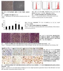
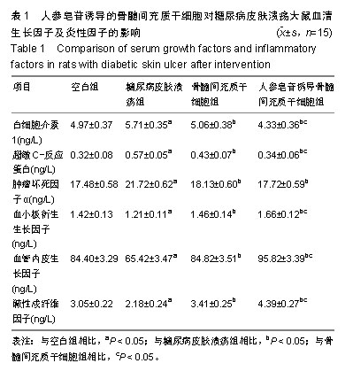
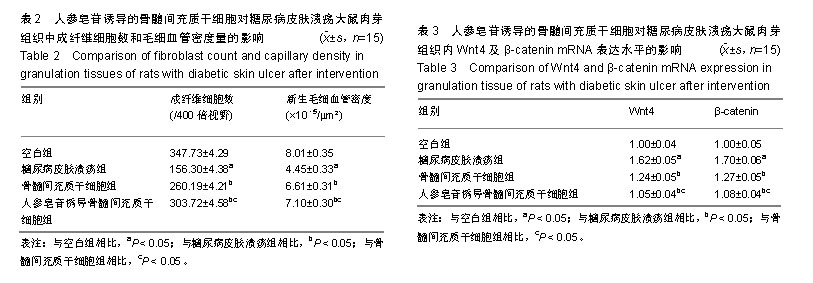
| [1]郭勇英,位庚,李红蓉,等.通心络联合外周血间充质干细胞移植对糖尿病足大鼠血管新生磷脂酰肌醇3_激酶/蛋白激酶B信号通路的影响研究[J].中国全科医学,2016,19(13):1602-1606.[2]柏力,李刚,李立,等. AICAR通过提高MSC移植存活率促进糖尿病溃疡创面愈合的研究[J].局解手术学杂志, 2017,26(10):719-723.[3]张婧婷,吴闽枫,马明华,等.中药治疗糖尿病难愈性创面愈合的研究进展[J].中国药师,2015,18(12):2138-2140.[4]褚月颉,王鹏华.糖尿病伤口愈合不良的相关机制[J].国际内分泌代谢杂志,2014,34(2):131-134.[5]于昕,程梅,张红,等.D-松醇对晚期糖基化终末产物诱导的血管平滑肌细胞增殖和迁移的保护机制[J].中华老年医学杂志, 2018,37(11): 1283-1286.[6]李晓光,方勇,姚敏.单核巨噬细胞集落刺激因子对创面愈合及血管新生调控的研究进展[J].医学综述,2010,16(18):2721-2724.[7]Immonen JA, Zagon IS, McLaughlin PJ. Selective blockade of the OGF-OGFr pathway by naltrexone accelerates fibroblast proliferation and wound healing. Exp Biol Med (Maywood). 2014; 239(10):1300-1309. [8]Secchiero P, Zorzet S, Tripodo C, et al. Human bone marrow mesenchymal stem cells display anti-cancer activity in SCID mice bearing disseminated non-Hodgkin's lymphoma xenografts. PLoS One. 2010;5(6):e11140. [9]Tong C, Hao H, Xia L, et al. Hypoxia pretreatment of bone marrow- derived mesenchymal stem cells seeded in a collagen-chitosan sponge scaffold promotes skin wound healing in diabetic rats with hindlimb ischemia.Wound Repair Regen.2016;24(1):45-56.[10]赵亚男,刘明,张玥,等.愈疡灵软膏对糖尿病溃疡大鼠Wnt/β-catenin信号通路的影响[J].中医杂志, 2016,57(10):879-883.[11]王玉中,张志国,孙朝辉,等.2型糖尿病大鼠模型的建立与评价[J].毒理学杂志,2009,23(6):476-478.[12]Rager TM, Olson JK, Zhou Y, et al. Exosomes secreted from bone marrow-derived mesenchymal stem cells protect the intestines from experimental necrotizing enterocolitis. J Pediatr Surg. 2016; 51(6):942-947. [13]华夏,李广兴,刘仲涛,等.人参皂苷Rh2下调Wnt/β-catenin信号通路调控SHG44细胞的增殖和凋亡[J].解剖学研究, 2018,7(3):218-223.[14]Pouriran R, Piryaei A, Mostafavinia A, et al. The Effect of Combined Pulsed Wave Low-Level Laser Therapy and Human Bone Marrow Mesenchymal Stem Cell-Conditioned Medium on Open Skin Wound Healing in Diabetic Rats. Photomed Laser Surg. 2016;34(8):345-354. [15]Shin C, Kim M, Han JA, et al. Human periodontal ligament stem cells suppress T-cell proliferation via down-regulation of non-classical major histocompatibility complex-like glycoprotein CD1b on dendritic cells. J Periodontal Res. 2017;52(1):135-146. [16]Gao L, Bird AK, Meednu N, et al. Bone Marrow-Derived Mesenchymal Stem Cells From Patients With Systemic Lupus Erythematosus Have a Senescence-Associated Secretory Phenotype Mediated by a Mitochondrial Antiviral Signaling Protein-Interferon-β Feedback Loop. Arthritis Rheumatol. 2017; 69(8):1623-1635. [17]韩夫,王耀军,王耘川,等.吲哚胺2,3-双加氧酶基因修饰BMSCs在大鼠同种异体复合组织移植免疫排斥反应中的作用研究[J].中国修复重建外科杂志,2014,28(6):745-751.[18]黄凤,叶永生,董雨,等.经穴注射BMSCs联合中药对下肢缺血大鼠促血管生成相关因子的影响[J].世界中医药, 2016,11(2):292-295,300.[19]李香丽,张小影,臧立,等.骨髓间充质干细胞在多发性骨髓瘤活动与缓解期具有不同的促血管生成能力[J].中国组织工程研究, 2014, 18(37):5935-5941.[20]李新喜,吾甫尔·依马尔,王护国.大鼠骨髓间充质干细胞诱导血管内皮细胞的实验研究[J].新疆医科大学学报, 2017,40(7):928-933.[21]魏英,余丽梅,王钰莹,等.人参总皂苷促进血管新生改善急性心肌梗死大鼠心功能[J].中国药理学通报,2016,32(4):559-564.[22]黄琦,邓雯雯,许家鸾,等.人参总皂苷对波动性高糖诱导的人脐静脉内皮细胞凋亡的影响[J].中药新药与临床药理, 2012,23(5): 500-504.[23]王会玲,李燕,田军,等.力竭运动对大鼠肾小管凋亡和缺氧诱导因子-1α表达的影响及人参总皂苷干预研究[J].解放军医学杂志, 2014, 39(2):161-166.[24]Ghoneim MA, Agina HA, Elbaz MA, et al. Differentiation of bone marrow-derived mesenchymal stem cells in diabetic patients into islet-like insulin-producing cells: a new era in the treatment of diabetes. Egypt J Pathol. 2016;36(1):87-94.[25]Zhu M, Nikolajczyk BS. Immune cells link obesity-associated type 2 diabetes and periodontitis. J Dent Res. 2014;93(4):346-352.[26]黄思洋,陈继尧,王逸安,等.人参皂苷Rg1对丙二醛抑制小鼠间充质干细胞成骨分化的保护作用[J].生命科学研究, 2016,20(2): 140-144.[27]Cao Y, Gang X, Sun C, et al. Mesenchymal Stem Cells Improve Healing of Diabetic Foot Ulcer. J Diabetes Res. 2017; 2017: 9328347. [28]鲁花,于露,甄欢欢,等.人参皂苷Rg1通过抑制Wnt/β-catenin通路促进退变人腰椎间盘髓核细胞的生长及胞外基质合成[J].中国病理生理杂志,2018,34(4):705-710.[29]Maione AG, Brudno Y, Stojadinovic O, et al. Three-dimensional human tissue models that incorporate diabetic foot ulcer-derived fibroblasts mimic in vivo features of chronic wounds. Tissue Eng Part C Methods. 2015;21(5): 499-508.[30]Maione AG, Smith A, Kashpur O,et al. Altered ECM deposition by diabetic foot ulcer-derived fibroblasts implicates fibronectin in chronic wound repair. Wound Repair Regen. 2016;24(4): 630-643.[31]刘如俊,赵文志,张路,等.表皮生长因子在糖尿病足溃疡创面愈合过程中的作用观察及其机制探讨[J].大连医科大学学报, 2014,(4): 322-327.[32]Zang S, Jin L, Kang S, et al. Periodontal Wound Healing by Transplantation of Jaw Bone Marrow-Derived Mesenchymal Stem Cells in Chitosan/Anorganic Bovine Bone Carrier Into One-Wall Infrabony Defects in Beagles. J Periodontol. 2016;87(8):971-981. [33]郭勇英,常丽萍,张军芳,等.通心络对糖尿病足大鼠缺血下肢移植外周血间充质干细胞的微环境影响[J].中国药理学通报, 2017,33(7): 1032-1033.[34]赵宁,程颖,刘永锋,等.真核表达载体血管内皮生长因子转染血管内皮细胞及对新生血管形成的影响[J].中国组织工程研究, 2008, 12(18):3571-3574.[35]边素艳,刘宏斌,刘宏伟,等.生长因子刺激的间充质干细胞释放具有很强促血管新生功能的外泌体[J].中国实验血液学杂志, 2018, 26(5):294-298.[36]唐乾利,李利青,李辉,等.MEBT/MEBO对大鼠糖尿病足创面组织TGF-β1、Smad3、P-smad3表达及形态学结构的影响[J].重庆医科大学学报,2017,8(3):41-46.[37]张丹丹,张涵,赵久凤,等.积雪草总苷对糖尿病皮肤溃疡大鼠的促愈合作用研究[J].中国药学杂志,2017,52(8):40-45.[38]杨少玲,孙蕾蕾,胡丽叶,等.创面负压治疗对糖尿病足创面肉芽组织碱性成纤维细胞生长因子的影响[J].中国糖尿病杂志, 2016,8(2): 103-106.[39]王妍,杨玉清.β链蛋白和淋巴增强因子-1在人脑胶质瘤中的表达及意义[J].中国药物与临床,2016,16(8):1146-1148.[40]孟佳林,徐雨辰,沈旭峰,等.人PKD2基因慢病毒的构建及其对常染色体显性遗传性多囊肾病小鼠肾脏上皮细胞多囊蛋白-2和Wnt/β-catenin信号通路的修复作用[J].中华泌尿外科杂志, 2018, 39(1):62-65. |