Chinese Journal of Tissue Engineering Research ›› 2026, Vol. 30 ›› Issue (36): 9572-9579.doi: 10.12307/2026.920
Previous Articles Next Articles
Inducing cuproptosis in pancreatic β cells: regulating homocysteine via targeting SLC39A14
Li Shujuan1, Ma Lingju2, 3, Li Xinru4, Sun Rui4, Ma Ben4, Qi Ning4, Zhang Qifan4, Liu Li4, Chai Yuee5, Ma Shengchao3, 4
- 1Emergency Department, 2Department of Geriatrics and Special Needs Medicine, General Hospital of Ningxia Medical University, Yinchuan 750004, Ningxia Hui Autonomous Region, China; 3Key Laboratory of Metabolic Cardiovascular Disease Research of National Health and Wellness Committee, Yinchuan 750004, Ningxia Hui Autonomous Region, China; 4Inspection College of Ningxia Medical University, Yinchuan 750004, Ningxia Hui Autonomous Region, China; 5College of Pharmacy, Guizhou Medical University, Guiyang 550000, Guizhou Province, China
-
Received:2025-11-06Revised:2026-03-25Online:2026-12-28Published:2026-05-25 -
Contact:Ma Shengchao, PhD, Associate professor, Key Laboratory of Metabolic Cardiovascular Disease Research of National Health and Wellness Committee, Yinchuan 750004, Ningxia Hui Autonomous Region, China; Inspection College of Ningxia Medical University, Yinchuan 750004, Ningxia Hui Autonomous Region, China Co-corresponding author: Chai Yuee, PhD, Associate professor, College of Pharmacy, Guizhou Medical University, Guiyang 550000, Guizhou Province, China -
About author:Li Shujuan, MS, Physician, Emergency Department of General Hospital of Ningxia Medical University, Yinchuan 750004, Ningxia Hui Autonomous Region, China -
Supported by:National Natural Science Foundation of China (Young Scientist Program), No. 81900273 (to MSC); National Natural Science Foundation of China (General Program), Nos. 82060139 and 82270492 (both to MSC); Ningxia Natural Science Foundation (Excellent Young Program), No. 2023AAC05035 (to MSC); Key Project of Ningxia Medical University, No. XZ2022004; Ningxia Medical University College Student Innovation and Entrepreneurship Training Program, No. S202210752015 (to TR [principal investigator] and MSC [advisor])

CLC Number:
Cite this article
Li Shujuan, Ma Lingju, Li Xinru, Sun Rui, Ma Ben, Qi Ning, Zhang Qifan, Liu Li, Chai Yuee, Ma Shengchao. Inducing cuproptosis in pancreatic β cells: regulating homocysteine via targeting SLC39A14[J]. Chinese Journal of Tissue Engineering Research, 2026, 30(36): 9572-9579.
share this article
Add to citation manager EndNote|Reference Manager|ProCite|BibTeX|RefWorks
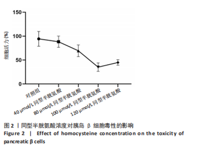
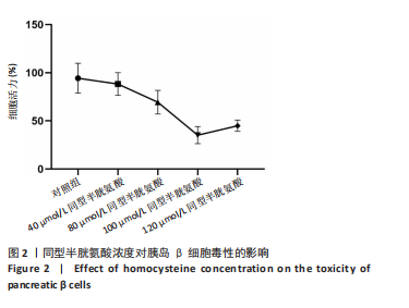
2.1 不同浓度同型半胱氨酸对胰岛β细胞毒性的影响 分别用0,40,80,100,120 μmol/L浓度同型半胱氨酸干预胰岛β细胞48 h 后,CCK-8实验筛选同型半胱氨酸对于胰岛β细胞的最适浓度区间,结果显示当采用100 μmol/L的同型半胱氨酸刺激后,胰岛β细胞的致死率最高(见图2),最终选取100 μmol/L的同型半胱氨酸干预胰岛β细胞。"
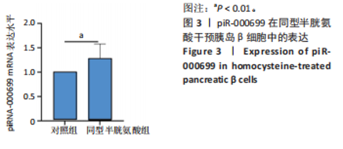
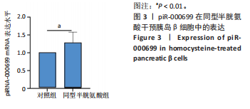
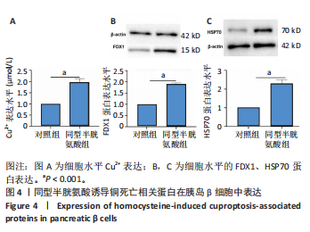
2.2 同型半胱氨酸诱导胰岛β细胞损伤与piR-000699表达情况 为了探讨piR-000699在同型半胱氨酸中表达情况,采用 qRT-PCR方法检测了piR-000699 mRNA的表达水平,结果发现与对照组相比,同型半胱氨酸组胰岛β细胞中piR-000699表达明显增高(P < 0.01),见图3。 2.3 同型半胱氨酸对胰岛β细胞铜死亡相关蛋白表达及Cu2+的影响 为了探讨同型半胱氨酸致胰岛β细胞铜死亡影响,采用铜离子试剂盒检测发现,与对照组相比,同型半胱氨酸组中Cu2+"
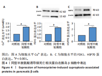
水平明显增高(P < 0.001),见图4A。采用Western blot方法检测铜死亡相关蛋白FDX1、HSP70,与对照组相比,同型半胱氨酸组中FDX1、HSP70蛋白水平明显增加(P < 0.001),见图4B,C。提示同型半胱氨酸可诱导胰岛β细胞发生铜死亡并损伤细胞。"
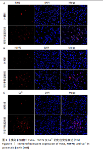
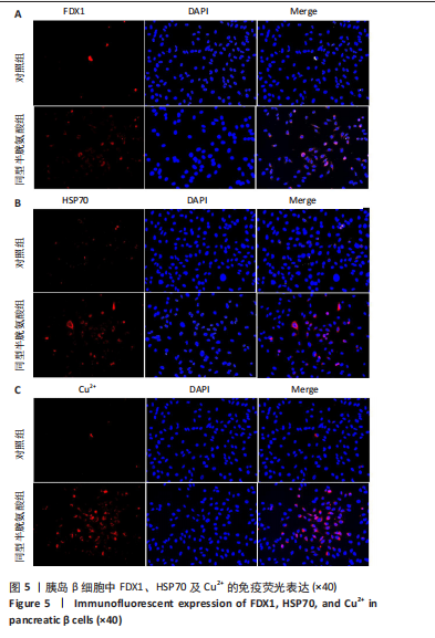
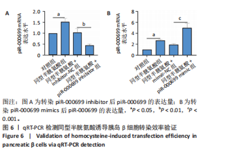
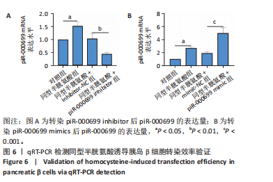
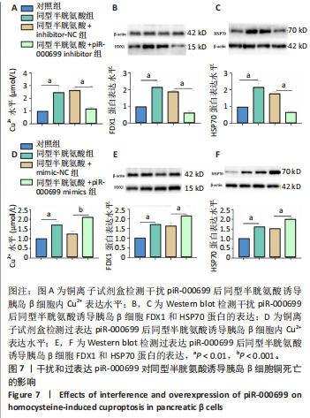
2.4 免疫荧光检测胰岛β细胞铜死亡相关蛋白荧光表达 通过免疫荧光检测FDX1、HSP70及Cu2+荧光表达。结果显示,与对照组相比,同型半胱氨酸组FDX1、HSP70及Cu2+荧光表达显著升高,提示同型半胱氨酸促进胰岛β细胞铜死亡。见图5。 2.5 干扰和过表达piR-000699在同型半胱氨酸诱导胰岛β细胞中的验证 为了明确piR-000699对同型半胱氨酸诱导后胰岛β细胞铜死亡的作用,分别将piR-000699 inhibitor和piR-000699 mimics转染至胰岛β细胞,采用qRT-PCR检测piR-000699的表达。实验结果显示,与对照组相比,同型半胱氨酸组piR-000699 mRNA的表达量明显上调(P < 0.05);与inhibitor-NC组相比,piR-000699 inhibitor组piR-000699 mRNA的表达显著下调(P < 0.01);而与mimics-NC组相比,piR-000699 mimics组piR-000699 mRNA的表达明显上调(P < 0.001);表明piR-000699干扰和过表达片段转染成功,并能在同型半胱氨酸诱导的胰岛β细胞中稳定表达,见图6。 2.6 干扰和过表达piR-000699对同型半胱氨酸诱导胰岛β细胞铜死亡的影响 为了进一步探讨piR-000699对同型半胱氨酸诱导后处理胰岛β细胞铜死亡的作用,采用铜离子试剂盒检测胰岛β细胞中Cu2+的表达量。结果显示,与对照组相比,同型半胱氨酸组胰岛β细胞中Cu2+表达水平增高(P < 0.01);转染piR-000699 inhibitor后与同型半胱氨酸+inhibitor-NC组相比,胰岛β细胞中Cu2+水平显著降低(P < 0.01);转染piR-000699 "
"
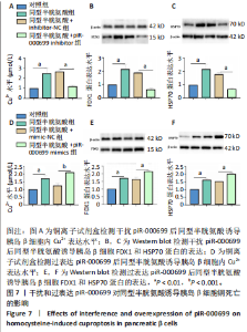
mimics与同型半胱氨酸+mimics-NC组相比,胰岛β细胞中Cu2+水平显著增高(P < 0.001)。采用Western blot检测铜死亡相关蛋白FDX1和HSP70的表达,结果显示,转染piR-000699 inhibitor后与同型半胱氨酸+inhibitor-NC组相比,FDX1和HSP70表达明显降低(P < 0.01);转染piR-000699 mimics后与同型半胱氨酸+mimics-NC组相比,FDX1和HSP70表达明显增高(P < 0.01),见图7。"
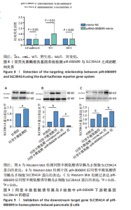
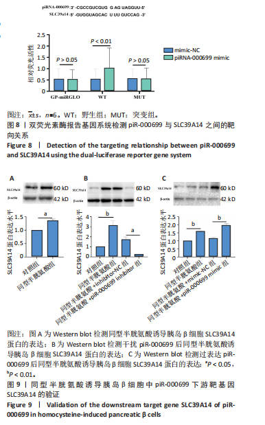
2.7 SLC39A14靶向结合piR-000699调控胰岛β细胞铜死亡损伤 为了验证SLC39A14是否存在靶向调控piR-000699的猜想,运用生物信息学网站预测发现SLC39A14与piR-000699存在潜在结合位点,为了阐明这种诱导的分子机制,并确定piR-000699和SLC39A14之间是否存在靶向结合关系,构建了SLC39A14的野生型和突变型双荧光素酶基因载体,分组为SLC39A14野生型质粒(mimics-NC野生型组)、SLC39A14野生型质粒(piR-000699 mimics野生型组)、SLC39A14突变型质粒(mimics-NC突变型组)、SLC39A14突变型质粒(piR-000699 mimics组),同时转染胰岛β细胞(100 μmol/L)48 h后进行荧光素酶强度测定,结果提示与mimics-NC比较,转染SLC39A14野生型质粒的胰岛β细胞中同时转染piR-000699 mimics后,其相对荧光素酶活性明显升高(P < 0.01);这些共转染组的荧光素酶活性显著高于mimics-NC组(图8)。综上所述,数据表明,piR-000699在胰岛β细胞中与其下游靶基因SLC39A14具有靶向性结合。 2.8 piR-000699靶向同型半胱氨酸诱导胰岛β细胞中的SLC39A14 为了验证SLC39A14与piR-000699靶向调控的猜想,转染piR-000699 inhibtior、piR-000699 mimic后采用Western blot方法检测SLC39A14蛋白水平表达,与对照组相比,同型半胱氨酸组胰岛β细胞中的SLC39A14蛋白表达水平明显升高(P < 0.01);转染piR-000699 inhibitor与同型半胱氨酸+inhibitor-NC组相比,胰岛β细胞中的SLC39A14蛋白水平明显降低(P < 0.05);转染piR-000699 mimics后与同型半胱氨酸+mimics-NC相比,胰岛β细胞中的SLC39A14蛋白水平明显增高(P < 0.01),见图9。"
| [1] MŁYNARSKA E, CZARNIK W, DZIEŻA N, et al. Type 2 Diabetes Mellitus: New Pathogenetic Mechanisms, Treatment and the Most Important Complications. Int J Mol Sci. 2025;26(3):1094. [2] BOUTARI C, DEMARSILIS A, MANTZOROS CS. Obesity and dia betes. Diabetes Res Clin Pract. 2023;20:110773. [3] ZHANG Y, YANG Y, HUANG Q, et al. The effectiveness of lifestyle interventions for diabetes remission on patients with type 2 diabetes mellitus: A systematic review and meta-analysis. Worldviews Evid Based Nurs. 2023;20(1):64-78. [4] LI J, INOUE R, TOGASHI Y, et al. Imeglimin Ameliorates β-Cell Apoptosis by Modulating the Endoplasmic Reticulum Homeostasis Pathway. Diabetes. 2022; 71(3):424-439. [5] MIAO R, FANG X, ZHANG Y, et al. Iron metabolism and ferroptosis in type 2 diabetes mellitus and complications: mechanisms and therapeutic opportunities. Cell Death Dis. 2023;14(3):186. [6] RUZE R, LIU T, ZOU X, et al. Obesity and type 2 diabetes mellitus:connections in epidemiology, pathogenesis, and treatments. Front Endocrinol. 2023;14:1161521. [7] FAN X, LIU Y, CHEN X, et al. Synergies between diabetes and hyperhomocysteinaemia: New insights to predict and prevent adverse cardiovascular effects. Diabetes Obes Metab. 2024:26(12):5776-5785. [8] LIN Z, HE J, YUAN S, et al. Glycemic control and cardiovascular outcomes in patients with diabetes and coronary artery disease according to triglyceride-glucose index: a large scale cohort study. Cardiovasc Diabetol. 2024;23(1):11. [9] KOREN D, KNUTSON KL, BURKE BK, et al. The association of self-reported sleep and circadian measures with glycemic control and diabetes complications among young adults with type 2 diabetes. Am J Physiol Heart Circ Physiol. 2024; 326(6):H1386-H1395. [10] SHEMESH S, LAKS A, COHEN I, et al. Diabetes mellitus and poor glycemic control are associated with a higher risk of lumbar spinal stenosis: an analysis of a large nationwide database. Spine(Phila Pa 1976). 2024;49(9):595-600. [11] YUAN D, CHU J, LIN H, et al. Mechanism of homocysteine-mediated endothelial injury and its consequences for atherosclerosis. Front Cardiovasc Med. 2023; 9:1109445. [12] D’SOUZA SW, GLAZIER JD. Homocysteine metabolism in pregnancy and developmental impacts. Front Cell Dev Biol. 2022;10:802285. [13] 马凌桔,汪乐新,迟宏扬,等. METTL3在同型半胱氨酸诱导小鼠胰岛β细胞自噬中的作用[J].中国组织工程研究,2024,28(26):4221-4225. [14] TSVETKOV P, COY S, PETROVA B, et al. Golub, Copper induces cell death by targeting lipoylated TCA cycle proteins. Science. 2022;375(6586):1254-1261. [15] COBINE PA, MOORE SA, LEARY SC. Getting out what you put in: Copper in mitochondria and its impacts on human disease. Biochim Biophys Acta Mol Cell Res. 2021;1868(1):118867. [16] OZATA DM, GAINETDINOV I, ZOCH A, et al. PIWI-interacting RNAs: small RNAs with big functions. Nat Rev Genet. 2019;20(2):89-108. [17] WANG X, RAMAT A, SIMONELIG M, et al. Emerging roles and functional mechanisms of PIWI-interacting RNAs. Nat Rev Mol Cell Biol. 2023;24(2):123-141. [18] HENAOUI IS, JACOVETTI C, GUERRA MOLLET I, et al. PIWI-interacting RNAs as novel regulators of pancreatic beta cell function. Diabetologia. 2017;60(10): 1977-1986. [19] ZHOU Y, FANG Y, DAI C, et al. PiRNA pathway in the cardiovascular system: a novel regulator of cardiac differentiation, repair and regeneration. J Mol Med (Berl). 2021;99(12):1681-1690. [20] LOVIC D, PIPERIDOU A, ZOGRAFOU I, et al. The Growing Epidemic of Diabetes Mellitus. Curr Vasc Pharmacol. 2020;18(2):104-109. [21] MOHAMMADI-MOTLAGH HR, SADEGHALVAD M, YAVARI N, et al. β Cell and Autophagy: What Do We Know? Biomolecules. 2023;13(4):649. [22] ZENG Q, CAI J, WAN H, et al. PIWI-interacting RNAs and PIWI proteins in diabetes and cardiovascular disease: Molecular pathogenesis and role as biomarkers. Clin Chim Acta. 2021;518:33-37. [23] MAXEL T, SMIDT K, PETERSEN CC, et al. The zinc transporter Zip14 (SLC39a14) affects Beta-cell Function: Proteomics, Gene expression, and Insulin secretion studies in INS-1E cells. Sci Rep. 2019;9(1):8589. [24] 周梦瑶,王浩,尚艳娜,等. 金属转运蛋白slc39a14 的生理功能 及作用机制研究进展[J]. 生命科学,2020,32(9):890-902 [25] AIERKEN Y, HE H, LI R, et al. Inhibition of slc39a14/ slc39a8 reduce vascular calcification via alleviating iron overload induced ferroptosis in vascular smooth muscle cells. Cardiovasc Diabetol. 2024;23(1):186 [26] HUNG YH, KIM Y, MITCHELL SB, et al. Absence of Slc39a14/Zip14 in mouse pancreatic beta cells results in hyperinsulinemia. Am J Physiol Endocrinol Metab. 2024;326(1):E92-E105. [27] SUN H, SAEEDI P, KARURANGA S, et al.Erratum to IDF Diabetes Atlas: Global,Regional and Country-Level Diabetes Prevalence Estimates for 2021 and Projections for 2045. Diabetes Res Clin Pract. 2023;204:110945. [28] JIANG Y, HUO Z, QI X, et al. Copper-induced tumor cell death mechanisms and antitumor theragnostic applications of copper complexes. Nanomedicine (Lond). 2022;17(5):303-324. [29] SONG C, WANG K, QIAN B, et al. Nrf-2/ROS/NF- κB pathway is modulated by cynarin in human mesenchymal stem cells in vitro from ankylosing spondylitis. Clin Transl Sci. 2024;17(3):e13748 [30] CHEN CH, CHOU YT, YANG YW, et al. High-dose copper activates p53-independent apoptosis through the induction of nucleolar stress in human cell lines. Apoptosis. 2021;26(11-12):612-627. [31] YANG X, LAN Q, WANG Q,et al. Association of homocysteine and uric acid with type 2 diabetes mellitus: a case-control study. Sci Rep. 2025;15(1):1914. [32] PICARD M, TAUVERON I, MAGDASY S, et al. Effect of exercise training on heart rate variability in type 2 diabetes mellitus patients: A systematic review and meta-analysis. PLoS One. 2021;16(5):e0251863. [33] 张晴,高春兰,于飞飞,等.同型半胱氨酸致胰岛β细胞凋亡中肝配蛋白A型受体2 DNA 甲基化升高[J].中国组织工程研究,2023,27(5):714-719. [34] 揭育祯,杨慧霞,周瑜瑾,等.TRPC6 DNA甲基化在同型半胱氨酸致胰岛β细胞焦亡中的作用研究[J].广东医学,2022,43(2):162-168. [35] HUSAIN N, MAHMOOD R. Copper(II) generates ROS and RNS, impairs antioxidant system and damages membrane and DNA in human blood cells. Environ Sci Pollut Res Int. 2019;26(20):20654-20668. [36] OMIDIAN A, POURFARZAM M, GHANADIAN SM, et al. Determination of plasma and erythrocyte levels of copper, magnesium and zinc by atomic absorption spectrometry in type-2 diabetes mellitus patients with metabolic syndrome. Res Pharm Sci. 2021;17(1):86-98. [37] LATHIYA N, AZIZ Q, FAWWAD A, et al. Change in pulmonary function tests and their relation to the serum levels of ceruloplasmin, copper and superoxide dismutase in patients suffering from Type-1 and Type-2 diabetes. Pak J Med Sci. 2023;39(3):880-884. [38] CUI Z, ZHOU H, LIU K, et al. Dietary Copper and Selenium Intakes and the Risk of Type 2 Diabetes Mellitus: Findings from the China Health and Nutrition Survey. Nutrients. 2022;14(10):2055. [39] BJØRKLUND G, DADAR M, PIVINA L, et al. The Role of Zinc and Copper in Insulin Resistance and Diabetes Mellitus. Curr Med Chem. 2020;27(39):6643-6657. [40] ELJAZZAR S, ABU-HIJLEH H, ALKHATIB D, et al. The Role of Copper Intake in the Development and Management of Type 2 Diabetes: A Systematic Review. Nutrients. 2023;15(7):1655. [41] ZHOU J, LIU C, FRANCIS M, et al. The Causal Effects of Blood Iron and Copper on Lipid Metabolism Diseases: Evidence from Phenome-Wide Mendelian Randomization Study. Nutrients. 2020;12(10):3174. [42] YU L, YAN J, ZHANG Q, et al. Association between Serum Ferritin and Blood Lipids: Influence of Diabetes and hs-CRP Levels.J Diabetes Res. 2020;2020:4138696. [43] FANG Y, XING C, WANG X, et al. Activation of the ROS/HO-1/NQO1 signaling pathway contributes to the copper-induced oxidative stress and autophagy in duck renal tubular epithelial cells. Sci Total Environ. 2021;757:143753. [44] 黎慧萍,李莹芳,黄济华,等. SLC39A14调控HepG2细胞胰岛素抵抗的机制研究[J].医学研究杂志,2025,54(8):108-113+128. [45] TANG D, CHEN X, KROEMER G. Cuproptosis: a copper-triggered modality of mitochondrial cell death. Cell Res. 2022;32(5):417-418. |
| [1] | Tao Xiangyu, Wang Shuang, Li Yuhan, Cao Jimin, Sun Teng. Effects of piRNA CFAPIR in doxorubicin-induced ferroptosis models of rat and human cardiomyocytes [J]. Chinese Journal of Tissue Engineering Research, 2026, 30(22): 5756-5769. |
| [2] | Sheng Siqi, Xie Lin, Zhao Xiangyu, Jiang Yideng, Wu Kai, Xiong Jiantuan, Yang Anning, Hao Yinju, Jiao Yun. Involvement of miR-144-3p in Cbs+/- mouse hepatocyte autophagy induced by high-methionine diet [J]. Chinese Journal of Tissue Engineering Research, 2024, 28(8): 1289-1294. |
| [3] | Ma Lingju, Wang Lexin, Chi Hongyang, Zhang Jingwen, Peng Hongjian, Gao Chunlan, Jiang Yideng, Huang Hui, Yang Li, Ma Shengchao. Role of METTL3 in homocysteine-induced autophagy in mouse islet beta cells [J]. Chinese Journal of Tissue Engineering Research, 2024, 28(26): 4221-4225. |
| [4] | Pei Jiansheng, Yang Wenjuan, He Jing, Yan Ru, Huang Hui, Jia Shaobin. Bone morphogenetic protein-2 mediated homocysteine promotes vascular calcification [J]. Chinese Journal of Tissue Engineering Research, 2024, 28(25): 4027-4033. |
| [5] | Kuang Yuanjun, Yu Sumei, Zhong Yingyi, Zhang Xuhong, Ma Shengchao, Yang Anning, Hao Yinju, Xiong Jiantuan, Jiao Yun, Jiang Yideng. Effect of cyclic RNA hsa-circ-0001360 on homocysteine-induced apoptosis of human umbilical vein endothelial cells [J]. Chinese Journal of Tissue Engineering Research, 2024, 28(25): 4060-4064. |
| [6] | Zhang Qing, Gao Chunlan, Yu Feifei, Zhang Zhenghao, Ma Fang, Gao Yuan, Li Guizhong, Jiang Yideng, Ma Shengchao. Ephrin A receptor 2 DNA methylation increases in pancreatic beta cell apoptosis induced by homocysteine [J]. Chinese Journal of Tissue Engineering Research, 2023, 27(5): 714-719. |
| [7] | Li Yuanyuan, Sun Yue, Bao Rui, Chang Sirong, Wang Meng, Yu Mengxue, Yang Anning, Liu Zhihong. Ferroptosis is involved in the pathogenesis of liver injury induced by high methionine diet in ApoE-/- mice [J]. Chinese Journal of Tissue Engineering Research, 2023, 27(17): 2681-2686. |
| [8] | Liu Kun, Xie Lin, Cao Jun, Ding Ning, Xu Lingbo, Ma Shengchao, Li Guizhong , Jiang Yideng, Lu Guanjun. Increased FoxO1 DNA methylation level in homocysteine-induced podocyte apoptosis [J]. Chinese Journal of Tissue Engineering Research, 2021, 25(2): 269-273. |
| [9] | Wu Kai, Liu Kun, Xie Lin, Lu Guanjun, Ma Shengchao, Li Guizhong, Cao Jun, Jie Yuzhen, Jiang Yideng, Hao Yinju. Mechanism of hyperhomocysteinemia induced renal injury in Cbs+/- mice [J]. Chinese Journal of Tissue Engineering Research, 2021, 25(11): 1728-1732. |
| [10] | Xie Xiufeng, Zhang Yue, Qu Ze. Clinical outcomes of drug-eluting balloons and drug-eluting stents for the treatment of in-stent restenosis [J]. Chinese Journal of Tissue Engineering Research, 2020, 24(4): 555-560. |
| [11] | Li Xiang, Wang Wei, Xia Shuyue, He Wei. Effects of N-acetylcysteine on oxidative stress in lung tissue of smoked rats [J]. Chinese Journal of Tissue Engineering Research, 2020, 24(2): 254-259. |
| [12] | Xing Hong-xia, Yuan Yan, Liu Sheng, Yin Chuang, Han Jin-hong, Zhou Hui-cong, Su Zhou, Guo Shuang-xi, Wang Yu-mei. Relationship between learning and memory capacities and levodopa in rat models of Parkinson’s disease [J]. Chinese Journal of Tissue Engineering Research, 2014, 18(7): 1069-1075. |
| [13] | Bi Jia-jia, Zhang Si-yun, Luo Wen-yuan, Guan Xing-qiu. Relationships of plasma homocysteine levels and other clinical indexes with the polymorphisms of MTHFR gene C677T among the young people in Shenzhen area [J]. Chinese Journal of Tissue Engineering Research, 2014, 18(42): 6871-6877. |
| Viewed | ||||||
|
Full text |
|
|||||
|
Abstract |
|
|||||