Chinese Journal of Tissue Engineering Research ›› 2026, Vol. 30 ›› Issue (36): 9526-9537.doi: 10.12307/2026.919
Previous Articles Next Articles
Mechanism and effect of aescin intervention against brain injury in a rat model of intracerebral hemorrhage
Ji Lingshan1, Qin Hewei2, Cheng Shuaifang1, Zhao Jing2
- 1Department of Encephalopathy, 2Department of Rehabilitation, Henan Provincial Hospital of Traditional Chinese Medicine (The Second Affiliated Hospital of Henan University of Chinese Medicine), Zhengzhou 450002, Henan Province, China
-
Received:2025-10-21Revised:2026-03-20Online:2026-12-28Published:2026-05-23 -
Contact:Qin Hewei, PhD, Associate chief physician, Master’s supervisor, Department of Rehabilitation, Henan Provincial Hospital of Traditional Chinese Medicine (The Second Affiliated Hospital of Henan University of Chinese Medicine), Zhengzhou 450002, Henan Province, China -
About author:Ji Lingshan, PhD, Associate chief physician, Department of Encephalopathy, Henan Provincial Hospital of Traditional Chinese Medicine (The Second Affiliated Hospital of Henan University of Chinese Medicine), Zhengzhou 450002, Henan Province, China -
Supported by:National Natural Science Foundation of China, No. 81704030 (to QHW); Henan Province Traditional Chinese Medicine Top-notch Talent Cultivation Project, No. 2021-24 (to QHW)

CLC Number:
Cite this article
Ji Lingshan, Qin Hewei, Cheng Shuaifang, Zhao Jing. Mechanism and effect of aescin intervention against brain injury in a rat model of intracerebral hemorrhage[J]. Chinese Journal of Tissue Engineering Research, 2026, 30(36): 9526-9537.
share this article
Add to citation manager EndNote|Reference Manager|ProCite|BibTeX|RefWorks
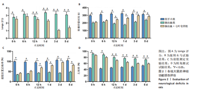
2.1 实验动物数量分析 实验选用大鼠54只,分成3组,实验过程无动物脱失,全部进入结果分析。 2.2 七叶皂苷对脑出血大鼠神经功能损伤的影响 2.2.1 Longa评分评估大鼠神经学功能 假手术组大鼠Longa评分均是0,无神经功能损伤;与假手术组相比,脑出血组大鼠Longa评分在术后均明显升高(P < 0.05);与脑出血组相比,七叶皂苷治疗组大鼠在术后12 h及1,3,5 d的Longa评分均显著降低(P < 0.05),见图2A。 2.2.2 前肢放置测试、转角实验以及旋转木马试验量化感觉运动功能 与假手术组相比,脑出血组大鼠旋转木马时间和同侧肢体"
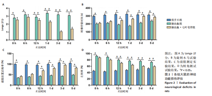
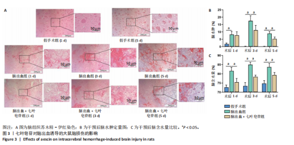
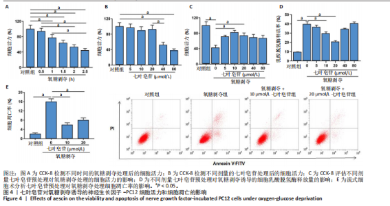
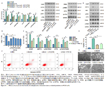
前肢放置次数比例均显著降低(P < 0.05),向左侧转角次数比例明显升高(P < 0.05);与脑出血组相比,七叶皂苷治疗组大鼠在术后12 h及1,3,5 d的旋转木马转棒停留时间和同侧肢体前肢放置次数比例均明显上升(P < 0.05),而向左侧转角次数比例明显下降(P < 0.05),见图2B-D。 上述结果说明,七叶皂苷可改善脑出血大鼠的神经功能缺损情况。 2.3 七叶皂苷改善脑组织病理损伤 2.3.1 苏木精-伊红染色结果 与假手术组相比,脑出血组大鼠脑组织在术后1,3,5 d明显出现出血灶,结构紊乱疏松,呈空泡状水肿,神经元可见大量核固缩等改变;七叶皂苷治疗组大鼠脑组织结构出血灶、胞浆内空泡以及核固缩等在第1天时有所减轻,术后3,5 d时损伤改善更为显著,见图3A。除术后第1天外,七叶皂苷处理在第3,5天可显著减少脑水肿体积,见图3B。 2.3.2 脑含水量测定结果 与假手术组相比,脑出血大鼠在术后1,3,5 d的脑组织含水量显著升高;而同一时间点七叶皂苷治疗组的脑含水量较脑出血组显著降低,见图3C。 2.4 七叶皂苷对氧糖剥夺诱导神经生长因子-PC12细胞损伤和凋亡的保护作用 2.4.1 CCK-8检测细胞活力结果 氧糖剥夺处理1,1.5,2,2.5 h后,神经生长因子-PC12细胞活力均显著降低(P < 0.05),其中以2.5 h处理的抑制效应最显著,见图4A。与对照组相比,浓度为5,10,20 μmol/L 的七叶皂苷处理均不影响神经生长因子-PC12细胞活力(P > 0.05),而浓度为40 μmol/L 和80 μmol/L 的七叶皂苷处理显著降低神经生长因子-PC12细胞活力(P < 0.05),见图4B。与对照组相比,氧糖剥夺组细胞活力降低(P < 0.05);与氧糖剥夺组相比,浓度为10 μmol/L和20 μmol/L的七叶皂苷处理可显著提高神经生长因子-PC12细胞活力(P < 0.05),而40 μmol/L和80 μmol/L的七叶皂苷处理则不改变细胞活力(P > 0.05),见图4C。 2.4.2 ELISA检测乳酸脱氢水平 乳酸脱氢酶是一种稳定的细胞质酶,当细胞膜受损时乳酸脱氢酶会迅速释放到培养基中。与氧糖剥夺组相比,10 μmol/L和20 μmol/L的七叶皂苷处理可降低培养基中乳酸脱氢酶的释放量(P < 0.05),而40 μmol/L和80 μmol/L的七叶皂苷处理则对乳酸脱氢酶的释放比没有显著影响(P > 0.05),见图4D。 基于这些结果,选择10 μmol/L和20 μmol/L的七叶皂苷剂量用于后续实验。 2.4.3 流式细胞术检测细胞凋亡情况 与对照组相比, 氧糖剥夺组细胞凋亡率显著升高(P < 0.05);与氧糖剥夺组相比,10 μmol/L和20 μmol/L的七叶皂苷处理可显著降低细胞凋亡率(P < 0.05),见图4E。 上述结果表明,七叶皂苷对氧糖剥夺诱导的神经生长因子-PC12细胞损伤和凋亡具有保护作用。 2.5 七叶皂苷逆转氧糖剥夺对PC12细胞内质网应激相关蛋白水平的促进作用 鉴于内质网应激是脑出血后诱导神经元凋亡的关键分子事件,随后探讨了七叶皂苷的保护作用是否与调控内质网应激及其下游未折叠蛋白反应(unfolded protein response,UPR)信号通路有关。内质网应激发生时,分子伴侣GRP78从UPR蛋白(IRE1、ATF6 和 PERK)中解离出来,进而激活这些通路以恢复稳态。然而,持续且强烈的内质网应激会导致UPR的促凋亡信号通路过度激活。已有研究表明,脑出血后的大面积缺氧会通过引发内质网应激和钙离子过载,导致神经元凋亡。靶向抑制IRE1/PERK通路对内质网应激介导的细胞凋亡具有较强的神经保护作用[20]。 2.5.1 七叶皂苷缓解内质网应激发挥保护作用 Western blot检测了内质网应激标志蛋白GRP78和促凋亡终末蛋白CHOP的表达,见图5A。结果显示,与对照组相比,氧糖剥夺处理显著上调了GRP78和CHOP的蛋白水平(P < 0.05),证实氧糖剥夺成功"

"

"

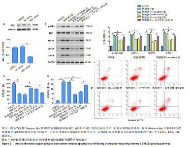
诱导了细胞内质网应激。然而,七叶皂苷预处理显著抑制了氧糖剥夺诱导的GRP78和CHOP蛋白的上调(P < 0.05),见图5A,提示七叶皂苷能够缓解氧糖剥夺诱导的内质网应激。 2.5.2 七叶皂苷对UPR三条核心分支通路的影响 结果显示,与对照组相比,氧糖剥夺处理显著上调了p-IRE1/IRE1、 p-PERK/PERK和p-eIF2α/eIF2α比值及ATF6表达水平(P < 0.05)。与氧糖剥夺组相比,七叶皂苷预处理显著降低了p-IRE1、p-PERK和p-eIF2α的表达水平(P < 0.05),但对ATF6的蛋白水平影响不大(P > 0.05),见图5A。 2.5.3 七叶皂苷对PC12细胞的保护作用依赖于其对内质网应激的抑制 采用内质网应激激动剂衣霉素建立内质网应激细胞模型,4-PBA被用作内质网应激抑制的阳性对照。与对照组相比,衣霉素处理可显著降低细胞活力(P < 0.05),而七叶皂苷处理能显著逆转这一效应(P < 0.05),见图5B。 此外,与对照组相比,衣霉素触发了强烈的内质网应激反应,显著上调了GRP78、CHOP、p-IRE1、p-PERK和ATF6的表达水平(P < 0.05);与衣霉素组相比,七叶皂苷处理显著下调CHOP、GRP78、p-IRE1及p-PERK蛋白水平(P < 0.05),对IRE1/PERK通路的抑制模式与4-PBA一致,但七叶皂苷对衣霉素诱导的ATF6表达没有影响(P > 0.05),见图5C,D。 流式细胞术结果显示:与对照组相比,衣霉素组细胞凋亡率显著升高(P < 0.05);与衣霉素组相比,七叶皂苷处理组的细胞凋亡率均显著降低(P < 0.05),这与4-PBA处理组的结果一致,见图5E,F。 透射电镜观察发现,衣霉素组细胞可见胞质内细胞器整体排布紊乱,局部区域因内质网扩张呈现疏松空泡样改变;七叶皂苷组与4-PBA组细胞胞质内疏松空泡样区域减少,细胞器排布更趋规整,见图5G。 上述结果说明,七叶皂苷可阻断IRE1/PERK信号通路缓解氧糖剥夺和衣霉素诱导的细胞内质网应激损伤。 2.6 七叶皂苷通过抑制IRE1和PERK信号通路缓解氧糖剥夺诱导的内质网应激和细胞损伤 为验证七叶皂苷的作用机制,分别利用IRE1或PERK的特异性siRNA进行基因敲低,并应用通路激动剂进行功能恢复实验。 2.6.1 IRE1信号通路方面 与NC siRNA组相比,敲低IRE1显著降低细胞中IRE1蛋白表达水平,见图6A。与氧糖剥夺组相比,氧糖剥夺+IRE1 siRNA组与氧糖剥夺+七叶皂苷组细胞中内质网应激标志物GRP78、CHOP以及p-IRE1及其下游效应分子XBP1s的蛋白水平均显著降低(P < 0.05),见图6B,提示敲低IRE1与七叶皂苷干预对IRE1-XBP1通路的抑制作用一致。"

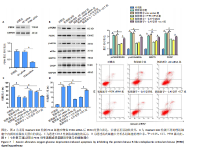
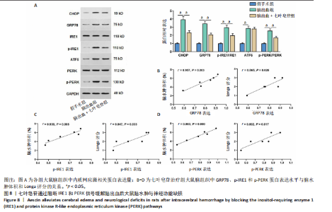
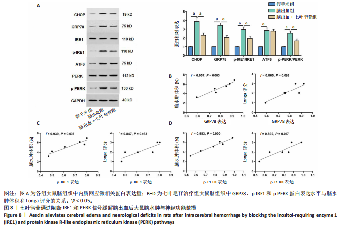
与氧糖剥夺+七叶皂苷组相比,加入IRE1激动剂IXA4后,细胞中GRP78、CHOP、p-IRE1与XBP1s蛋白水平显著升高(P < 0.05),见图6B,表明IXA4可通过激活IRE1通路,逆转七叶皂苷对该通路的抑制作用。此外,与氧糖剥夺组相比,氧糖剥夺+IRE1 siRNA组与氧糖剥夺+七叶皂苷组的细胞活力均明显升高(P < 0.05)、细胞凋亡率均降低(P < 0.05),见图6C,D,提示IRE1的基因敲低与七叶皂苷的保护效应一致。 与氧糖剥夺+七叶皂苷组相比,氧糖剥夺+七叶皂苷+ IXA4组的细胞活力显著下降(P < 0.05),凋亡率显著升高(P < 0.05),见图6C,D,表明IRE1的激活逆转了七叶皂苷对细胞损伤的保护作用。 2.6.2 PERK信号通路方面 观察到了与1IRE1信号通路高度一致的结果。与NC siRNA组相比,敲低PERK显著降低了细胞中PERK蛋白表达水平,见图7A。敲低PERK与七叶皂苷处理同样有效地抑制了氧糖剥夺诱导的GRP78、CHOP表达以及p-PERK及其下游靶点eIF2α的磷酸化(P < 0.05)(图7B),并改善了细胞存活(P < 0.05)(图7C)、抑制了凋亡(P < 0.05)(图7D)。同样地,PERK激动剂CCT020312的共处理也成功恢复了PERK-eIF2α通路的活性、并逆转了七叶皂苷对细胞的保护作用(P < 0.05)(图7B-D)。 以上结果表明,七叶皂苷可通过特异性抑制IRE1-XBP1与PERK-eIF2α信号通路,缓解氧糖剥夺诱导的内质网应激,提高细胞活力、减少细胞凋亡。 2.7 七叶皂苷通过阻断IRE1和PERK信号缓解脑出血后脑水肿与神经功能缺损 为验证七叶皂苷对脑出血大鼠内质网应激的保护作用,构建了脑出血大鼠模型并检测其对脑出血大鼠脑组织中内质网应激相关蛋白表达水平的影响。 2.7.1 Western blot实验结果 与假手术组相比,脑出血大鼠脑组织中CHOP、GRP78、p-IRE1、p-PERK和ATF6表达水平显著升高(P < 0.05);与脑出血组相比,七叶皂苷治疗组大鼠脑组织中CHOP、GRP78、p-IRE1和p-PERK表达水平显著降低(P < 0.05),而ATF6表达水平无显著变化(P > 0.05),见图8A。 2.7.2 相关性分析结果 为直接验证七叶皂苷的疗效与其机制的相关性,对七叶皂苷治疗组大鼠进行了相关性分析。结果显示,GRP78、p-IRE1和PERK的蛋白表达水平与脑水肿体积呈显著正相关(r=0.957,P=0.003;r=0.939, P=0.005;r=0.983,P=0.000),同时与神经功能Longa评分亦呈显著正相关(r=0.865,P=0.026;r=0.847,P=0.033;r=0.892,P=0.017),见图8B-D。这表明,七叶皂苷对p-IRE1和PERK通路的抑制作用越强,其减轻脑水肿和改善神经功能的效果越显著。"

"
| [1] MORRIS NA, SIMARD JM, CHATURVEDI S. Surgical Management for Primary Intracerebral Hemorrhage. Neurology. 2024;103(4):e209714. [2] HANLEY DF, ZIAI WC, VAHIDY FS, et al. Intracerebral Hemorrhage: Keep It Simple. Stroke. 2024;55(12):2942-2945. [3] LIANG Y, FU W, TANG Y, et al. Selective Activation of G Protein-Coupled Estrogen Receptor 1 (GPER1) Reduces ER Stress and Pyroptosis via AMPK Signaling Pathway in Experimental Subarachnoid Hemorrhage. Mol Neurobiol. 2025;62 (1): 871-884. [4] TIAN Q, LIU S, HAN SM, et al. The mechanism and relevant mediators associated with neuronal apoptosis and potential therapeutic targets in subarachnoid hemorrhage. Neural Regen Res. 2023;18(2):244-252. [5] JIN MH, LIU XD, SUN HN, et al. Peroxiredoxin II exerts neuroprotective effects by inhibiting endoplasmic reticulum stress and oxidative stress-induced neuronal pyroptosis. Mol Biol Rep. 2024;51(1):607. [6] WEI P, HAN Y, CHEN H, et al. Ang-1 Inhibited Endoplasmic Reticulum Stress and Apoptosis of VECs in Rats with aSAH-induced CVS Through the Regulated PI3K/Akt Pathway. Curr Neurovasc Res. 2023;20(1):140-148. [7] ALMARGHALANI DA, BAHADER GA, ALI M, et al. Cofilin Inhibitor Improves Neurological and Cognitive Functions after Intracerebral Hemorrhage by Suppressing Endoplasmic Reticulum Stress Related-Neuroinflammation. Pharmaceuticals (Basel). 2024;17(1):114. [8] YANG Z, ZHOU C, SHI H, et al. Heme Induces BECN1/ATG5-Mediated Autophagic Cell Death via ER Stress in Neurons. Neurotox Res. 2020;38(4):1037-1048. [9] GAO X, YANG H, SU J, et al. Aescin Protects Neuron from Ischemia-Reperfusion Injury via Regulating the PRAS40/mTOR Signaling Pathway. Oxid Med Cell Longev. 2020;2020:7815325. [10] SUN X, LIU Z, ZHOU L, et al. Escin avoids hemorrhagic transformation in ischemic stroke by protecting BBB through the AMPK/Cav-1/MMP-9 pathway. Phytomedicine. 2023;120(1):155071. [11] XIAO GM, WEI J. Effects of beta-Aescin on the expression of nuclear factor-kappaB and tumor necrosis factor-alpha after traumatic brain injury in rats. J Zhejiang Univ Sci B. 2005;6(1):28-32. [12] ZHANG L, FEI M, WANG H, et al. Sodium aescinate provides neuroprotection in experimental traumatic brain injury via the Nrf2-ARE pathway. Brain Res Bull. 2020;157(1):26-36. [13] BHARDWAJ S, JINDAL A, SINGH S, et al. Pharmacological Evaluation of Aescin for Neuroprotection in Intracerebroventricular Streptozotocin Model of Alzheimer’s Disease in Experimental Rats. Assay Drug Dev Technol. 2025;23(6):299-312. [14] SELVAKUMAR GP, JANAKIRAMAN U, ESSA MM, et al. Escin attenuates behavioral impairments, oxidative stress and inflammation in a chronic MPTP/probenecid mouse model of Parkinson’s disease. Brain Res. 2014;1585(1):23-36. [15] SUN Y, JIANG X, PAN R, et al. Escins Isolated from Aesculus chinensis Bge. Promote the Autophagic Degradation of Mutant Huntingtin and Inhibit its Induced Apoptosis in HT22 cells. Front Pharmacol. 2020;11:116. [16] CHENG P, KUANG F, JU G. Aescin reduces oxidative stress and provides neuroprotection in experimental traumatic spinal cord injury. Free Radic Biol Med. 2016;99(1):405-417. [17] 姬令山, 秦合伟, 程率芳, 等. 七叶皂苷通过抑制TXNIP_MLXIPL信号通路改善脑出血大鼠神经性损伤[J]. 辽宁中医杂志,2023,50(4):194-198+229. [18] CHEN Q, SONG W, TANG Y, et al. Electroacupuncture Reduces Cerebral Hemorrhage Injury in Rats by Improving Cerebral Iron Metabolism. Mediators Inflamm. 2022;2022:6943438. [19] QU J, ZONG H F, SHAN Y, et al. Piezo1 suppression reduces demyelination after intracerebral hemorrhage. Neural Regen Res. 2023;18(8):1750-1756. [20] HUANG Q, LAN T, LU J, et al. DiDang Tang Inhibits Endoplasmic Reticulum Stress-Mediated Apoptosis Induced by Oxygen Glucose Deprivation and Intracerebral Hemorrhage Through Blockade of the GRP78-IRE1/PERK Pathways. Front Pharmacol. 2018;9:1423. [21] CAO Y, LIU C, LI G, et al. Metformin Alleviates Delayed Hydrocephalus after Intraventricular Hemorrhage by Inhibiting Inflammation and Fibrosis. Transl Stroke Res. 2023;14(3):364-382. [22] ZHAO Q, XU J, XIA Z, et al. Ghrelin attenuates metabolic dysfunction-associated steatotic liver disease by activating glutamine metabolism via inhibition of the IRE1α-XBP-1 pathway. Biochem Pharmacol. 2025;240:117089. [23] YANG C, ZHAO M, CHEN Y, et al. Dietary bitter ginger-derived zerumbone improved memory performance during aging through inhibition of the PERK/CHOP-dependent endoplasmic reticulum stress pathway. Food Funct. 2024;15(18):9070-9084. [24] 黄婷, 陈波 , 曾昭明. VEGF基因修饰人骨髓间充质干细胞对脑出血大鼠早期脑水肿的影响[J]. 四川大学学报(医学版),2020,51(5):622-629. [25] WAN Y, HOLSTE K G, HUA Y, et al. Brain edema formation and therapy after intracerebral hemorrhage. Neurobiol Dis. 2023;176:105948. [26] DING YX, EERDUNA GW, DUAN SJ, et al. Escin ameliorates the impairments of neurological function and blood brain barrier by inhibiting systemic inflammation in intracerebral hemorrhagic mice. Exp Neurol. 2021;337(1):113554. [27] YU N, LI X, WANG B, et al. Unraveling the Role of α2δ-1 in Cerebral Hemorrhage: Calcium Overload, Endoplasmic Reticulum Stress, and Microglial Apoptosis. Brain Behav. 2025;15(5):e70499. [28] CHEN X, SHI C, HE M, et al. Endoplasmic reticulum stress: molecular mechanism and therapeutic targets. Signal Transduct Target Ther. 2023;8(1):352. [29] ZHENG J, LI Y, ZHANG T, et al.Endoplasmic reticulum stress and autophagy in cerebral ischemia/reperfusion injury: PERK as a potential target for intervention. Neural Regen Res. 2025;20(5):1455-1466. [30] LIU X, HONG E, XIE J, et al. Txnrd2 Attenuates Early Brain Injury by Inhibition of Oxidative Stress and Endoplasmic Reticulum Stress via Trx2/Prx3 Pathway after Intracerebral Hemorrhage in Rats. Neuroscience. 2024;545(1):158-170. [31] YIN H, RAN Z, LUO T, et al. BCL-3 Promotes Intracerebral Hemorrhage Progression by Increasing Blood-Brain Barrier Permeability, Inflammation, and Cell Apoptosis via Endoplasmic Reticulum Stress. Mediators Inflamm. 2023;2023:1420367. [32] CHEN G, GAO C, YAN Y, et al. Inhibiting ER Stress Weakens Neuronal Pyroptosis in a Mouse Acute Hemorrhagic Stroke Model. Mol Neurobiol. 2020;57(12):5324-5335. [33] LIU Y, CUI F, XU A, et al. Interaction Between the PERK/ATF4 Branch of the Endoplasmic Reticulum Stress and Mitochondrial One-Carbon Metabolism Regulates Neuronal Survival After Intracerebral Hemorrhage. Int J Biol Sci. 2024; 20 (11):4277-4296. [34] 吴宇. 香叶醇通过PERK-ATF4-CHOP途径抑制内质网应激保护脑缺血再灌注损伤研究[D]. 重庆:重庆医科大学,2023. [35] 徐晨阳, 苑兵舰. PERK通路对大鼠脑出血继发性脑损伤及神经细胞凋亡的作用[J].河南大学学报(医学版),2022,41(2):110-116. [36] CHEN Y, CHEN Z, CHENG W, et al. The Role of the GRP78/PERK/ATF4 Pathway in the Ability of Gua Lou Gui Zhi Decoction to Attenuate Apoptosis by Inhibiting Endoplasmic Reticulum Stress after Ischemia-Reperfusion Injury. Front Biosci (Landmark Ed). 2022;27(10):296. [37] HETZ C, PAPA FR. The Unfolded Protein Response and Cell Fate Control. Mol Cell. 2018;69(2):169-181. [38] GLEMBOTSKI CC, ROSARDA JD,WISEMAN RL. Proteostasis and Beyond: ATF6 in Ischemic Disease. Trends Mol Med. 2019;25(6):538-550. [39] 张倩. 七叶皂苷钠通过调控ERK_STAT3信号轴促进胰腺腺泡细胞凋亡进而影响急性胰腺炎转归[D]. 济南:山东中医药大学,2022. [40] 汪先凯, 周大臣, 喻宗繁, 等. 七叶皂苷钠通过降低4EBP1的磷酸化水平抑制梗阻性黄疸大鼠小胆管增生的研究[J]. 安徽医科大学学报,2021,56(10): 1521-1526. [41] LEI Y, YU H, DING S, et al. Molecular mechanism of ATF6 in unfolded protein response and its role in disease. Heliyon. 2024;10(5):e25937. [42] LI M, TANG S, VELKOV T, et al. Copper exposure induces mitochondrial dysfunction and hepatotoxicity via the induction of oxidative stress and PERK/ATF4 -mediated endoplasmic reticulum stress. Environ Pollut. 2024;352:124145. [43] PAN E Z, XIN Y, LI X Q, et al. Ameliorative effects of silybin against avermectin-triggered carp spleen mitochondrial dysfunction and apoptosis through inhibition of PERK-ATF4-CHOP signaling pathway. Fish Physiol Biochem. 2023;49(5): 895-910. [44] QURESHI S, LEE S, STEIDL W, et al. Endoplasmic Reticulum Stress Disrupts Mitochondrial Bioenergetics, Dynamics and Causes Corneal Endothelial Cell Apoptosis. Invest Ophthalmol Vis Sci. 2023;64(14):18. [45] ALI F E M, AHMED S F, ELTRAWY A H, et al. Pretreatment with Coenzyme Q10 Combined with Aescin Protects against Sepsis-Induced Acute Lung Injury. Cells Tissues Organs. 2021;210(3):195-217. [46] MEI T, MA L, KONG F. Sodium aescinate improve behavioral performance by inhibiting dorsal raphe nucleus NLRP3 inflammasome in Post-traumatic stress disorder Rat Model. Biochem Biophys Res Commun. 2023;671:166-172. [47] SUN Y, ZHANG R, PAN Y, et al. Anti-Apoptotic Effects of Escin on Porphyromonas gingivalis-Derived Lipopolysaccharide-Induced Injury in SH-SY5Y Cells. Brain Behav. 2025;15(9):e70810. [48] 徐飞, 郭东凯, 季士亮, 等. 七叶皂苷钠的药理作用及临床药物联用[J]. 中国医药科学,2022,10(10):51-55. [49] 孙静,林丽桥. 甘露醇、七叶皂苷钠联合应用治疗脑出血致脑水肿的疗效及对神经功能的影响[J]. 医学信息,2025,38(12):82-85+90. [50] 张奇, 郭彦俊, 汪宁, 等. 七叶皂苷钠联合依达拉奉、氨氯地平治疗对高血压性脑出血患者脑血管功能恢复和预后的影响[J]. 贵州医药,2024,48(12): 1884-1886. |
| [1] | Chen Haojie, Wang Dai, Shen Shan. Immune inflammatory microenvironment mechanisms in peri-implantitis [J]. Chinese Journal of Tissue Engineering Research, 2026, 30(8): 2054-2062. |
| [2] | Hu Xiongke, Liu Shaohua, Tan Qian, Liu Kun, Zhu Guanghui. Shikonin intervention with bone marrow mesenchymal stem cells improves microstructure of femur in aged mice [J]. Chinese Journal of Tissue Engineering Research, 2026, 30(7): 1609-1615. |
| [3] | Yuan Xiaoshuang, Yang Xu, Yang Bo, Chen Xiaoxu, Tian Ting, Wang Feiqing, Li Yanju, Liu Yang, Yang Wenxiu. Effect of conditioned medium of diffuse large B-cell lymphoma cells on proliferation and apoptosis of human bone marrow mesenchymal stem cells [J]. Chinese Journal of Tissue Engineering Research, 2026, 30(7): 1632-1640. |
| [4] | Han Nianrong, Huang Yifei, Akram · Osman, Liu Yanlu, Hu Wei . Programmed cell death receptor-1 suppresses osteogenic differentiation of rat bone marrow mesenchymal stem cells in a high-glucose microenvironment [J]. Chinese Journal of Tissue Engineering Research, 2026, 30(7): 1649-1657. |
| [5] | He Jiale, Huang Xi, Dong Hongfei, Chen Lang, Zhong Fangyu, Li Xianhui. Acellular dermal matrix combined with adipose-derived stem cell exosomes promotes burn wound healing [J]. Chinese Journal of Tissue Engineering Research, 2026, 30(7): 1699-1710. |
| [6] | Liu Anting, Lu Jiangtao, Zhang Wenjie, He Ling, Tang Zongsheng, Chen Xiaoling. Regulation of AMP-activated protein kinase by platelet lysate inhibits cadmium-induced neuronal apoptosis [J]. Chinese Journal of Tissue Engineering Research, 2026, 30(7): 1800-1807. |
| [7] | Chen Ju, Zheng Jinchang, Liang Zhen, Huang Chengshuo, Lin Hao, Zeng Li. Effect and mechanism of beta-caryophyllene in mice with osteoarthritis [J]. Chinese Journal of Tissue Engineering Research, 2026, 30(6): 1341-1347. |
| [8] | Lyu Guoqing, Aizimaitijiang·Rouzi, Xiong Daohai. Irisin inhibits ferroptosis in human articular chondrocytes: roles and mechanisms [J]. Chinese Journal of Tissue Engineering Research, 2026, 30(6): 1359-1367. |
| [9] | Peng Zhiwei, Chen Lei, Tong Lei. Luteolin promotes wound healing in diabetic mice: roles and mechanisms [J]. Chinese Journal of Tissue Engineering Research, 2026, 30(6): 1398-1406. |
| [10] | Zhu Kuicheng, Du Chunyan, Zhang Jintao. Mechanism by which hairless gene mutation promotes white adipose tissue browning in hairless mice [J]. Chinese Journal of Tissue Engineering Research, 2026, 30(6): 1424-1430. |
| [11] | Jia Jinwen, Airefate·Ainiwaer, Zhang Juan. Effects of EP300 on autophagy and apoptosis related to allergic rhinitis in rats [J]. Chinese Journal of Tissue Engineering Research, 2026, 30(6): 1439-1449. |
| [12] | Li Hao, Tao Hongcheng, Zeng Ping, Liu Jinfu, Ding Qiang, Niu Chicheng, Huang Kai, Kang Hongyu. Mitogen-activated protein kinase signaling pathway regulates the development of osteoarthritis: guiding targeted therapy with traditional Chinese medicine [J]. Chinese Journal of Tissue Engineering Research, 2026, 30(6): 1476-1485. |
| [13] | Liu Yu, Lei Senlin, Zhou Jintao, Liu Hui, Li Xianhui. Mechanisms by which aerobic and resistance exercises improve obesity-related cognitive impairment [J]. Chinese Journal of Tissue Engineering Research, 2026, 30(5): 1171-1183. |
| [14] | Wang Zhengye, Liu Wanlin, Zhao Zhenqun. Advance in the mechanisms underlying miRNAs in steroid-induced osteonecrosis of the femoral head [J]. Chinese Journal of Tissue Engineering Research, 2026, 30(5): 1207-1214. |
| [15] | Chen Yixian, Chen Chen, Lu Liheng, Tang Jinpeng, Yu Xiaowei. Triptolide in the treatment of osteoarthritis: network pharmacology analysis and animal model validation [J]. Chinese Journal of Tissue Engineering Research, 2026, 30(4): 805-815. |
| Viewed | ||||||
|
Full text |
|
|||||
|
Abstract |
|
|||||