Chinese Journal of Tissue Engineering Research ›› 2026, Vol. 30 ›› Issue (15): 3855-3861.doi: 10.12307/2026.592
Previous Articles Next Articles
Correlation between quantitative indicators of three-dimensional deformity of the first metatarsal bone and functional prognosis after osteotomy and orthopedic treatment in patients with hallux valgus deformity
Sun Meilan1, Zhao Xiaoliang1, Yan Tianyuan1, Zhang Shizhe2, Niu Guochang1, Guan Yulong1, Li Hua1
- 1Department of Hand and Foot Surgery, Hengshui People’s Hospital, Hengshui 053000, Hebei Province, China; 2Department of Surgery, Hengshui Third People’s Hospital, Hengshui 053000, Hebei Province, China
-
Accepted:2024-12-06Online:2026-05-28Published:2025-11-06 -
Contact:Li Hua, MD, Chief physician, Department of Hand and Foot Surgery, Hengshui People’s Hospital, Hengshui 053000, Hebei Province, China -
About author:Sun Meilan, MS, Attending physician, Department of Hand and Foot Surgery, Hengshui People’s Hospital, Hengshui 053000, Hebei Province, China -
Supported by:2023 Hebei Medical Science Research Project Plan, No. 20232186 (to ZXL)
CLC Number:
Cite this article
Sun Meilan, Zhao Xiaoliang, Yan Tianyuan, Zhang Shizhe, Niu Guochang, Guan Yulong, Li Hua. Correlation between quantitative indicators of three-dimensional deformity of the first metatarsal bone and functional prognosis after osteotomy and orthopedic treatment in patients with hallux valgus deformity[J]. Chinese Journal of Tissue Engineering Research, 2026, 30(15): 3855-3861.
share this article
Add to citation manager EndNote|Reference Manager|ProCite|BibTeX|RefWorks
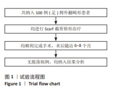
2.1 参与者数量分析 纳入拇外翻畸形患者100例(足),全部进入结果分析,无脱落。所有患者均未出现植入物过敏反应、免疫反应、周围感染等,植入物生物相容性良好。 2.2 试验流程图 见图1。"
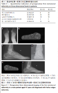
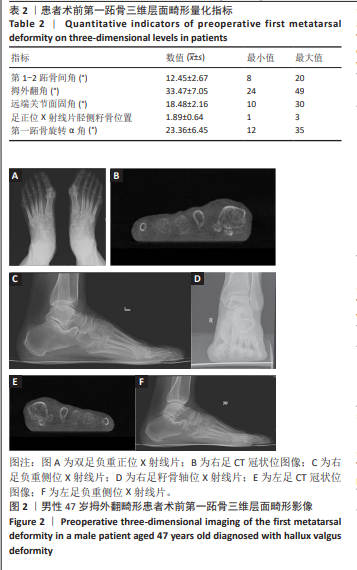
2.3 第一跖骨三维层面畸形量化指标 此组拇外翻畸形患者100例(100足)均完成负重位足部正侧位X射线片及模拟负重位CT片拍摄,第一跖骨三维层面畸形量化指标及图像见表2及图2。"
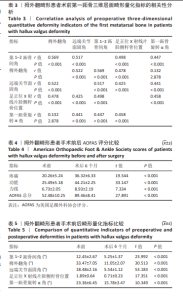
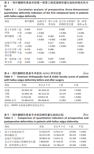
2.4 拇外翻畸形患者术前第一跖骨三维层面畸形量化指标的相关性分析 拇外翻角与第一跖骨旋转α角之间无相关性(P > 0.05),与远端关节面固角、第1-2跖骨间角、足正位X射线片胫侧籽骨位置之间呈正相关(P < 0.05);远端关节面固角、第1-2跖骨间角、第一跖骨旋转α角、足正位X射线片胫侧籽骨位置之间均呈正相关(P < 0.05)。见表3。 2.5 拇外翻畸形患者术前术后AOFAS评分比较 相较于术前,患者术后6个月AOFAS评分显著升高(P < 0.05),见表4。 2.6 拇外翻畸形患者手术前后第一跖骨三维层面畸形量化指标比较 相较于术前,术后6个月足正位X射线片远端关节面固角、胫侧籽骨位置分级、拇外翻角、第1-2跖骨间角、第一跖骨旋转α角均减小(P < 0.05)。见表5。"
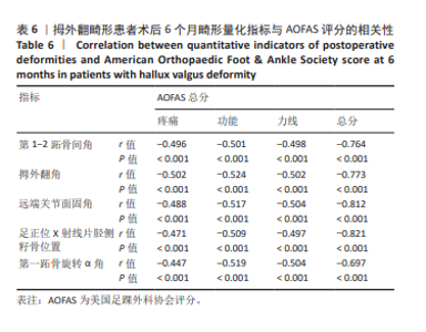
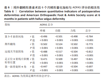
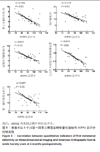
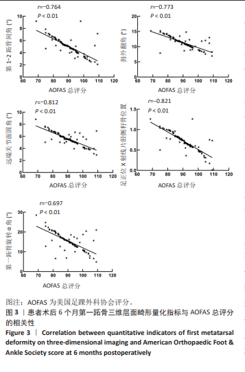
2.7 第一跖骨三维层面畸形量化指标与AOFAS评分的相关性 术后6个月患者足正位X射线片胫侧籽骨位置、拇外翻角、第1-2跖骨间角、第一跖骨旋转α角、远端关节面固角与AOFAS疼痛、力线、功能及总评分均呈负相关(P < 0.05),见表6及图3。"
"
| [1] SUH DH, KIM HJ, PARK JH, et al. Relationship between Hallux Valgus and Pes Planus in Adult Patients. J Foot Ankle Surg. 2021;60(2):297-301. [2] MIRANDA MAM, MARTINS C, CORTEGANA IM, et al. Complications on Percutaneous Hallux Valgus Surgery:A Systematic Review. J Foot Ankle Surg. 2021;60(3):548-554. [3] CAI Y, SONG Y, HE M, et al. Global prevalence and incidence of hallux valgus: a systematic review and meta-analysis. J Foot Ankle Res. 2023; 20;16(1):63. [4] SALET E, LEGGHE B, BAROUK P, et al. Imaging of the post-operative hallux valgus: what do radiologists need to know? Skeletal Radiol. 2023;52(9):1629-1637. [5] KORWIN-KOCHANOWSKA K, POTIÉ A, EL-BOGHDADLY K, et al. PROSPECT guideline for hallux valgus repair surgery:a systematic review and procedure-specific postoperative pain management recommendations. Reg Anesth Pain Med. 2020;45(9):702-708. [6] SHI GG, WHALEN JL, TURNER NS 3RD, et al. Operative Approach to Adult Hallux Valgus Deformity:Principles and Techniques. J Am Acad Orthop Surg. 2020;28(10):410-418. [7] KACZMARCZYK K, BARTON GJ, WISZOMIRSKA I, et al. Women after Bilateral Surgical Correction of Hallux Valgus Do Not Show Improvement in Spatiotemporal Gait Parameters at 18 Weeks Postoperatively. J Clin Med. 2021;5;10(4):608. [8] 黄丽先, 董红, 彭琪, 等. Akin截骨术联合第一跖骨基底截骨术治疗中重度拇外翻畸形的疗效及安全性分析[J]. 实用医院临床杂志, 2022,19(6):75-78. [9] 张惠, 李威. 第一跖骨基底部楔形截骨联合改良Mcbride手术治疗中重度拇外翻[J]. 中国临床研究,2020,33(1):62-65. [10] CHRABAŃSKI O, GOŁĄB T. First Metatarsal Bone Metastasis From Clear Cell Renal Cell Carcinoma on SPECT/CT. Clin Nucl Med. 2022;47(1):91-94. [11] JIAO X, GAN Y, LI Y, et al. Outcomes of V-cut Osteotomy on the First Metatarsal Head Combined with Fixation in Mortise-shaped Bone Groove-Plasty and Akin Osteotomy on the First Toe for Hallux Valgus Correction. Orthop Surg. 2022;14(11):3070-3077. [12] ZAMBELLI R, BAUMFELD D. Intraoperative and Postoperative Evaluation of Hallux Valgus Correction:What Is Important. Foot Ankle Clin. 2020; 25(1):127-139. [13] KADAKIA AR, ALSHOULI MT, BARBOSA MP, et al. Turf Toe, Traumatic Hallux Valgus, and Hallux Rigidus -What Can I Do After an Metatarsophalangeal Fusion. Clin Sports Med. 2020;39(4):801-818. [14] SALET E, LEGGHE B, BAROUK P, et al. Imaging of the post-operative hallux valgus: what do radiologists need to know? Skeletal Radiol. 2023;52(9):1629-1637. [15] MEYR AJ. Multivariate Analysis of Hallux Valgus Radiographic Parameters. J Foot Ankle Surg. 2022;61(4):776-779. [16] BU P, LI C, PU L, et al. Radiographic Assessment of Relationship Between Medial Cuneiform Obliquity and Hallux Valgus. J Foot Ankle Surg. 2023;62(3):583-589. [17] 杨艳军, 白子兴, 曹旭含, 等. 改良中西医结合微创术联合Akin截骨术治疗中重度拇外翻的疗效观察[J]. 实用临床医药杂志,2022, 26(17):81-86. [18] 李大成, 王青松, 宋传航, 等. 改良Lapidus截骨术第1跖楔关节单平面截骨联合第1跖骨下移治疗拇外翻的疗效分析[J]. 生物骨科材料与临床研究,2024,21(3):53-57,63. [19] ZHONG Z, ZHANG P, DUAN H, et al. A Comparison Between X-ray Imaging and an Innovative Computer-aided Design Method Based on Weightbearing CT Scan Images for Assessing Hallux Valgus. J Foot Ankle Surg. 2021;60(1):6-10. [20] 杨勤梦, 付小勇, 林国杰, 等. 可吸收螺钉在拇外翻畸形微创截骨术中的应用分析[J]. 中国骨伤,2022,35(9):836-842. [21] KIMURA T, KUBOTA M, SUZUKI N, et al. Weightbearing Computed Tomography and 3-Dimensional Analysis of Mobility Changes of the First Ray After Proximal Oblique Osteotomy for Hallux Valgus. Foot Ankle Int. 2021;42(3):333-339. [22] LOTAN R, SHLOMOV B, DOTAN A, et al. Hallux Valgus Repair with Chevron Osteotomy Significantly Narrows Forefoot Width. J Clin Med. 2023;12(7):2607. [23] THEVER Y, YONGQIANG JC, CHUIN TR, et al. Scarf osteotomy for hallux valgus surgery: determining indications for an additional Akin osteotomy. J Orthop Surg Res. 2023;18(1):438. [24] PALMANOVICH E, OHANA N, TAVDI A, et al. A modified minimally invasive osteotomy for hallux valgus enables reduction of malpositioned sesamoid bones. Arch Orthop Trauma Surg. 2023;143(10):6105-6112. [25] 王文成,张兴飞,许亚军. Scarf截骨横行截骨线倾斜角度与拇外翻矫形力度关系的3D骨骼重建分析[J]. 中国组织工程研究,2021, 25(27):4265-4270. [26] 纪霖锋,丁声龙,张明珠. 第一跖楔关节矢状不稳与拇外翻合并转移性跖骨痛的相关性[J]. 中华医学杂志,2023,103(1):25-31. [27] SEGAL NA, ANDERSON DD. Editorial commentary on Fritz et al. article entitled ‘Three-dimensional analysis for quantification of knee joint space width with weight-bearing CT: comparison with non-weight-bearing CT and weight-bearing radiography’. Osteoarthritis Cartilage. 2022;30(5):629-632. [28] STEADMAN J, BARG A, SALTZMAN CL. First Metatarsal Rotation in Hallux Valgus Deformity Foot Ankle Int. 2021;42(4):510-522. [29] CONTI MS, PATEL TJ, ZHU J, et al. Association of First Metatarsal Pronation Correction With Patient-Reported Outcomes and Recurrence Rates in Hallux Valgus. Foot Ankle Int. 2022;43(3):309-320. [30] 谢坤铭, 陈兆军, 李昕宇, 等. Chevron联合Akin术与Scarf联合Akin术矫正不同年龄拇外翻术后影像学参数的比较研究[J]. 海南医学院学报,2021,27(13):993-999. [31] LALEVÉE M, BARBACHAN MANSUR NS, LEE HY, et al. Distal Metatarsal Articular Angle in Hallux Valgus Deformity. Fact or Fiction? A 3-Dimensional Weightbearing CT Assessment. Foot Ankle Int. 2022; 43(4):495-503. [32] MOSCA M, CARAVELLI S, VOCALE E, et al. Hallux valgus associated to osteoarthritis: Clinical-radiological outcomes of modified SERI technique at mid- to long-term follow-up. A retrospective analysis. Foot Ankle Surg. 2022;28(1):49-55. [33] 朱楠,张硕,刘伟,等. 微创截骨单螺钉固定结合外侧软组织松解治疗轻中度拇外翻[J]. 生物骨科材料与临床研究,2023,20(4):61-65. [34] SOARES S, MOTA GOMES T, CAMPOS G, et al. Vascular anatomy of the first metatarsal bone and surgical implications according to the severity of hallux valgus deformity: A cadaveric study. Foot Ankle Surg. 2021;27(5):567-576. [35] MANSUR NSB, LALEVEE M, SCHMIDT E, et al. Correlation between indirect radiographic parameters of first metatarsal rotation in hallux valgus and values on weight-bearing computed tomography. Int Orthop. 2021;45(12):3111-3118. [36] WEBER C, WAIZY H. Distale plantarisierende Osteotomie des Os metatarsale I zur Behandlung des Hallux limitus bei Metatarsus primus elevatus Distal osteotomy of the first metatarsal bone with plantarization for the treatment of hallux limitus due to metatarsus primus elevatus. Oper Orthop Traumatol. 2021;33(6):487-494. [37] KIMURA T, KUBOTA M, SUZUKI N, et al. Weightbearing Computed Tomography and 3-Dimensional Analysis of Mobility Changes of the First Ray After Proximal Oblique Osteotomy for Hallux Valgus. Foot Ankle Int. 2021;42(3):333-339. [38] SIDDIQUI NA, FINK JN, SHARMA P, et al. Mechanical Axis Method to Determine First Intermetatarsal Angle and Tibial Sesamoid Position. J Foot Ankle Surg. 2023;62(1):55-60. [39] RAMACHANDRAN SS, REINE S, ARCHER H, et al. Interreader reliability assessment of hallux valgus evaluation on dorsoplantar weightbearing radiographs from a prospective multi-center trial and correlation with patient-reported outcome measures. Skeletal Radiol. 2023;52(12):2419-2425. [40] 马俊安. 第1跖骨基底部楔型截骨联合软组织松解手术治疗中重度足拇外翻的临床疗效[J]. 山西医药杂志,2021,50(10):1665-1667. [41] 俞艳, 姜淑云, 李阳, 等. 基于三维步态分析技术对拇外翻儿童步态变化的研究[J]. 中国运动医学杂志,2021,40(4):259-264. [42] OZTURK AM, SUER O, COBAN I, et al. Three-Dimensional Printed Anatomical Models Help in Correcting Foot Alignment in Hallux Valgus Deformities. Indian J Orthop. 2020;54(1):199-209. |
| [1] | Haonan Yang, Zhengwei Yuan, Junpeng Xu, Zhiqi Mao, Jianning Zhang. Preliminary study on the mechanisms and efficacy of deep brain stimulation in treating depression [J]. Chinese Journal of Tissue Engineering Research, 2026, 30(在线): 1-9. |
| [2] | Cheng Qisheng, Julaiti·Maitirouzi, Xiao Yang, Zhang Chenwei, Paerhati·Rexiti. Finite element analysis of novel variable-diameter screws in modified cortical bone trajectory of lumbar vertebrae [J]. Chinese Journal of Tissue Engineering Research, 2026, 30(9): 2162-2171. |
| [3] | Wu Hongxu, Liu Xuanyu, Wang Taoyu, Wang Shiyao, Cheng Jingyi, Zhang Mingwen, Zhang Yinxia, Liu Zhihua, Wang Xiaojie. Finite element simulation of scoliosis with muscle unit introduction: verification of correction effect under bidirectional load [J]. Chinese Journal of Tissue Engineering Research, 2026, 30(9): 2172-2181. |
| [4] | Chen Huiting, Zeng Weiquan, Zhou Jianhong, Wang Jie, Zhuang Congying, Chen Peiyou, Liang Zeqian, Deng Weiming. Tail anchoring technique of vertebroplasty in treatment of osteoporotic vertebral compression fractures with intravertebral cleft: a finite element analysis [J]. Chinese Journal of Tissue Engineering Research, 2026, 30(9): 2145-2152. |
| [5] | Cai Qirui, Dai Xiaowei, Zheng Xiaobin, Jian Sili, Lu Shaoping, Liu Texi, Liu Guoke, Lin Yuanfang. Mechanical effects of Long’s traction orthopedic method on cervical functional units: quantitative analysis of biomechanical model of head and neck [J]. Chinese Journal of Tissue Engineering Research, 2026, 30(9): 2208-2216. |
| [6] | Rao Jingcheng, Li Yuwan, Zheng Hongbing, Xu Zhi, Zhu Aixiang, Shi Ce, Wang Bing, Yang Chun, Kong Xiangru, Zhu Dawei. Biomechanical differences between the new proximal femoral stable intramedullary nail and traditional intramedullary nail#br# [J]. Chinese Journal of Tissue Engineering Research, 2026, 30(9): 2217-2225. |
| [7] | Zhou Daobin, Wang Kehao, Xie Yang, Ning Rende. Biomechanical characteristics of volar locking plate only versus combined dorsal mini-plate fixation of distal radius fractures with dorsal ulnar fragment [J]. Chinese Journal of Tissue Engineering Research, 2026, 30(9): 2255-2261. |
| [8] | Li Zhifei, Han Bin, Liu Qiuli, Zhang Zhanming, Wei Haokai, Zuo Kuangshi, Zhang Yisheng. Cervical motion characteristics in patients with cervical spondylotic radiculopathy based on motion capture technology [J]. Chinese Journal of Tissue Engineering Research, 2026, 30(9): 2286-2293. |
| [9] | Li Sa, Sun Ning, Sun Zhaozhong, Feng Zhimeng, Li Xuedong. Evaluation parameters and specific region of C6 nerve oppression by uncinate process degeneration [J]. Chinese Journal of Tissue Engineering Research, 2026, 30(9): 2294-2302. |
| [10] | Wang Bokai, Wang Zhiqiang, Zhou Hongyan, Li Junran, Wu Yiheng, Zhao Hongbo. Fracture mapping and imaging analysis of triplane fracture of distal tibia in adolescents [J]. Chinese Journal of Tissue Engineering Research, 2026, 30(9): 2248-2254. |
| [11] | Wang Nan, Chen Shuang, Xi Zhipeng, Qian Yuzhang, Zhang Xiaoyu, Gu Jun, Kang Ran, Xie Lin. MRI evaluation of nerve root subsidence sign affecting efficacy of percutaneous endoscopic decompression in lumbar spinal stenosis [J]. Chinese Journal of Tissue Engineering Research, 2026, 30(9): 2262-2268. |
| [12] | Jiang Xianglong, Li Zhongshan, Che Tongtong. Application effects and mechanisms of low-frequency pulsed electromagnetic fields in muscle repair and growth [J]. Chinese Journal of Tissue Engineering Research, 2026, 30(9): 2350-2360. |
| [13] | Zhu Xiaolong, Zhang Wei, Yang Yang. Visualization analysis of research hotspots and cutting-edge information in the field of intervertebral disc regeneration and repair [J]. Chinese Journal of Tissue Engineering Research, 2026, 30(9): 2391-2402. |
| [14] | Zhang Nan, Meng Qinghua, Bao Chunyu. Characteristics and clinical application of ankle joint finite element models [J]. Chinese Journal of Tissue Engineering Research, 2026, 30(9): 2343-2349. |
| [15] | Wen Fayan, Li Yan, Qiang Tianming, Yang Chen, Shen Linming, Li Yadong, Liu Yongming. Unilateral biportal endoscopic technology for treatment of lumbar degenerative diseases: global research status and changing trends [J]. Chinese Journal of Tissue Engineering Research, 2026, 30(9): 2380-2390. |
| Viewed | ||||||
|
Full text |
|
|||||
|
Abstract |
|
|||||