Chinese Journal of Tissue Engineering Research ›› 2026, Vol. 30 ›› Issue (31): 8253-8263.doi: 10.12307/2026.337
Previous Articles Next Articles
Synaptic Schwann cells promote neuromuscular junction regeneration and function maintenance
Fu Zhenyi1, Yang Xiao1, He Yunkai1, Zhang Yating1, Liu Jiaxin1, Yao Zhihui2, Yang Junying2, Zhao Yao2
- 1School of Medicine, Kunming University of Science and Technology, Kunming 650500, Yunnan Province, China; 2Department of Burns and Plastic Surgery, No. 926 Hospital of the PLA Joint Logistic Support Forces, School of Medicine, Kunming University of Science and Technology, Kaiyuan 661600, Yunnan Province, China
-
Received:2025-05-19Accepted:2025-08-18Online:2026-11-08Published:2026-05-26 -
Contact:Yao Zhihui, PhD, Associate chief physician, Department of Burns and Plastic Surgery, No. 926 Hospital of the PLA Joint Logistic Support Forces, School of Medicine, Kunming University of Science and Technology, Kaiyuan 661600, Yunnan Province, China. Co-corresponding author: Yang Junying, MS, Technologist in charge, Department of Burns and Plastic Surgery, No. 926 Hospital of the PLA Joint Logistic Support Forces, School of Medicine, Kunming University of Science and Technology, Kaiyuan 661600, Yunnan Province, China. Co-corresponding author: Zhao Yao, Attending physician, Department of Burns and Plastic Surgery, No. 926 Hospital of the PLA Joint Logistic Support Forces, School of Medicine, Kunming University of Science and Technology, Kaiyuan 661600, Yunnan Province, China -
About author:Fu Zhenyi, School of Medicine, Kunming University of Science and Technology, Kunming 650500, Yunnan Province, China -
Supported by:Yunnan Provincial Department of Science and Technology Basic Research Project, No. 202401AT070153 (to YZH); Luo Gaoxing Expert Workstation of Yunnan Province, No. 202405AF140056 (to YZH); Science and Technology Strengthening Project of No. 926 Hospital of the PLA Joint Logistic Support Forces, No. 926YY22XK02 (to YZH)

CLC Number:
Cite this article
Fu Zhenyi, Yang Xiao, He Yunkai, Zhang Yating, Liu Jiaxin, Yao Zhihui, Yang Junying, Zhao Yao. Synaptic Schwann cells promote neuromuscular junction regeneration and function maintenance[J]. Chinese Journal of Tissue Engineering Research, 2026, 30(31): 8253-8263.
share this article
Add to citation manager EndNote|Reference Manager|ProCite|BibTeX|RefWorks
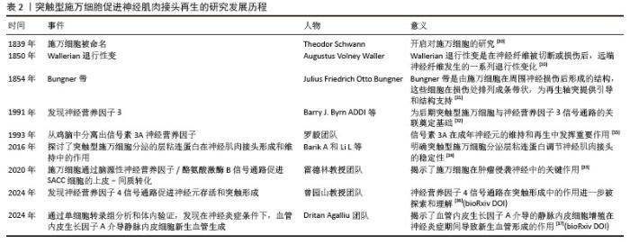
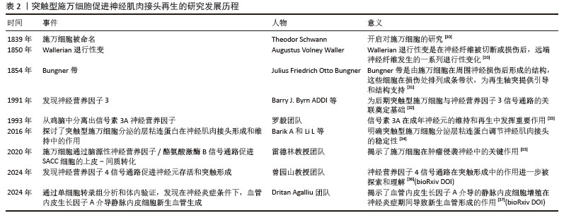
2.1 突触型施万细胞研究的里程碑事件 在神经科学领域,施万细胞自1839年Theoder Schwann发现以来,得到深入研究并获得里程碑式的重大突破,明确了施万细胞的生理功能以及促进神经再生的能力,为后续临床治疗应用奠定了坚实基础,见表2[10,30-37]。 "
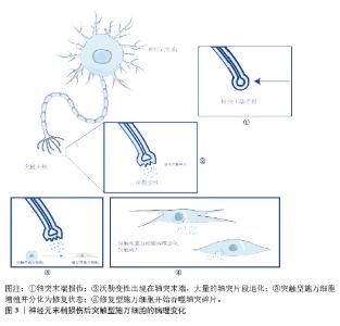
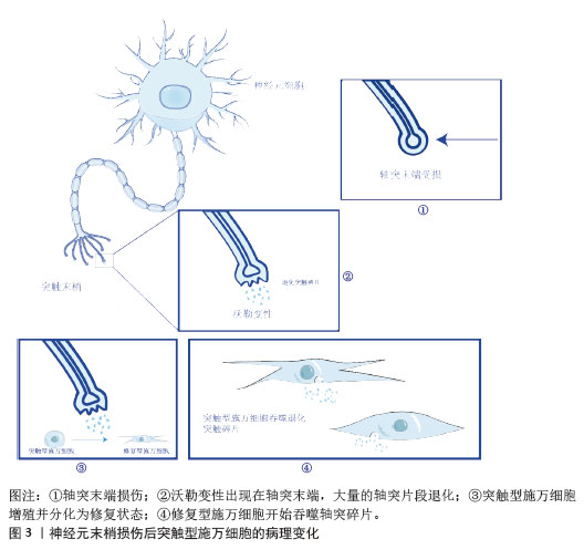
2.2 突触型施万细胞起源与分化 突触型施万细胞是施万细胞的一种特殊亚型,主要分布在神经肌肉接头处。突触型施万细胞起源于神经嵴细胞,神经嵴细胞是一种多能干细胞,能够在胚胎发育过程中迁移到不同的部位,分化成多种细胞类型,包括神经内膜成纤维细胞、副交感神经元细胞、黑色素细胞以及施万细胞等。神经嵴干细胞表达FoxD3(Forkhead box D3)和Sox10(SRY-box10)基因,在向施万细胞分化成熟过程中至关重要。在胚胎发育过程中,神经嵴细胞从神经管的背侧迁移出来,并向周围组织迁移分化成前体施万细胞,随着发育的深入,前体施万细胞进入分化阶段晚期,开始进行形态和功能的特化。在形态方面,细胞伸出细长且高度分支的突起,逐渐与神经元轴突建立紧密的联系[38-55]。这个过程依赖于细胞表面的多种黏附分子和信号分子,例如神经细胞黏附分子和L1细胞黏附分子通过介导细胞间的相互作用,引导施万细胞突起准确地包裹轴突,形成突触型施万细胞的经典形态结构。突触型施万细胞开始表达与突触功能相关的蛋白——连接蛋白43,它在突触型施万细胞之间以及施万细胞与神经元之间形成细胞间通道,允许小分子物质和离子的交换,从而实现细胞间的通讯和信号传递[39]。 2.3 突触型施万细胞的生理功能 2.3.1 感应动作电位与调节终板电位的振幅 当动作电位到达神经末梢时,会引起突触前膜去极化,突触型施万细胞通过其细胞膜上的电压门控通道感应来自运动神经元的动作电位[40-41],这种变化触发突触型施万细胞内部的信号传导路径,通过局部信号分子或细胞外基质成分影响突触小泡融合,调节突触前膜乙酰胆碱等神经递质的释放量[8,42-43]。突触型施万细胞还可以通过分泌神经营养因子影响突触后膜上的乙酰胆碱受体的密度和敏感性进而调节终板电位的振幅。此外,突触型施万细胞还可以通过调节突触间隙中钾离子浓度影响突触后膜的静息电位和终板电位的振幅[44]。 2.3.2 神经肌肉接头的形成与维持 突触型施万细胞通过包裹神经肌肉接头的突触前和突触后部分,为神经肌肉接头提供物理支持和保护。这种包裹作用不仅维持了神经肌肉接头的结构稳定性,还防止了外界因素(如炎症递质、氧化应激等)对神经肌肉接头的损伤。此外,突触型施万细胞可通过分泌集聚蛋白[45]、低密度脂蛋白受体相关蛋白4[46]、骨骼肌特异性酪氨酸受体激酶等多种因子[47],形成复合物,参与神经肌肉接头突触后膜乙酰胆碱受体的聚集和稳定性,从而维持神经肌肉接头的功能;突触型施万细胞还可通过抑制肌肉释放凝血酶、15-前列腺素脱氢酶等负性因子,减少乙酰胆碱受体的降解和碎片化,间接维持神经肌肉接头的稳定性[48]。 2.3.3 神经再生与修复 在神经损伤后,突触型施万细胞能够去分化为修复型施万细胞,这种细胞具有更高的增殖能力和分泌活性[49]。修复型施万细胞能够分泌单核细胞趋化蛋白1、白细胞介素6、白血病抑制因子等趋化因子招募巨噬细胞,协同吞噬损伤后的髓鞘碎片[40];分泌神经生长因子、胶质细胞源性神经营养因子、脑源性神经营养因子等多种神经营养因子以及胶原蛋白、层粘连蛋白等细胞外基质成分,引导再生轴突到达去神经支配的神经肌肉接头[50]。此外,突触型施万细胞分泌突触受体关联蛋白,该蛋白液-液相分离形成的凝聚体为乙酰胆碱受体聚集提供平台,将乙酰胆碱受体募集到突触后膜[51]。 2.4 神经肌肉接头损伤后的病理表现 2.4.1 神经肌肉接头变化 神经肌肉接头损伤后,由于神经肌肉接头的退化造成轴突与肌肉纤维间的纤维连接障碍,引发运动轴突的去神经支配,造成严重肌肉功能障碍[51]。神经肌肉接头中的神经元末梢受到损伤后(图3①),引发沃勒变性[52],即神经肌肉接头远端轴突末梢和髓鞘出现一系列断裂和退化的过程(图3②) [31-53]。 电镜下沃勒变性的轴突细胞骨架出现颗粒状崩解,髓鞘出现断裂。巨噬细胞与施万细胞是参与沃勒变性的主要细胞[54],但轴突退化过程中决定退化速度的是轴突细胞本身而不是施万细胞和巨噬细胞[55],其机制类似于细胞凋亡,多见于轴突横断和轴突运输破坏。 2.4.2 施万细胞形态及功能改变 神经损伤后,施万细胞基因发生变化,开始快速分化增殖,转变为修复样状态,即修复型施万细胞[56],细胞形态伸长而多支,具有吞噬和分泌神经营养因子功能(图3③)。为确保去神经支配的顺利进行,突触型施万细胞会吞噬远端神经损伤和退化的突触部分(图3④) [57],整个过程中,突触型施万细胞依据自身迁移和扩展能力[58],从去神经支配的终板延伸至临近神经支配的神经肌肉接头[59]。为防止神经肌肉接头出现进一步损伤,修复型施万细胞会立即形成伪足,沿细胞索覆盖乙酰胆碱受体,但这部分突触区域失去生理功能,不再进行神经的重新支配,导致突触结构和乙酰胆碱受体的丢失。正常情况下神经末梢通过释放集聚蛋白激活骨骼肌特异性酪氨酸受体激酶信号通路,使乙酰胆碱受体在突触后膜聚集[60-61],而运动神经元损伤后,乙酰胆碱受体在突触后膜形成密集的聚集簇,与神经末梢形成有效的突触连接,但仍有部分肌肉纤维处于病理状态,轴突变薄,突触后膜扁平化,突触区域的受体簇丢失[62]。在神经损伤后,突触型施万细胞不仅吞噬远端突触碎片,部分突触型施万细胞还转变为修复状态,开始伸长并形成分支[63],形成长而平行的结构。在覆盖乙酰胆碱受体后,还会沿着基底层聚集形成Büngner带[31-64],为神经轴突末梢的再生提供方向引导以及物理支持,从而促进神经再生和功能恢复。 2.4.3 乙酰胆碱及其受体下降 在生理状态下,为了保障有效的神经肌肉信号传递,乙酰胆碱受体在运动神经末梢表面的聚集密度较高[64]。轴突中断导致神经末梢乙酰胆碱的释放减少[65],与之对应的突触后膜(肌膜)的乙酰胆碱受体密度下降,由聚集状态变为弥散状态;非突触区域乙酰胆碱受体进行重新分布且密度上升,但并不发挥神经信号传递的功能[66]。同时由于去神经支配,运动终板表面的基底层成分(如层粘连蛋白)短时间内与肌管结合[67],为神经提供机械支持以促进重建,增强表面受体的重排,使得运动终板基底层的乙酰胆碱受体从聚集状逐渐向斑块状转变。在肌萎缩侧索硬化症导致的神经肌肉接头功能障碍中,突触型施万细胞发生丢失;在形态学上,神经肌肉接头突触后膜发生皱缩,乙酰胆碱受体排列出现紊乱[68]。 "
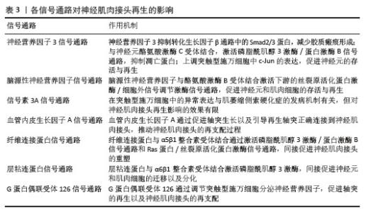
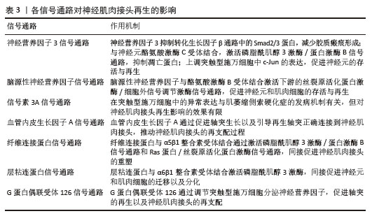
2.5 神经肌肉接头再生的作用机制 截至目前研究结果,有多种信号通路在神经肌肉接头的再生和功能修复中发挥作用,此文章选择了神经营养因子3、脑源性神经营养因子、信号素3A、血管内皮生长因子A、纤维连接蛋白、层粘连蛋白和G蛋白偶联受体126经典信号通路的功能及其相互作用进行阐述。这些信号分子与相应受体结合通过抑制神经元凋亡、减少胶质瘢痕形成、调节细胞骨架重组、促进轴突生长和突触可塑性等多种机制共同促进神经肌肉接头的再生和功能恢复,见表3。"
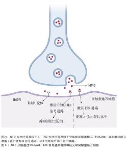
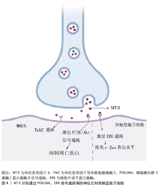
2.5.1 神经营养因子3信号通路 神经营养因子3通过与神经元上的神经营养因子受体酪氨酸激酶C受体结合,激活下游的磷脂酰肌醇3激酶(phosphoinositide 3 kinase,PI3K)/蛋白激酶 B(protein kinase B,Akt)信号通路,通过抑制凋亡蛋白(如BAD),减少神经元的凋亡。此外,神经营养因子3还可通过抑制转化生长因子β信号通路中的Smad2/3蛋白表达,减少胶质瘢痕的形成,对神经元的存活与轴突再生起促进作用[69]。XU等[70]研究发现,在慢性失神经支配后,神经营养因子3与突触型施万细胞上的酪氨酸激酶C受体结合,通过细胞外调节蛋白激酶/转录因子c-Jun信号通路,维持远端失神经支配中c-Jun的高表达水平,使突触型施万细胞处于修复状态[71],进而促进周围神经再生和神经肌肉接头的功能恢复(图4)。通过siRNA的腺相关病毒介导的c-Jun 沉默则会显著削弱神经营养因子3所发挥的促进作用。由此可见,神经营养因子3对于神经肌肉接头的形成与功能维持具有至关重要的作用。 "
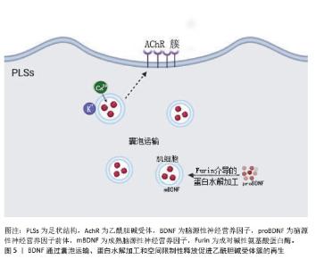
2.5.2 脑源性神经营养因子信号通路 当脑源性神经营养因子在低浓度状态下与神经元高亲和力酪氨酸激酶B受体结合后,诱导酪氨酸激酶B受体自磷酸化,激活下游的磷脂酰肌醇3激酶和细胞外调节蛋白激酶1/2信号通路,从而促进神经元的存活与轴突生长[72]。脑源性神经营养因子浓度进一步升高时,与低亲和力受体p75NTR结合,可以调节Rho家族小GTP酶的活性,如RAC1和CDC42。激活RAC1和CDC42可以促进微管的组装和轴突的延伸,有助于神经肌肉接头的再生[73]。此外,与p75NTR结合还可以促进神经干细胞的自我更新和增殖,进一步促进神经肌肉接头的再生和功能维持[73]。ZHANG等[74]的研究揭示了骨骼肌细胞中的脑源性神经营养因子通过囊泡运输、蛋白水解加工和空间限制性释放的细胞生物学机制以促进乙酰胆碱受体簇的生成。见图5。尽管脑源性神经营养因子信号通路具有显著作用,但其在人体内的生物利用度低,易在体内降解,可能导致其在临床应用中效果有限[75]。 "
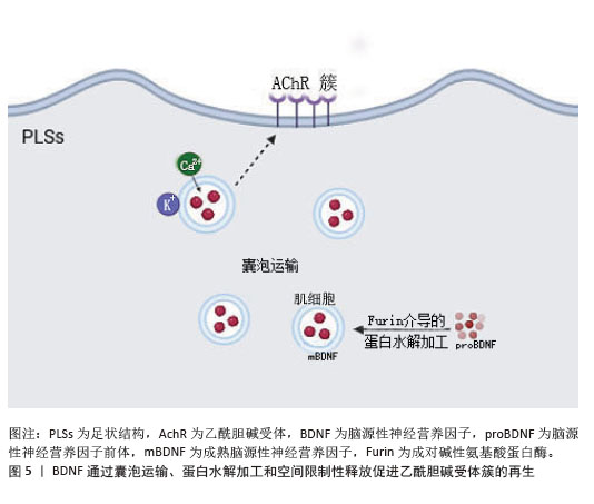
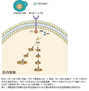
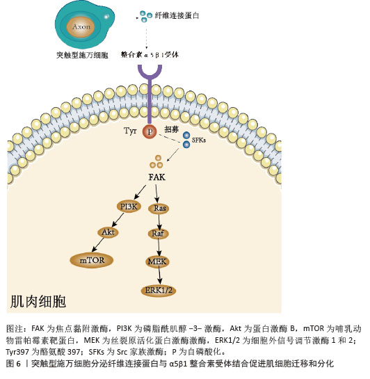
(1)囊泡运输:脑源性神经营养因子在肌细胞中的亚细胞定位情况如下:脑源性神经营养因子在骨骼肌细胞中与足状结构的肌动蛋白富集核心区域存在高度共定位现象[74]。这些足状结构在无神经支配的乙酰胆碱受体簇中发挥着关键作用,引导含脑源性神经营养因子囊泡的运输以及表面靶向过程。含脑源性神经营养因子囊泡在肌细胞中的运输与释放过程受到严格调控,其释放依赖钙离子的内流[76],并且呈现出活动依赖性特征。 (2)蛋白水解加工:脑源性神经营养因子的前体蛋白(proBDNF)在肌细胞中通过Furin介导的蛋白水解加工转化为成熟脑源性神经营养因子(mBDNF)。成熟脑源性神经营养因子能够激活酪氨酸激酶B受体,从而调节突触结构和功能,这一转化过程对于脑源性神经营养因子的功能发挥具有决定性意义。BAO等[77]实验表明,通过药物抑制Furin的活性,可以减少成熟脑源性神经营养因子的生成量,进而显著减少乙酰胆碱受体簇的形成。 (3)空间限制性释放:ZHANG等[74]的研究发现,脑源性神经营养因子在乙酰胆碱受体簇中的释放与足状结构相关联,受神经活动的调节并依赖钙离子内流。通过活细胞时间序列成像,研究者观察到脑源性神经营养因子囊泡被运输到足状结构,并在乙酰胆碱受体簇中进行空间限制性释放,这一过程确保了脑源性神经营养因子在特定区域的高效释放。 (4)脑源性神经营养因子信号通路与谷氨酸信号通路正反馈机制:脑源性神经营养因子信号通路与谷氨酸介导的信号通路相互作用形成正反馈,脑源性神经营养因子与酪氨酸激酶B受体结合,激活下游的磷脂酶C/蛋白激酶C通路,促进谷氨酸的释放[78]。谷氨酸作为主要的兴奋性神经递质,通过激活N-甲基-D-天冬氨酸受体,增加脑源性神经营养因子的合成和释放。此外,谷氨酸还可以通过激活Ca2+/CaM依赖性蛋白磷酸酶通路,促进cAMP反应元件结合蛋白的磷酸化,从而增强脑源性神经营养因子的转录[79]。这种正反馈机制有助于增强神经元的存活和突触可塑性[80]。 2.5.3 信号素3A 信号通路 信号素3A是一种神经营养因子,其在突触型施万细胞中的表达增强与肌萎缩侧索硬化的发病机制密切相关。肌萎缩侧索硬化是一种神经退行性疾病,其主要特征是运动神经元逐渐退化,导致肌肉无力和萎缩[81-82]。在MOLONEY等[83]的研究中,研究者采用了一种表达突变信号素3A蛋白(K108N-SEMA3A)的小鼠模型,这种突变蛋白的信号传导能力显著降低。研究者通过两种去神经支配模型来探究信号素3A信号通路的作用:肌萎缩侧索硬化模型(G93A-hSOD1肌萎缩侧索硬化小鼠)和损伤模型(BotoxA 诱导的腓肠肌瘫痪)。研究结果显示,在肌萎缩侧索硬化小鼠中,无论是表达1个或2个突变信号素3A等位基因的小鼠,其运动行为功能与野生型肌萎缩侧索硬化小鼠相比并没有显著差异,这表明突变信号素3A信号传导能力降低,并未使肌萎缩侧索硬化相关的运动功能下降得到改善;在 BotoxA 诱导的腓肠肌瘫痪模型中,观察到突变信号素3A蛋白的存在没有增强神经肌肉接头可塑性。雄性小鼠:TypeIIb纤维:突变型信号素3A小鼠(杂合/纯合)的神经突触出芽能力与野生型无显著差异;TypeI/IIa纤维:突变型信号素3A小鼠的出芽能力显著低于野生型(杂合P=0.01,纯合P=0.02)。雌性小鼠:TypeIIb纤维:野生型雌性小鼠的出芽能力显著高于野生型雄性小鼠(25% vs. 9.5%,P=0.048);TypeI/IIa纤维:突变型信号素3A雌性小鼠的出芽能力与野生型无显著差异。因此,突变型信号素3A蛋白小鼠在肌萎缩侧索硬化模型和BotoxA诱导瘫痪模型中并未显著增强神经肌肉接头的可塑性,甚至可能抑制TypeI/IIa纤维中的神经肌肉接头可塑性。通过下调信号素3A蛋白信号传导能力促进神经肌肉接头功能恢复效果甚微。 2.5.4 血管内皮生长因子A信号通路 在神经损伤之后,巨噬细胞会迅速浸润到神经肌肉接头区域,并显著上调血管内皮生长因子A的表达量。巨噬细胞分泌的血管内皮生长因子A在促进神经肌肉接头再支配方面发挥着重要作用。血管内皮生长因子A与血管内皮生长因子受体2结合,激活磷脂酰肌醇3激酶/蛋白激酶B通路,调节细胞骨架的重组,以及促进轴突生长并引导再生轴突正确连接到神经肌肉接头,推动神经肌肉接头的再支配过程[84-85]。当血管内皮生长因子受体2被抑制时,受损的神经肌肉接头处分泌血管内皮生长因子A 的巨噬细胞数量减少,阻碍突触型施万细胞的延伸以及神经肌肉接头神经再支配[75]。而通过基因敲除血管内皮生长因子A或者使用血管内皮生长因子A中和抗体,发现血管内皮生长因子A的缺失会显著降低神经肌肉接头的再支配效率,进而导致肌肉功能恢复出现延迟[85]。血管内皮生长因子A不仅能够促进轴突生长,还能够激活炎症细胞,炎症反应在早期有助于清除损伤后的碎片,但过度的炎症反应会导致组织损伤,延缓神经肌肉接头的再生。肿瘤坏死因子α和白细胞介素1β等细胞因子可以抑制血管内皮生长因子A的活性,减少其对轴突生长的促进作用[75]。此外,炎症细胞释放的蛋白酶可以降解血管内皮生长因子A,进一步降低其生物利用度[84]。 2.5.5 纤维连接蛋白信号通路 突触型施万细胞分泌的纤维连接蛋白通过与肌细胞上的α5β1整合素受体结合,受体在酪氨酸 397位点发生自磷酸化。磷酸化的酪氨酸397位点能够招募Src家族激酶,该激酶与焦点黏附激酶结合后进一步激活焦点黏附激酶[86]。活化的焦点黏附激酶可以激活下游的磷脂酰肌醇3激酶/蛋白激酶B和丝裂原活化蛋白激酶(mitogen-activated protein kinase,MAPK)信号通路[87]。磷脂酰肌醇3激酶/蛋白激酶B信号通路:焦点黏附激酶激活磷脂酰肌醇3激酶,磷脂酰肌醇3激酶进一步激活蛋白激酶B,蛋白激酶B通过调节哺乳动物雷帕霉素靶蛋白信号通路,促进细胞的生长以及存活[88]。丝裂原活化蛋白激酶信号通路:焦点黏附激酶激活Ras蛋白,Ras蛋白进一步激活Raf激酶和丝裂原活化蛋白激酶激酶(mitogen-activated protein kinase kinase,MEK),最终激活细胞外信号调节激酶1和2 (Extracellular signal-regulated kinases 1 and 2,ERK1/2)。活化的细胞外调节蛋白激酶1/2能够促进细胞的增殖以及迁移,推动神经肌肉接头的重塑过程[89],见图6。 "
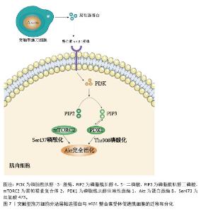
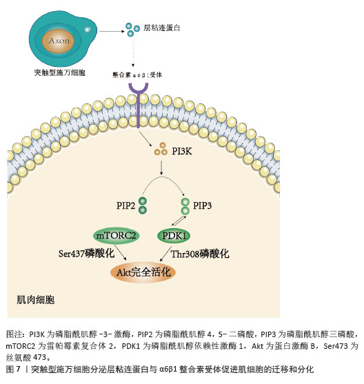
2.5.6 层粘连蛋白信号通路 层粘连蛋白通过与肌肉细胞上的α6β1 整合素受体结合,激活磷脂酰肌醇3激酶。磷脂酰肌醇3激酶将磷脂酰肌醇4,5-二磷酸(PIP2)转化为磷脂酰肌醇3,4,5-三磷酸(PIP3)。磷脂酰肌醇3,4,5-三磷酸作为第二信使,招募并激活磷脂酰肌醇依赖性激酶1(PDK1),磷脂酰肌醇依赖性激酶1进一步磷酸化蛋白激酶B的苏氨酸308 位点。蛋白激酶B的丝氨酸473 位点被雷帕霉素复合体 2磷酸化,完成蛋白激酶B的激活[90],见图7。蛋白激酶B 通过磷酸化并抑制促凋亡蛋白、磷酸化CDK抑制剂、激活雷帕霉素复合体1、磷酸化细胞骨架蛋白的方式,分别促进细胞存活和细胞周期进程[91]、促进蛋白质合成以及细胞生长、调节细胞的迁移以及侵袭能力,最终促进肌肉细胞的迁移和分化。 "
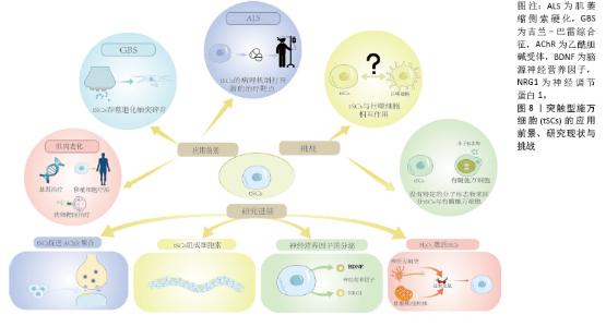
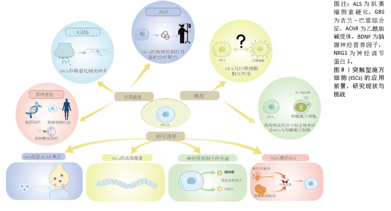
2.5.7 G蛋白偶联受体126信号通路 JABLONKA-SHARIFF等[92]的研究发现,G蛋白偶联受体126 在突触型施万细胞中表达,并且在神经肌肉接头修复过程中发挥着重要作用。G蛋白偶联受体126的缺失会导致神经肌肉接头再支配出现延迟,表现为突触型施万细胞的细胞质扩展减少。G蛋白偶联受体126通过调节突触型施万细胞分泌脑源性神经营养因子、神经营养因子3等神经营养因子,促进再生轴突的生长以及神经肌肉接头的再支配[93]。G蛋白偶联受体126的缺失还会影响突触型施万细胞分泌趋化因子水平[94],进而影响巨噬细胞的招募以及神经损伤之后的炎症反应。 2.5.8 其他影响因素 促进神经肌肉接头修复和再生的机制除上述信号通路外,T-box 转录因子21基因表达也发挥着重要作用,T-box 转录因子21主要在免疫细胞中发挥作用。但JABLONKA-SHARIFF 等[92]的研究发现,T-box 转录因子21也在突触型施万细胞中表达,并且参与神经肌肉接头的形成以及维持,同时还涉及相关的免疫调节。此外,T-box 转录因子21的表达可能与神经肌肉接头的炎症反应以及疾病进展有着密切关系。研究发现T-box 转录因子21在突触型施万细胞中呈现高表达状态,其表达水平比神经和肌肉组织高出 9 倍以上。免疫荧光标记结果显示T-box 转录因子21、S100-增强型绿色荧光蛋白以及α-银环蛇毒素(标记突触后乙酰胆碱受体)共定位。通过基因敲除实验进一步发现,T-box 转录因子21的缺失导致神经肌肉接头的形成以及维持出现缺陷,这表明该因子在促进神经肌肉接头修复过程中发挥着重要作用。 2.6 应用前景 突触型施万细胞在神经肌肉接头损伤后的修复和再生中发挥着关键作用,在肌萎缩侧索硬化、老化相关肌肉功能障碍、吉兰-巴雷综合征等疾病治疗中具有广阔的临床应用前景[95]。 2.6.1 肌萎缩侧索硬化症 肌萎缩侧索硬化症即俗称的渐冻症,是一种去神经性疾病[96]。突触型施万细胞在肌萎缩侧索硬化中表现出早期和持续的功能异常,PERE2-GONZALEZ等[97]研究显示,在肌萎缩侧索硬化模型小鼠的早期阶段,突触型施万细胞表现出异常的解码能力,其对神经递质释放的反应增强,在SOD1G37R小鼠模型中,突触型施万细胞中的乙酰胆碱受体活性增加,导致其对突触传递的解码能力异常,而这种异常的解码能力可能与突触型施万细胞在神经肌肉接头稳定性和修复中的功能受损有关。随着疾病的进展,突触型施万细胞在神经肌肉接头的修复机制中表现出缺陷,包括在去神经支配的神经肌肉接头上未能有效引导神经末梢再生和清除突触碎片。这些缺陷可能与突触型施万细胞的乙酰胆碱受体活性增加有关,从而影响其向修复状态的转变。此外,突触型施万细胞在肌萎缩侧索硬化模型中还表现出对嘌呤能信号的敏感性降低,进一步影响它在神经肌肉接头修复中的功能[98]。这些变化不仅在疾病早期出现,而且在未出现症状前已持续存在,表明突触型施万细胞的功能障碍可能在肌萎缩侧索硬化的发病机制中起重要作用。基于小鼠模型的肌萎缩侧索硬化研究提示突触型施万细胞可能成为潜在的治疗靶点。但目前对肌萎缩侧索硬化中的神经胶质细胞研究较少。明确突触型施万细胞在肌萎缩侧索硬化中的作用机制对缓解症状、延缓病程起到重要作用,见图8。 2.6.2 老化相关肌肉功能障碍 随着年龄增长,突触型施万细胞数量增加但功能异常,结构上表现为部分覆盖突触前区域,突起侵入突触裂隙或延伸至相邻的神经肌肉接头。这些变化导致突触后膜折叠度降低,乙酰胆碱受体表达量下降,进而影响神经信号传递[99]。同时,突触型施万细胞中的多个信号通路发生改变,如神经调节蛋白-表皮生长因子受体、CD44、S100和转化生长因子β等,这些通路的改变影响了突触型施万细胞的正常功能,导致神经肌肉接头的退行性变化[100]。此外,在衰老的突触型施万细胞中,与炎症和细胞间信号传导相关的基因和通路发生改变,如Ccl7、Ccl4、Adgre1(F4/80)和Cxcl16等与炎症和免疫细胞招募相关的基因表达增加,加剧神经肌肉接头的退行性变化[101]。针对这些突触型施万细胞相关的机制,未来治疗可采用药物干预、细胞治疗和基因治疗等方式。药物干预可以通过调节突触型施万细胞中的信号通路,如使用神经调节蛋白1靶向治疗促进突触型施万细胞的正常功能并维持神经肌肉接头的稳定性,或使用芬戈莫德磷酸盐来调节突触型施万细胞的炎症反应,从而减缓神经肌肉接头的退行性变化[99]。 细胞治疗则可以通过移植突触型施万细胞或诱导突触型施万细胞的前体细胞来替代受损的突触型施万细胞,恢复神经肌肉接头的正常功能[102]。基因治疗则通过基因编辑技术,如应用成簇规律间隔短回文重复序列/CRISPR相关蛋白9(Clustered Regularly Interspaced Short Palindromic Repeats/CRISPR-Associated Protein 9,CRISPR-Cas9)技术来修复或替换突触型施万细胞中的缺陷基因,恢复其正常功能,进而改善神经肌肉接头的退行性变化[103-104]。这些策略为治疗与年龄相关的肌肉功能障碍提供了新的途径,见图8。 2.6.3 吉兰-巴雷综合征 吉兰-巴雷综合征患者自身抗体靶向攻击神经节苷脂,导致运动神经轴突损伤。在吉兰-巴雷综合征小鼠模型中,CUNNINGHAM等[57]发现,突触型施万细胞在神经末梢损伤后迅速发挥吞噬作用,清除神经末梢碎片。通过降低抗神经节苷脂抗体和补体的剂量,可提高小鼠损伤后存活率,从而能够在更长时间内观察免疫病理变化,结果显示,突触型施万细胞在损伤后4 h内开始吞噬轴突碎片,而中性粒细胞和巨噬细胞并未显著浸润损伤部位。这表明在吉兰-巴雷综合征模型中突触型施万细胞是主要的清除细胞,而不是炎症细胞[105]。这一发现有助于理解吉兰-巴雷综合征中神经肌肉接头损伤的病理生理机制,以及突触型施万细胞在其中的重要作用,并为针对吉兰-巴雷综合征新的治疗策略提供了研究靶点,见图8。 "
| [1] 陈波,陈振兵,杜远立.去神经支配骨骼肌萎缩变化及康复治疗研究进展[J].中国康复医学杂志,2011,26(8):792-795. [2] 宋彬彬,张巍,徐畅,等.去神经支配引起的骨骼肌萎缩及其机制[J].医学研究杂志,2018, 47(3):157-160+180. [3] VAN NES JJ. An introduction to clinical neuromuscular electrophysiology. Vet Q. 1986; 8(3):233-239. [4] LARSSON L, DEGENS H, LI M, et al. Sarcopenia: Aging-Related Loss of Muscle Mass and Function. Physiol Rev. 2019;99(1):427-511. [5] 杨滨瑞,赵琳琳,汪煜楠,等. YAP蛋白在神经肌肉接头中作用机制的研究[J].中国实验诊断学,2023,27(5):603-607. [6] RIGOARD P, BUFFENOIR K, BAUCHE S, et al. Tools and techniques dedicated to neuromuscular junction observation. Neurochirurgie. 2009; 55 Suppl 1:S43-48. [7] THESLEFF S. Highlights from 40 years’ research on the neuromuscular junction. Physiol Res. 1991; 40(4):381-394. [8] XING G, JING H, YU Z, et al. Membraneless condensates by Rapsn phase separation as a platform for neuromuscular junction formation. Neuron. 2022;110(2):350. [9] VAUTRIN J, MAMBRINI J. Synaptic current between neuromuscular junction folds. J Theor Biol. 1989;140(4):479-498. [10] HASTINGS RL, AVILA MF, SUNEBY E, et al. Cellular and molecular evidence that synaptic Schwann cells contribute to aging of mouse neuromuscular junctions. Aging Cell. 2023;22(11):e13981. [11] CHU XL, SONG XZ, LI Q, et al. Basic mechanisms of peripheral nerve injury and treatment via electrical stimulation. Neural Regen Res. 2022;17(10):2185-2193. [12] JABLONKA-SHARIFF A, BALTA E, SANTOSA KB, et al. Terminal Schwann Cells Are Essential for Neuromuscular Junction Function and Recovery after Nerve Injury. Plast Reconstr Surg. 2023;151(4):792-803. [13] NICOLETTI C, WEI X, ETXANIZ U, et al. Muscle denervation promotes functional interactions between glial and mesenchymal cells through NGFR and NGF. iScience. 2023;26(7):107114. [14] LUNDT S, DING S. Non-cell autonomous effect of neuronal nicotinamide phosphoribosyl transferase on the function of neuromuscular junctions. Neural Regen Res. 2021;16(2):302-303. [15] SUN S, SHEN Y, ZHANG X, et al. The MuSK agonist antibody protects the neuromuscular junction and extends the lifespan in C9orf72-ALS mice. Mol Ther J Am Soc Gene Ther. 2024;32(7):2176-2189. [16] ZHANG W, BAI L, XU W, et al. Sirt6 Mono-ADP-Ribosylates YY1 to Promote Dystrophin Expression for Neuromuscular Transmission. Adv Sci (Weinh). 2024;11(44):e2406390. [17] LI L, YOKOYAMA H, KABURAGI H, et al. Remnant neuromuscular junctions in denervated muscles contribute to functional recovery in delayed peripheral nerve repair. Neural Regen Res. 2020; 15(4):731-738. [18] CHANG M, CAI Y, GAO Z, et al. Duchenne muscular dystrophy: pathogenesis and promising therapies. J Neurol. 2023;270(8):3733-3749. [19] EL OUAAMARI Y, VAN DEN BOS J, WILLEKENS B, et al. Neurotrophic Factors as Regenerative Therapy for Neurodegenerative Diseases: Current Status, Challenges and Future Perspectives. Int J Mol Sci. 2023;24(4):3866. [20] WEISSMILLER AM, WU C. Current advances in using neurotrophic factors to treat neurodegenerative disorders. Transl Neurodegener. 2012;1(1):14. [21] SOCIALI G, GROZIO A, CAFFA I, et al. SIRT6 deacetylase activity regulates NAMPT activity and NAD(P)(H) pools in cancer cells. FASEB J. 2019;33(3):3704-3717. [22] ANCEL S, MICHAUD J, MIGLIAVACCA E, et al. Nicotinamide and pyridoxine stimulate muscle stem cell expansion and enhance regenerative capacity during aging. J Clin Invest. 2024;134(24):e163648. [23] HWANG ES, SONG SB. Possible Adverse Effects of High-Dose Nicotinamide: Mechanisms and Safety Assessment. Biomolecules. 2020;10(5):687. [24] FUJITANI M, TARIF AMM, OTANI Y. Regeneration mechanisms and therapeutic strategies for neuromuscular junctions in aging and diseases. Neural Regen Res. 2025;20(1):193-194. [25] SAKUMA M, GORSKI G, SHEU SH, et al. Lack of motor recovery after prolonged denervation of the neuromuscular junction is not due to regenerative failure. Eur J Neurosci. 2016;43(3):451-462. [26] ZANOTELI E, FRANÇA MCJ, MARQUES WJ. Gene-based therapies for neuromuscular disorders. Arq Neuropsiquiatr. 2024;82(6):1-10. [27] NUSSBAUM EL, HOUGHTON P, ANTHONY J, et al. Neuromuscular Electrical Stimulation for Treatment of Muscle Impairment: Critical Review and Recommendations for Clinical Practice. Physiother Can Physiother Can. 2017;69(5):1-76. [28] HANKOV N, CABAN M, DEMESMAEKER R, et al. Augmenting rehabilitation robotics with spinal cord neuromodulation: A proof of concept. Sci Robot. 2025;10(100): eadn5564. [29] EVANCHO A, TYLER WJ, MCGREGOR K. A review of combined neuromodulation and physical therapy interventions for enhanced neurorehabilitation. Front Hum Neurosci. 2023;17:1151218. [30] RIOS R, JABLONKA-SHARIFF A, BROBERG C, et al. Macrophage roles in peripheral nervous system injury and pathology: Allies in neuromuscular junction recovery. Mol Cell Neurosci. 2021;111: 103590. [31] BOSCH-QUERALT M, FLEDRICH R, STASSART RM. Schwann cell functions in peripheral nerve development and repair. Neurobiol Dis. 2023;176: 105952. [32] SUDA T, TAKAHASHI T, GOLSTEIN P, et al. Molecular cloning and expression of the Fas ligand, a novel member of the tumor necrosis factor family. Cell. 1993;75(6):1169-1178. [33] 邢国奕,孙乐刚,马向瑞,等. Sema3A对口腔间充质干细胞功能的调控[J].中国组织工程研究,2023,27(10):1626-1633. [34] HEREDIA DJ, DE ANGELI C, FEDI C, et al. Calcium Signaling in Schwann cells. Neurosci Lett. 2020; 729:134959. [35] 单春.施万细胞通过BDNF/TrkB信号通路促进SACC嗜神经侵袭机制的研究[D].西安:中国人民解放军空军军医大学,第四军医大学,2016. [36] KONG P, YANG M, WANG Y, et al. Ferroptosis triggered by STAT1- IRF1-ACSL4 pathway was involved in radiation-induced intestinal injury. Redox Biol. 2023;66:102857. [37] LI G, HUANG LJ, ZHANG B, et al. The LINGO-1-deficient neural stem cell-derived neural tissueoid showed enhanced retention and neuronal relay in the transected spinal cord. Chem Eng J. 2024; 497:155032. [38] COLOMBO F, MELDOLESI J. L1-CAM and N-CAM: From Adhesion Proteins to Pharmacological Targets. Trends Pharmacol Sci. 2015;36(11): 769-781. [39] TAVEGGIA C, FELTRI ML. Beyond Wrapping: Canonical and Noncanonical Functions of Schwann Cells. Annu Rev Neurosci. 2022;45:561-580. [40] 蔺海燕,刘芳,许家军,等.周围神经损伤与再生中施万细胞可塑性的研究进展[J].解剖学杂志,2021,44(5):430-433. [41] MICHEVA KD, KIRALY M, PEREZ MM, et al. Conduction Velocity Along the Local Axons of Parvalbumin Interneurons Correlates With the Degree of Axonal Myelination. Cereb Cortex N Y N 1991. 2021;31(7):3374-3392. [42] 王雁,潘华.神经肌肉接头动态改变在肌萎缩侧索硬化中的研究价值与进展[J].中国神经精神疾病杂志,2022,48(6):380-384. [43] 崔梦鸽,张斌.JPH2参与神经肌肉接头处乙酰胆碱受体簇的形成与维持的作用与机制研究[D].武汉:华中科技大学,2021. [44] GARCIA-SEGURA LM, MCCARTHY MM. Minireview: Role of glia in neuroendocrine function. Endocrinology. 2004;145(3):1082-1086. [45] CHEN J, CHEN H, DONG X, et al. Deficiency of skeletal muscle Agrin contributes to the pathogenesis of age-related sarcopenia in mice. Cell Death Dis. 2024;15(3):201. [46] CHEN BH, LIN ZY, ZENG XX, et al. LRP4-related signalling pathways and their regulatory role in neurological diseases. Brain Res. 2024;1825: 148705. [47] OURY J, GAMALLO-LANA B, SANTANA L, et al. Agonist antibody to MuSK protects mice from MuSK myasthenia gravis. Proc Natl Acad Sci U S A. 2024;121(39):e2408324121. [48] BAKOOSHLI MA, WANG YX, MONTI E, et al. Regeneration of neuromuscular synapses after acute and chronic denervation by inhibiting the gerozyme 15-prostaglandin dehydrogenase. Sci Transl Med. 2023;15(717):eadg1485. [49] 肖雨,翁秋燕,邵磊,等.周围神经损伤后再生与修复机制研究进展[J].生物化学与生物物理进展,2022,49(7):1243-1250. [50] CAI A, ZHENG ZM, HIMMLER M, et al. Schwann Cells Promote Myogenic Differentiation of Myoblasts and Adipogenic Mesenchymal Stromal Cells on Poly-ɛ-Caprolactone-Collagen I-Nanofibers. Cells. 2022;11(9):1436. [51] CHEN TJ, KUKLEY M. Glutamate receptors and glutamatergic signalling in the peripheral nerves. Neural Regen Res. 2020;15(3):438-447. [52] TIAN R, ZHOU Y, REN Y, et al. Wallerian degeneration: From mechanism to disease to imaging. Heliyon. 2025;11(1):e40729. [53] NEGRO S, PIRAZZINI M, RIGONI M. Models and methods to study Schwann cells. J Anat. 2022; 241(5):1235-1258. [54] VARGAS ME, BARRES BA. Why is Wallerian degeneration in the CNS so slow? Annu Rev Neurosci. 2007;30:153-179. [55] FELTRI ML, POITELON Y, PREVITALI SC. How Schwann Cells Sort Axons: New Concepts. Neuroscientist. 2016;22(3):252-265. [56] DEININGER S, SCHUMACHER J, BLECHSCHMIDT A, et al. Nerve injury converts Schwann cells in a long-term repair-like state in human neuroma tissue. Exp Neurol. 2024;382:114981. [57] CUNNINGHAM ME, MEEHAN GR, ROBINSON S, et al. Perisynaptic Schwann cells phagocytose nerve terminal debris in a mouse model of Guillain-Barré syndrome. J Peripher Nerv Syst. 2020;25(2):143-151. [58] CHENG Z, ZHANG Y, TIAN Y, et al. Cyr61 promotes Schwann cell proliferation and migration via αvβ3 integrin. BMC Mol Cell Biol. 2021;22(1):21. [59] MOSS KR, SAXENA S. Schwann Cells in Neuromuscular Disorders: A Spotlight on Amyotrophic Lateral Sclerosis. Cells. 2025; 14(1):47. [60] DELERS P, SAPALY D, SALMAN B, et al. A link between agrin signalling and Ca(v)3.2 at the neuromuscular junction in spinal muscular atrophy. Sci Rep. 2022;12(1):18960. [61] XIE T, XU G, LIU Y, et al. Structural insights into the assembly of the agrin/LRP4/MuSK signaling complex. Proc Natl Acad Sci U S A. 2023;120(23): e2300453120. [62] DING Q, KESAVAN K, LEE KM, et al. Impaired signaling for neuromuscular synaptic maintenance is a feature of Motor Neuron Disease. Acta Neuropathol Commun. 2022;10(1):61. [63] GITIK M, ELBERG G, REICHERT F, et al. Deletion of CD47 from Schwann cells and macrophages hastens myelin disruption/dismantling and scavenging in Schwann cells and augments myelin debris phagocytosis in macrophages. J Neuroinflammation. 2023;20(1):243. [64] SHIMIZU K, KASSAI H, KAMEI Y, et al. Alignment of Skeletal Muscle Cells Facilitates Acetylcholine Receptor Clustering and Neuromuscular Junction Formation with Co-Cultured Human iPSC-Derived Motor Neurons. Cells. 2022;11(23):3760. [65] WINTHER JB, MORGEN JJ, SKOV M, et al. Role of recovery of acetylcholine release in compromised neuromuscular junction function. Neuromuscul Disord. 2024;36:48-59. [66] HUANG X, JIANG J, XU J. Denervation-Related Neuromuscular Junction Changes: From Degeneration to Regeneration. Front Mol Neurosci. 2021;14:810919. [67] NGUYEN B, BIX G, YAO Y. Basal lamina changes in neurodegenerative disorders. Mol Neurodegener. 2021;16(1):81. [68] ALHINDI A, SHAND M, SMITH HL, et al. Neuromuscular junction denervation and terminal Schwann cell loss in the hTDP-43 overexpression mouse model of amyotrophic lateral sclerosis. Neuropathol Appl Neurobiol. 2023;49(4):e12925. [69] CHEN T, HE X, WANG J, et al. NT-3 Combined with TGF-β Signaling Pathway Enhance the Repair of Spinal Cord Injury by Inhibiting Glial Scar Formation and Promoting Axonal Regeneration. Mol Biotechnol. 2024;66(6):1484-1495. [70] XU X, SONG L, LI Y, et al. Neurotrophin-3 promotes peripheral nerve regeneration by maintaining a repair state of Schwann cells after chronic denervation via the TrkC/ERK/c-Jun pathway. J Transl Med. 2023;21(1):733. [71] WAGSTAFF LJ, GOMEZ-SANCHEZ JA, FAZAL SV, et al. Failures of nerve regeneration caused by aging or chronic denervation are rescued by restoring Schwann cell c-Jun. Elife. 2021;10:e62232. [72] 张佳,姚凯.视网膜神经节细胞生存与凋亡分子机制的研究进展[J].生命科学,2022,34(7): 838-847. [73] SANDHYA VK, RAJU R, VERMA R, et al. A network map of BDNF/TRKB and BDNF/p75NTR signaling system. J Cell Commun Signal. 2013;7(4):301-307. [74] ZHANG J, KWAN HR, CHAN CB, et al. Localized release of muscle-generated BDNF regulates the initial formation of postsynaptic apparatus at neuromuscular synapses. Cell Death Differ. 2025;32(3):546-560. [75] LU CY, SANTOSA KB, JABLONKA-SHARIFF A, et al. Macrophage-Derived Vascular Endothelial Growth Factor-A Is Integral to Neuromuscular Junction Reinnervation after Nerve Injury. J Neurosci Off J Soc Neurosci. 2020;40(50):9602-9616. [76] BOSE D, BERA M, NORMAN CA, et al. Minimal presynaptic protein machinery governing diverse kinetics of calcium-evoked neurotransmitter release. Nat Commun. 2024;15(1):10741. [77] BAO Z, CUI C, LIU C, et al. Prevention of age-related neuromuscular junction degeneration in sarcopenia by low-magnitude high-frequency vibration. Aging Cell. 2024;23(7):e14156. [78] JIN W. Regulation of BDNF-TrkB Signaling and Potential Therapeutic Strategies for Parkinson’s Disease. J Clin Med. 2020; 9(1):257. [79] MARTIN JL, FINSTERWALD C. Cooperation between BDNF and glutamate in the regulation of synaptic transmission and neuronal development. Commun Integr Biol. 2011;4(1):14-16. [80] 王世涛,龙锦,周亮,等.BDNF-TrkB信号通路基因及其编码蛋白在癫痫中的作用研究进展[J].中华神经医学杂志,2020,19(12):1269-1272. [81] FISCHER LR, CULVER DG, TENNANT P, et al. Amyotrophic lateral sclerosis is a distal axonopathy: evidence in mice and man. Exp Neurol. 2004;185(2):232-240. [82] KILLIAN JM, WILFONG AA, BURNETT L, et al. Decremental motor responses to repetitive nerve stimulation in ALS. Muscle Nerve. 1994;17(7): 747-754. [83] MOLONEY EB, HOBO B, DE WINTER F, et al. Expression of a Mutant SEMA3A Protein with Diminished Signalling Capacity Does Not Alter ALS-Related Motor Decline, or Confer Changes in NMJ Plasticity after BotoxA-Induced Paralysis of Male Gastrocnemic Muscle. PloS One. 2017; 12(1):e0170314. [84] 龙倩,王悬峰,肖海.脑卒中诱导VEGF-A保护神经血管单元作用的研究进展[J].赣南医学院学报,2021,41(2):115-120. [85] IDRISOVA KF, ZEINALOVA AK, MASGUTOVA GA, et al. Application of neurotrophic and proangiogenic factors as therapy after peripheral nervous system injury. Neural Regen Res. 2022;17(6):1240-1247. [86] 范丽丽.整合素α5β1、整合素αvβ3和FAK在上皮性卵巢肿瘤组织中的表达及其临床意义[D].洛阳:河南科技大学,2010. [87] 刘红岩.整合素激活FAK介导的信号传导途径研究进展[J].国外医学(免疫学分册),2000, 23(1):3-7. [88] 李树裕,王志钢.粘附斑激酶(FAK)及其信号通路研究进展[J].生物技术通报,2009(12):6-10. [89] KATOH K. Signal Transduction Mechanisms of Focal Adhesions: Src and FAK-Mediated Cell Response. Front Biosci (Landmark Ed). 2024;29(11):392. [90] HASHEMOLHOSSEINI S, GESSLER L. Crosstalk among canonical Wnt and Hippo pathway members in skeletal muscle and at the neuromuscular junction. Neural Regen Res. 2025; 20(9):2464-2479. [91] PAN J, SHANG F, MA R, et al. Advances of the regulatory mechanism of cyclin, cyclin- dependent kinases and related kinase inhibitors in cell cycle progression. Sheng Wu Gong Cheng Xue Bao. 2023;39(4):1525-1547. [92] JABLONKA-SHARIFF A, LU CY, CAMPBELL K, et al. Gpr126/Adgrg6 contributes to the terminal Schwann cell response at the neuromuscular junction following peripheral nerve injury. Glia. 2020;68(6):1182-1200. [93] LI Q, HUO A, LI M, et al. Structure, ligands, and roles of GPR126/ADGRG6 in the development and diseases. Genes Dis. 2024;11(1):294-305. [94] KÖSTERS P, CAZORLA-VÁZQUEZ S, KRÜGER R, et al. Adhesion G Protein-Coupled Receptor Gpr126 (Adgrg6) Expression Profiling in Diseased Mouse, Rat, and Human Kidneys. Cells. 2024;13(10):874. [95] HASTINGS RL, VALDEZ G. Origin, identity, and function of terminal Schwann cells. Trends Neurosci. 2024;47(6):432-446. [96] ALFAHEL L, GSCHWENDTBERGER T, KOZAREVA V, et al. Targeting low levels of MIF expression as a potential therapeutic strategy for ALS. Cell Rep Med. 2024;5(5):101546. [97] PEREZ-GONZALEZ AP, PROVOST F, ROUSSE I, et al. Functional adaptation of glial cells at neuromuscular junctions in response to injury. Glia. 2022;70(9):1605-1629. [98] VERMA S, KHURANA S, VATS A, et al. Neuromuscular Junction Dysfunction in Amyotrophic Lateral Sclerosis. Mol Neurobiol. 2022;59(3):1502-1527. [99] CHEN J, XU Q, WANG X, et al. Cullin-3 intervenes in muscle atrophy in the elderly by mediating the degradation of nAchRs ubiquitination. Exp Gerontol. 2023;183:112318. [100] SU Q, NASSER MI, HE J, et al. Engineered Schwann Cell-Based Therapies for Injury Peripheral Nerve Reconstruction. Front Cell Neurosci. 2022;16: 865266. [101] YAN S, ZHENG X, LIN Y, et al. Cas9-mediated replacement of expanded CAG repeats in a pig model of Huntington’s disease. Nat Biomed Eng. 2023;7(5):629-646. [102] FUERTES-ALVAREZ S, IZETA A. Terminal Schwann Cell Aging: Implications for Age-Associated Neuromuscular Dysfunction. Aging Dis. 2021; 12(2):494-514. [103] HORODECKA K, DÜCHLER M. CRISPR/Cas9: Principle, Applications, and Delivery through Extracellular Vesicles. Int J Mol Sci. 2021;22(11): 6072. [104] AUBERTIN-LEHEUDRE M, PION CH, VALLÉE J, et al. Improved Human Muscle Biopsy Method To Study Neuromuscular Junction Structure and Functions with Aging. J Gerontol A Biol Sci Med Sci. 2020; 75(11):2098-2102. [105] MCGONIGAL R, CUNNINGHAM ME, SMYTH D, et al. The endogenous calpain inhibitor calpastatin attenuates axon degeneration in murine Guillain-Barré syndrome. J Peripher Nerv Syst. 2023;28(1):4-16. [106] TODD KJ, ROBITAILLE R. Purinergic modulation of synaptic signalling at the neuromuscular junction. Pflugers Arch. 2006;452(5):608-614. [107] SINGH T, ROBLES D, VAZQUEZ M. Neuronal substrates alter the migratory responses of nonmyelinating Schwann cells to controlled brain-derived neurotrophic factor gradients. J Tissue Eng Regen Med. 2020;14(4):609-621. [108] GUZMAN SD, BROOKS SV. Skeletal muscle innervation: Reactive oxygen species as regulators of neuromuscular junction dynamics and motor unit remodeling. Free Radic Biol Med. 2025; 230:58-65. [109] TANG H, ZHANG Q, YANG L, et al. Reprint of “GPR30 mediates estrogen rapid signaling and neuroprotection”. Mol Cell Endocrinol. 2014;389(1-2):92-98. [110] IONESCU A, ALTMAN T, PERLSON E. Looking for answers far away from the soma-the (un)known axonal functions of TDP-43, and their contribution to early NMJ disruption in ALS. Mol Neurodegener. 2023;18(1):35. [111] CASTRO R, TAETZSCH T, VAUGHAN SK, et al. Specific labeling of synaptic schwann cells reveals unique cellular and molecular features. Elife. 2020;9:e56935. [112] LU YZ, NAYER B, SINGH SK, et al. CGRP sensory neurons promote tissue healing via neutrophils and macrophages. Nature. 2024;628(8008):604-611. [113] JABLONKA-SHARIFF A, BROBERG C, SNYDER-WARWICK AK. Absence of T-box transcription factor 21 limits neuromuscular junction recovery after nerve injury in T-bet-knockout mice. Front Cell Dev Biol. 2025;13:1535323. [114] JABLONKA-SHARIFF A, BROBERG C, RIOS R, et al. T-box transcription factor 21 is expressed in terminal Schwann cells at the neuromuscular junction. Muscle Nerve. 2021;64(1):109-115. [115] OKANO H, MORIMOTO S. iPSC-based disease modeling and drug discovery in cardinal neurodegenerative disorders. Cell Stem Cell. 2022;29(2):189-208. [116] SINCLAIR F, BEGUM AA, DAI CC, et al. Recent advances in the delivery and applications of nonviral CRISPR/Cas9 gene editing. Drug Deliv Transl Res. 2023;13(5):1500-1519. [117] TAMURA R. Current Understanding of Neurofibromatosis Type 1, 2, and Schwannomatosis. Int J Mol Sci. 2021;22(11): 5850. [118] LIU J, ZHANG Y, CHEN L, et al. Polyphyllin I induces G2/M phase arrest and apoptosis in U251 human glioma cells via mitochondrial dysfunction and the JNK signaling pathway. Acta Biochim Biophys Sin (Shanghai). 2017; 49(6):479-486. [119] ASSOCIATION CMB.干细胞来源伦理评估指南[J].中国医药生物技术,2022,17(3):271-288. [120] KING NM, PERRIN J. Ethical issues in stem cell research and therapy. Stem Cell Res Ther. 2014;5(4):85. [121] ASSEN LS, JONGSMA KR, ISASI R, et al. Recognizing the ethical implications of stem cell research: A call for broadening the scope. Stem Cell Rep. 2021;16(7):1656-1661. |
| [1] | Wang Tao, Min Youjiang, Wang Min, Wang Shunpu, Li Le, Zhang Chen, Xiao Weiping, Yu Yiping. Causal relationship between gut microbiota and amyotrophic lateral sclerosis: sample analysis from the IEU Open GWAS Database [J]. Chinese Journal of Tissue Engineering Research, 2026, 30(12): 3182-3189. |
| [2] | Jiang Qianping, Yang Dan, , , Wan Shilei, Xu Dandan, , , , Cao Lu, , Zhou Jing, , , . Role of O-linked N-acetylglucosamine glycosylation in neurodegenerative diseases and its clinical application prospects [J]. Chinese Journal of Tissue Engineering Research, 2025, 29(26): 5704-5712. |
| [3] | Zhao Wen, Bi Yulin, Fu Xuyang, Duan Hongmei, Yang Zhaoyang, Li Xiaoguang. Stem cell therapy for amyotrophic lateral sclerosis: cell source, number, modification, and administration route [J]. Chinese Journal of Tissue Engineering Research, 2025, 29(19): 4083-4090. |
| [4] | Wang Shaona, Gao Chen, Fan Feiyan, Liu Feixiang, Zhang Yunke. Action mechanism of traditional Chinese medicine and mesenchymal stem cells regulating immune response in treatment of amyotrophic lateral sclerosis [J]. Chinese Journal of Tissue Engineering Research, 2024, 28(25): 4087-4093. |
| [5] | Lin Yanchen, Li Jingjing, Lu Guangxu, Dai Erqing, Chen Jing, Zhong Shijiang. Effects of neural stem cell transplantation in different ways on neuroinflammation in amyotrophic lateral sclerosis [J]. Chinese Journal of Tissue Engineering Research, 2023, 27(15): 2363-2370. |
| [6] | Zou Yu, Li Yan-jun Yang Shuang, Zhang Qing-wen. Knowledge base, research front and hot spot analysis of Cu-Zn superoxide dismutase [J]. Chinese Journal of Tissue Engineering Research, 2015, 19(42): 6861-6867. |
| [7] | Sun Yu-jiao, Gong Chun-mei, Hao Jian-zhong, Sun Yan, Liu Xiao-ying. Transcription factor Runx2 induces matrix extracellular phosphoglycoprotein promoter expression in preosteoblasts [J]. Chinese Journal of Tissue Engineering Research, 2015, 19(37): 5905-5910. |
| [8] | Qu Zhe, Zhang Jing-ying, Guo Ying, Ma Wei-dong, Ma Lan. Influence of different concentrations of enamel matrix proteins on bioactivity of human periodontal ligament cells [J]. Chinese Journal of Tissue Engineering Research, 2015, 19(3): 399-404. |
| [9] | Jiang Lai, Zhang Jin-ning, Chai Yuan, Li Fu-chun, Qu Yan-ping, Ma Xue-ling . The relationship of Slit2 and bone marrow mesenchymal stem cells with the angiogenesis [J]. Chinese Journal of Tissue Engineering Research, 2014, 18(37): 6034-6039. |
| [10] | Yang Zhen-cun, Wang Zeng-liang, Dang Mu-ren, Lin Ling-chao, Serick Duysenb. Differential expression of matrix structural proteins in cerebrovascular malformations [J]. Chinese Journal of Tissue Engineering Research, 2014, 18(24): 3785-3790. |
| [11] | Cai Bin, Fan Dong-sheng. Germline degradation of a mouse model of familial amyotrophic lateral sclerosis when breeding [J]. Chinese Journal of Tissue Engineering Research, 2013, 17(24): 4521-4528. |
| [12] | Ye Xue, Ma Chun-chao, Yao Yang, Zang Da-wei. Synapsin expression of endogenous neural stem cell membrane in a mouse model of amyotrophic lateral sclerosis [J]. Chinese Journal of Tissue Engineering Research, 2013, 17(19): 3527-3532. |
| [13] | Xiong Jie, Ning Li-na, Wang Zai-ling, Shi Xue-min. Prospects and problems of stem cells in the treatment of neurodegenerative diseases [J]. Chinese Journal of Tissue Engineering Research, 2013, 17(19): 3573-3580. |
| [14] | Zhou Yu-feng, Zhang Fan-xi, Zhang Jian-min. Cell transplantation for the treatment of neurodegenerative diseases: “Transdifferentiation” appears? [J]. Chinese Journal of Tissue Engineering Research, 2013, 17(18): 3397-3404. |
| Viewed | ||||||
|
Full text |
|
|||||
|
Abstract |
|
|||||