Chinese Journal of Tissue Engineering Research ›› 2026, Vol. 30 ›› Issue (31): 8264-8271.doi: 10.12307/2026.853
Previous Articles Next Articles
An innovative strategy for engineered exosomal long non-coding RNAs in treatment of osteoporosis
Zhang Chuanlin, Wu Yuhuai, Zhou Haixiang, Liu Jianping, Xu Qian, Zhang Lili, Li Cuie
- Sixth Affiliated Hospital of Kunming Medical University/People's Hospital of Yuxi City, Yuxi 653100, Yunnan Province, China
-
Received:2025-11-13Accepted:2026-01-22Online:2026-11-08Published:2026-05-26 -
Contact:Li Cuie, MS, Associate chief physician, Sixth Affiliated Hospital of Kunming Medical University/People's Hospital of Yuxi City, Yuxi 653100, Yunnan Province, China -
About author:Zhang Chuanlin, Chief physician, Sixth Affiliated Hospital of Kunming Medical University/People's Hospital of Yuxi City, Yuxi 653100, Yunnan Province, China -
Supported by:Yunnan Provincial Department of Science and Technology Science and Technology Plan Project - Basic Research Program - General Program, No. 202301AY070001-133 (to LCE)

CLC Number:
Cite this article
Zhang Chuanlin, Wu Yuhuai, Zhou Haixiang, Liu Jianping, Xu Qian, Zhang Lili, Li Cuie. An innovative strategy for engineered exosomal long non-coding RNAs in treatment of osteoporosis[J]. Chinese Journal of Tissue Engineering Research, 2026, 30(31): 8264-8271.
share this article
Add to citation manager EndNote|Reference Manager|ProCite|BibTeX|RefWorks
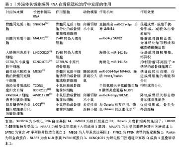
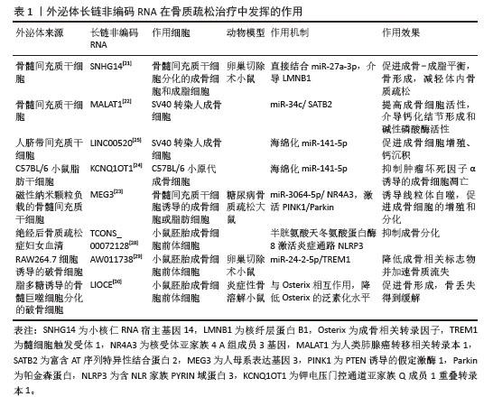
2.1 外泌体长链非编码RNA作用于骨质疏松 外泌体可以主动通过miRNA、蛋白质和mRNA向靶细胞传输和传递信息,影响细胞功能甚至改变整个微环境[11]。长链非编码RNA是一组长度超过200个核苷酸的转录本,参与一系列的生物学过程和疾病进展。研究表明,长链非编码RNA参与调节破骨细胞、成骨细胞和间充质干细胞的信号转导,调节破骨细胞和成骨细胞之间的通讯[12]。长链非编码RNA通过募集转录因子、作用于RNA聚合酶或沉默mRNA表达来调节骨相关基因表达[13]。研究表明,在骨质疏松情况下,有1 594个长链非编码RNA差异表达,包括482个上调的长链非编码RNA和1 112个下调的长链非编码RNA[14]。长链非编码RNA MEG3在卵巢切除小鼠或骨质疏松患者骨髓间充质干细胞中表达上调,通过下调miR-133a-3p表达抑制成骨分化[15]。Lnc-NTF3-5通过海绵化miR-93-3p促进上颌窦膜干细胞的成骨分化和骨再生[16]。低骨密度绝经后女性外周血单核细胞中长链非编码RNA拮抗非蛋白编码RNA水平上调,通过释放肿瘤坏死因子α和白细胞介素6促进骨吸收[17]。使用长链非编码RNA作为治疗手段已成为一个重要的研究领域。采集患或不患骨质疏松症个体的外周血,使用超速离心法提取血清外泌体,分离外泌体总RNA,采用RNA-Seq进行长链非编码RNA分析,共鉴定出393个差异表达长链非编码RNA,其中296个上调、97个下调;与差异表达长链非编码RNA共定位的mRNA在骨质疏松相关通路中高度富集,包括调节胰岛素分泌、激活丝裂原活化蛋白激酶活性、细胞对金属离子的反应、聚焦化和蛋白水解[18]。尽管在分子领域取得了进展,但人们对外泌体释放的长链非编码RNA在骨质疏松症中的作用机制知之甚少。此综述总结不同来源外泌体长链非编码RNA对骨质疏松症相关骨细胞的作用,为骨质疏松症提供潜在的治疗靶点[19]。 2.1.1 间充质干细胞来源外泌体 间充质干细胞具有多细胞系分化潜力,包括分化成软骨细胞、成骨细胞和脂肪细胞等。间充质干细胞在维持骨吸收和骨形成之间的稳态方面起着重要作用。外泌体可调节间充质干细胞分化为成骨细胞谱系[20]。全转录组分析研究表明,长链非编码RNA与成骨分化过程密切相关,间充质干细胞向成骨谱系分化的能力下降是骨形成不足的原因之一。因此,探索与临床应用相关的长链非编码RNA至关重要[13]。 将骨髓间充质干细胞诱导为成骨细胞和脂肪细胞,然后与骨髓间充质干细胞外泌体共培养,成骨细胞中钙沉积面积显著增大,碱性磷酸酶水平升高,与成骨分化相关的矮小相关转录因子2和人骨钙蛋白表达显著上调,脂肪细胞中脂滴形成减少,与成脂分化相关的过氧化物酶体增殖物激活受体γ和增强子结合蛋白C/EBPα表达显著下调,表明骨髓间充质干细胞外泌体可促进骨髓间充质干细胞的成骨-成脂平衡,进一步研究结果显示,骨髓间充质干细胞外泌体是通过递送长链非编码RNA小核仁RNA宿主基因14介导miR-27a-3p/核纤层蛋白B1轴调节骨髓间充质干细胞的成骨和成脂分化[21]。研究表明,骨髓间充质干细胞衍生外泌体中人类肺腺癌转移相关转录本1可能通过充当miR-34c海绵上调富含AT序列特异性结合蛋白2表达,缓解骨质疏松症症状。根据生物信息学网站的预测结果,确定了miR-34c与人类肺腺癌转移相关转录本1或miR-34c与富含AT序列特异性结合蛋白2 3’UTR之间的假定结合位点,RNA下拉实验进一步证明miR-34c和人类肺腺癌转移相关转录本1可以相互结合并促进富含AT序列特异性结合蛋白2的表达。将过表达人类肺腺癌转移相关转录本1的骨髓间充质干细胞外泌体与SV40转染的人成骨细胞共培养可提高成骨细胞活性。注射过表达人类肺腺癌转移相关转录本1的骨髓间充质干细胞外泌体可促进骨质疏松小鼠股骨远端骨小梁增厚,小梁间连通性和成骨细胞数量增加,皮质骨分离减少[22]。磁性纳米粒子已逐渐成为潜在的药物载体,利用载有磁性纳米粒子的骨髓间充质干细胞衍生外泌体可以放大靶区的药物浓度,提高治疗效果。负载磁性纳米粒子的骨髓间充质干细胞衍生外泌体通过递送长链非编码RNA人母系表达基因抑制miR-3064-5p的表达,促进线粒体自噬、成骨细胞增殖和分化,缓解大鼠糖尿病骨质疏松症[23]。 肿瘤坏死因子α诱导的脂肪干细胞来源外泌体剂量依赖性阻断原代成骨细胞凋亡。在脂肪干细胞中转染钾电压门控通道亚家族Q成员1重叠转录本1慢病毒载体,其释放的外泌体可抑制肿瘤坏死因子α诱导的成骨细胞凋亡,表明长链非编码RNA-钾电压门控通道亚家族Q成员1重叠转录本1可能成为脂肪干细胞来源外泌体治疗骨质疏松症的潜在靶点[24]。 敲低LINC00520的人脐带间充质干细胞外泌体抑制SV40转染的人成骨细胞存活和钙沉积,加速细胞凋亡,而过表达LINC00520的人脐带间充质干细胞外泌体促进SV40转染的人成骨细胞增殖和钙沉积,延长细胞周期,表明人脐带间充质干细胞外泌体LINC00520可作为钙沉积的调节剂治疗骨质疏松[25]。 2.1.2 组织细胞来源外泌体 有学者选取骨量正常者、骨量减少者和骨质疏松症者的血液样本进行长链非编码RNA测序研究,筛选出与骨质疏松症发生发展密切相关的长链非编码RNA MIR22宿主基因,结果显示,绝经后骨质疏松症患者长链非编码RNA MIR22宿主基因水平显著降低[26]。成肌细胞来源外泌体配对相关同源盒蛋白2通过海绵miR-128参与长链非编码RNA MIR22宿主基因的转录激活,它可激活河马通路促进骨髓间充质干细胞的成骨分化[27]。从健康人和绝经后骨质疏松症患者的外泌体中鉴定出26个差异表达的长链非编码RNA,其中长链非编码RNA TCONS_00072128表达显著下调,过表达长链非编码RNA TCONS_00072128可正向调控半胱氨酸天冬氨酸蛋白酶8,促进碱性磷酸酶表达,从而增强骨髓间充质干细胞的成骨分化能力,延缓绝经后骨质疏松症的进展[28]。研究表明,含有长链非编码RNA AW011738的破骨细胞衍生外泌体可降低卵巢切除小鼠的成骨细胞活性,加速骨质流失,在破骨细胞中敲降AW011738后提取的外泌体可显著促进成骨细胞分化。卵巢切除小鼠尾静脉注射敲降AW011738后的外泌体可缓解骨质疏松,表明破骨细胞外泌体中长链非编码RNA AW011738可通过海绵化miR-24-2-5p增加髓样细胞触发受体1表达,抑制小鼠胚胎成骨细胞前体细胞的成骨分化[29]。炎性破骨细胞外泌体通过Eph相关受体酪氨酸激酶配体6/Eph受体A2特异性靶向成骨细胞,Eph受体A2敲除后,进入小鼠胚胎成骨细胞前体细胞的炎性破骨细胞外泌体数量显著减少,促进成骨细胞产生更多的钙化结节。长链非编码RNA LIOCE富集于炎性破骨细胞外泌体中,被小鼠胚胎成骨细胞前体细胞摄取后提高了成骨细胞活性,注射包封长链非编码RNA LIOCE的破骨细胞外泌体后,炎症性骨溶解小鼠的骨质流失得到缓解[30](表1)。 "
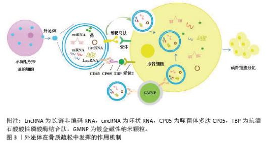
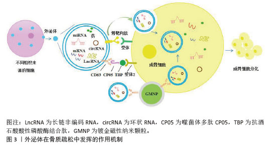
2.2 外泌体在骨质疏松症中的创新应用 尽管多种来源外泌体对骨质疏松症有一定的治疗作用,但它们的低产量、有限的功能性和不稳定的生物活性一直是阻碍外泌体实际应用的关键。除了天然分泌的外泌体外,近年来还研究了能够实现细胞特异性货物递送的转基因或化学修饰外泌体。例如,基因工程修饰的树突状细胞衍生外泌体通过将miR-140靶向递送至软骨深部区域,改善骨关节炎症状[31]。外泌体通过靶向表面修饰、分子封装机制发挥药物递送的协同作用,同时,外泌体与生物活性材料相结合是目前骨质疏松治疗研究中的热门方向。近年来,研究人员试图通过构建工程纳米囊泡来实现外泌体的大规模生产和精确靶向递送[32](图3)。经过修饰或材料加载的外泌体在治疗骨质疏松症方面显示出巨大的应用前景。 "
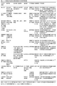
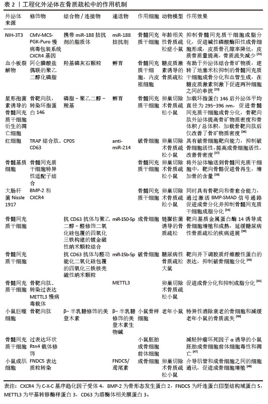
2.3 工程化外泌体的创新应用 2.3.1 靶向表面修饰 外泌体的表面修饰是指在外泌体表面设计功能配体以靶向受体细胞并激活免疫,一般分为外泌体提取前修饰和提取后修饰。提取前修饰涉及通过基因工程方法编辑细胞的蛋白质表达,通常诱导靶向膜蛋白的高表达,使分泌的外泌体间接高表达靶膜蛋白。例如,有研究构建高表达C-X-C基序趋化因子受体4的基因工程NIH-3T3细胞,提取其外泌体,然后将C-X-C基序趋化因子受体4阳性外泌体与加载miR-188拮抗剂的脂质体挤出形成混合纳米颗粒,经Balb/c小鼠尾部静脉注射发现混合纳米颗粒特异性聚集在骨髓中并释放miR-188拮抗剂,促进碱性磷酸酶阳性成骨细胞形成并抑制骨髓间充质干细胞成脂分化,降低骨髓miR-188水平和皮质骨孔隙率并挽救与衰老相关的骨质流失,另外,注射后48 h小鼠肝肾功能保持稳定,表明混合纳米颗粒在体内无短期或长期细胞毒性[33]。有研究将骨形态发生蛋白2和C-X-C基序趋化因子受体4重组质粒与大肠杆菌细胞融合后提取外泌体用于骨质疏松治疗,该外泌体在体外可促进成骨细胞分化,抑制骨髓间充质干细胞成脂分化,通过激活骨形态发生蛋白/Smad信号通路改善骨质疏松症[34]。 提取后修饰是通过脂质插入修饰、共价交联修饰或化学修饰等方式直接将脂质分子插入到外泌体表面。ZHENG等[35]从血小板裂解物中分离出外泌体,随后将外泌体与阿仑膦酸钠接枝的聚乙二醇化磷脂结合,从而构建出一种骨靶向的血小板裂解物外泌体。血小板裂解物外泌体富集血小板衍生生长因子,经阿仑膦酸盐修饰的外泌体可逆转地塞米松对骨髓间充质干细胞成骨分化的抑制作用,提高内皮祖细胞的血管生成能力。骨靶向肽通过磷脂聚乙二醇羧基连接在骨髓间充质干细胞衍生的凋亡细胞外泌体表面,在卵巢切除小鼠模型中发挥明显的骨靶向和促进骨生成的作用[36]。将双功能肽(抗酒石酸酸性磷酸酶结合肽-噬菌体多肽CP05)与红细胞外泌体结合后产生破骨细胞靶向红细胞外泌体,静脉注射后该外泌体富集于骨质疏松小鼠骨骼中,有效递送miR-214至体内破骨细胞并抑制其活性,提高成骨细胞活性,改善骨密度[37]。LUO等[38]将骨髓基质细胞衍生外泌体与骨髓间充质干细胞特异性适配体结合,骨髓基质细胞外泌体被递送至骨髓间充质干细胞中,此复合物可增加卵巢切除术小鼠的骨量,加速骨折小鼠的骨愈合。磁性纳米粒子能够将外泌体与靶向药物输送系统相结合。研究人员通过在磁性氧化铁二氧化硅纳米粒子以及硅质外壳的表面覆盖聚乙二醇-醛来构建金涂层磁性纳米粒子,将骨髓间充质干细胞外泌体负载在金涂层磁性纳米粒子上,该外泌体可通过递送miR-150-5p靶向调节基质金属蛋白酶14诱导成骨细胞增殖和成熟,延缓糖尿病性骨质疏松的疾病进展[39]。在骨髓间充质干细胞中过表达miR-15b-5p,提取外泌体,体外将聚乙二醇化羧基修饰的介孔二氧化硅包裹超顺磁性四氧化三铁纳米颗粒与外泌体结合,大鼠破骨细胞可成功摄取该外泌体,将miR-15b-5p传递给破骨细胞,靶向抑制胶质纤维酸性蛋白表达缓解糖尿病性大鼠骨质疏松[40]。 2.3.2 外泌体功能分子封装 功能分子包封是指将指定的小分子药物、核酸和蛋白质装入外泌体中,将其转化为功能载体,用于递送治疗,包封方法主要有电穿孔、质粒转染或渗透剂孵育[41]。外泌体作为天然递送载体,具有优异的生物相容性和内在组织趋向性,靶向递送系统可以通过外泌体表面修饰进行优化[42]。例如,骨靶向肽的掺入可以提高外泌体的递送效率,这种修饰使封装的miRNA具有优越的稳定性,降低免疫原性,提高生物利用度。LI等[43]通过装载丰富的甲基转移酶样3 mRNA并用骨靶向肽修饰来改造骨髓间充质干细胞外泌体,修饰后的外泌体表现出卓越的骨靶向效率,可显著减轻小鼠卵巢切除术引起的骨量损失,促进小鼠骨髓间充质干细胞成骨分化,抑制脂质分化。将间充质干细胞外泌体与骨靶向肽结合形成的骨靶向修饰外泌体可特异性将siRNA递送给成骨细胞并介导成骨细胞Shn3基因沉默,促进成骨分化和H型血管形成,抑制破骨细胞形成[44]。骨髓间充质干细胞外泌体递送的miR-17促进成骨分化,并通过BARX同源盒蛋白2促进骨愈合[45]。在巨噬细胞外泌体中加入骨靶向肽6,包裹半乳糖修饰的美登素,可以破坏微管蛋白聚合,诱导衰老的骨细胞凋亡,对老年小鼠施用骨靶向外泌体可以减轻骨质流失,而不会产生明显的毒性[46]。circ-Rtn4修饰的骨髓间充质干细胞来源外泌体通过充当miR-146a海绵减轻肿瘤坏死因子α诱导的小鼠胚胎成骨细胞前体细胞毒性和凋亡[47]。用纤连蛋白Ⅲ型结构域蛋白5表达质粒转染成肌细胞,待分化成肌管后提取外泌体,该外泌体可促进成骨细胞分化[48]。采用超声波法将整合素连锁激酶的靶向抑制剂QLT0267转入到从健康女性血清提取的外泌体中,绝经期骨质疏松女性骨髓间充质干细胞矿化结节显著减少,抑制了成骨细胞分化[49]。有研究设计了一种可注射的基于淫羊藿的细胞外纳米囊泡负载羟基磷灰石纳米复合物,该复合物成功修复了雌激素剥夺诱导的骨质疏松大鼠下颌骨缺损,为中医药的应用提供了新的视角[50]。然而,外泌体从实验规模到临床生产的过渡等方面的挑战可能会限制它们的发展和临床应用(表2)。 "
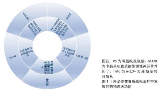
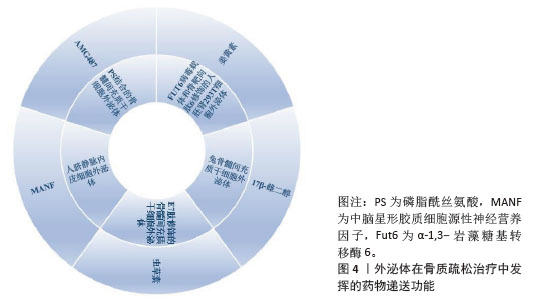
2.4 外泌体作为药物递送系统发挥骨质疏松治疗的协同效应 外泌体具有天然来源、介导细胞间通讯以及在脂质双层膜或管腔内包裹各种生物分子(如药物、蛋白质和核酸)的潜力[51],同时外泌体能够在细胞外液中安全移动并高效地将货物运输到目标细胞,因此外泌体可在体内提供更好的货物运输途径,发挥药物递送系统的作用(图4)。例如,磷脂酰丝氨酸是一种具有免疫抑制和吞噬信号传导功能的膜脂质,用薄膜水合结合挤出方法制备磷脂酰丝氨酸掺入的外泌体模拟物,该模拟物可抑制破骨细胞分化。此外,将趋化因子受体3拮抗剂AMG487封装于磷脂酰丝氨酸修饰的外泌体模拟物中,可有效防止卵巢切除小鼠的骨质流失[52]。将中脑星形胶质细胞源性神经营养因子用超声封装到人脐静脉内皮细胞外泌体中,封装后的外泌体经尾静脉注射到卵巢切除的骨质疏松小鼠体内,小鼠股骨干骺端的骨小梁体积、厚度和数量均显著增加,抑制破骨细胞分化,减少骨质流失[53]。外泌体可以通过亲和肽修饰来靶向特定细胞,E7肽是一种用于骨髓间充质干细胞靶向给药的亲和肽[54]。虫草素通过直接孵育或超声处理加载到E7肽修饰的外泌体中,研究人员通过小鼠电离辐射联合尾悬模拟航天诱发骨质疏松症,加载虫草素的E7肽修饰的外泌体可显著改善骨质疏松小鼠的骨质流失,成骨细胞活性增加[55]。将17β-雌二醇通过超声处理负载到骨髓间充质干细胞衍生外泌体中,与对照组相比,负载雌二醇的外泌体显著提高了骨髓间充质干细胞的存活率[56]。α-1,3-岩藻糖基转移酶6是一种与前列腺癌骨转移相关的关键蛋白质,将编码α-1,3-岩藻糖基转移酶6的反转录病毒载体与反转录病毒包装辅助质粒一起转染293T细胞,可生成表达α-1,3-岩藻糖基转移酶6的外泌体,用骨靶向肽6对这些外泌体进行修饰,合成α-1,3-岩藻糖基转移酶6-骨靶向肽6-外泌体,用超声处理使姜黄素负载于合成外泌体表面上,结果显示,α-1,3-岩藻糖基转移酶6的引入增加了骨髓内皮细胞对外泌体的摄取,在小鼠体内表现出更高的骨靶向效率,负载姜黄素的骨靶向外泌体恢复了吸烟相关骨质疏松小鼠的骨髓间充质干细胞成骨分化潜力,减轻骨质流失[57]。因此,当外泌体作为药物载体时,特异性靶向骨组织,很大程度上提高了药物的利用效率,增强了药物治疗骨质疏松的潜力。 "
| [1] LI P, XU TY, YU AX, et al. The Role of Ferroptosis in Osteoporosis and Advances in Chinese Herbal Interventions. Biology (Basel). 2025;14(4):367. [2] TABATABAEI-MALAZY O, SALARI P, KHASHAYAR P, et al. New horizons in treatment of osteoporosis. Daru. 2017;25(1):2. [3] BUDHIA S, MIKYAS Y, TANG M, et al. Osteoporotic fractures: a systematic review of U.S. healthcare costs and resource utilization. Pharmacoeconomics. 2012;30(2):147-170. [4] PATIL S, DANG K, ZHAO X, et al. Role of LncRNAs and CircRNAs in Bone Metabolism and Osteoporosis. Front Genet. 2020;11:584118. [5] PARK-MIN KH, MUN SH, BOCKMAN R, et al. New Horizons: Translational Aspects of Osteomorphs. J Clin Endocrinol Metab. 2024;109(5):e1373-e1378. [6] ERIKSEN EF, DÍEZ-PÉREZ A, BOONEN S. Update on long-term treatment with bisphosphonates for postmenopausal osteoporosis: a systematic review. Bone. 2014;58:126-135. [7] LU J, ZHANG Y, LIANG J, et al. Role of Exosomal MicroRNAs and Their Crosstalk with Oxidative Stress in the Pathogenesis of Osteoporosis. Oxid Med Cell Longev. 2021;2021:6301433. [8] HU Y, WANG Y, CHEN T, et al. Exosome: Function and Application in Inflammatory Bone Diseases. Oxid Med Cell Longev. 2021;2021:6324912. [9] ZHANG X, MENG S, XU Y, et al. New Qiangguyin activates Wnt/β-catenin pathway by down-regulating Notum to improve osteoporosis in rats. J Ethnopharmacol. 2025;347:119745. [10] HU Y, HOU Z, LIU Z, et al. Oyster mantle-derived exosomes alleviate osteoporosis by regulating bone homeostasis. Biomaterials. 2024; 311:122648. [11] CORRADO C, RAIMONDO S, CHIESI A, et al. Exosomes as intercellular signaling organelles involved in health and disease: basic science and clinical applications. Int J Mol Sci. 2013;14(3): 5338-5366. [12] LIU W, LI Z, CAI Z, et al. LncRNA-mRNA expression profiles and functional networks in osteoclast differentiation. J Cell Mol Med. 2020;24(17): 9786-9797. [13] SILVA AM, MOURA SR, TEIXEIRA JH, et al. Long noncoding RNAs: a missing link in osteoporosis. Bone Res. 2019;7:10. [14] LIU S, HUANG H, CHAI S, et al. Expression profile analysis of long non-coding RNA in skeletal muscle of osteoporosis by microarray and bioinformatics. J Biol Eng. 2019;13:50. [15] WANG Q, LI Y, ZHANG Y, et al. LncRNA MEG3 inhibited osteogenic differentiation of bone marrow mesenchymal stem cells from postmenopausal osteoporosis by targeting miR-133a-3p. Biomed Pharmacother. 2017;89: 1178-1186. [16] PENG W, ZHU SX, WANG J, et al. Lnc-NTF3-5 promotes osteogenic differentiation of maxillary sinus membrane stem cells via sponging miR-93-3p. Clin Implant Dent Relat Res. 2018;20(2):110-121. [17] TONG X, GU PC, XU SZ, et al. Long non-coding RNA-DANCR in human circulating monocytes: a potential biomarker associated with postmenopausal osteoporosis. Biosci Biotechnol Biochem. 2015;79(5):732-737. [18] TENG Z, ZHU Y, ZHANG X, et al. Osteoporosis Is Characterized by Altered Expression of Exosomal Long Non-coding RNAs. Front Genet. 2020;11:566959. [19] ZHAO ZM, DING JM, LI Y, et al. Human umbilical cord mesenchymal stem cell-derived exosomes promote osteogenesis in glucocorticoid-induced osteoporosis through PI3K/AKT signaling pathway-mediated ferroptosis inhibition. Stem Cells Transl Med. 2025;14(3):szae096. [20] XIE X, XIONG Y, PANAYI AC, et al. Exosomes as a Novel Approach to Reverse Osteoporosis: A Review of the Literature. Front Bioeng Biotechnol. 2020;8:594247. [21] TANG JS, YU HX, RUAN RX, et al. LncRNA SNHG14 Delivered by Bone Marrow Mesenchymal Stem Cells-Secreted Exosomes Regulates Osteogenesis and Adipogenesis in Osteoporosis by Mediating the miR-27a-3p/LMNB1 Axis. Kaohsiung J Med Sci. 2025;41(5):e70004. [22] YANG X, YANG J, LEI P, et al. LncRNA MALAT1 shuttled by bone marrow-derived mesenchymal stem cells-secreted exosomes alleviates osteoporosis through mediating microRNA-34c/SATB2 axis. Aging (Albany NY). 2019;11(20): 8777-8791. [23] XU C, WANG Z, LIU YJ, et al. Harnessing GMNP-loaded BMSC-derived EVs to target miR-3064-5p via MEG3 overexpression: Implications for diabetic osteoporosis therapy in rats. Cell Signal. 2024;118:111055. [24] WANG SZ, JIA J, CHEN CH. lncRNA-KCNQ1OT1: A Potential Target in Exosomes Derived from Adipose-Derived Stem Cells for the Treatment of Osteoporosis. Stem Cells Int. 2021;2021:7690006. [25] WU J, ZHANG L, LIU H, et al. Exosomes promote hFOB1.19 proliferation and differentiation via LINC00520. J Orthop Surg Res. 2023;18(1):546. [26] 霍凯伦.LncRNA MIR22HG调节绝经后骨质疏松症破骨细胞异常活化的机制研究[D].银川: 宁夏医科大学,2024. [27] LI Y, WANG X, PAN C, et al. Myoblast-derived exosomal Prrx2 attenuates osteoporosis via transcriptional regulation of lncRNA-MIR22HG to activate Hippo pathway. Mol Med. 2023;29(1):54. [28] YANG Y, MIAO L, CHANG S, et al. Exosome-Derived LncRNA TCONS_00072128 Mediated Osteogenic Differentiation and Inflammation by Caspase 8 Regulation. Front Genet. 2022;12:831420. [29] LIU J, WANG B, CHEN H, et al. Osteoclast-derived exosomes influence osteoblast differentiation in osteoporosis progression via the lncRNA AW011738/ miR-24-2-5p/ TREM1 axis. Biomed Pharmacother. 2024;178:117231. [30] REN L, ZENG F, DENG J, et al. Inflammatory osteoclasts-derived exosomes promote bone formation by selectively transferring lncRNA LIOCE into osteoblasts to interact with and stabilize Osterix. FASEB J. 2022;36(2):e22115. [31] LIANG Y, XU X, LI X, et al. Chondrocyte-Targeted MicroRNA Delivery by Engineered Exosomes toward a Cell-Free Osteoarthritis Therapy. ACS Appl Mater Interfaces. 2020;12(33):36938-36947. [32] TRAN PHL, XIANG D, TRAN TTD, et al. Exosomes and Nanoengineering: A Match Made for Precision Therapeutics. Adv Mater. 2020;32(18):e1904040. [33] HU Y, LI X, ZHANG Q, et al. Exosome-guided bone targeted delivery of Antagomir-188 as an anabolic therapy for bone loss. Bioact Mater. 2021;6(9):2905-2913. [34] LIU H, SONG P, ZHANG H, et al. Synthetic biology-based bacterial extracellular vesicles displaying BMP-2 and CXCR4 to ameliorate osteoporosis. J Extracell Vesicles. 2024;13(4):e12429. [35] ZHENG G, MA HW, XIANG GH, et al. Bone-targeting delivery of platelet lysate exosomes ameliorates glucocorticoid-induced osteoporosis by enhancing bone-vessel coupling. J Nanobiotechnology. 2022;20(1):220. [36] GUI L, YE Q, YU L, et al. Bone-Targeting Peptide and RNF146 Modified Apoptotic Extracellular Vesicles Alleviate Osteoporosis. Int J Nanomedicine. 2024;19:471-488. [37] XU L, XU X, LIANG Y, et al. Osteoclast-targeted delivery of anti-miRNA oligonucleotides by red blood cell extracellular vesicles. J Control Release. 2023;358:259-272. [38] LUO ZW, LI FX, LIU YW, et al. Aptamer-functionalized exosomes from bone marrow stromal cells target bone to promote bone regeneration. Nanoscale. 2019;11(43):20884-20892. [39] XU C, WANG Z, LIU Y, et al. Extracellular vesicles derived from bone marrow mesenchymal stem cells loaded on magnetic nanoparticles delay the progression of diabetic osteoporosis via delivery of miR-150-5p. Cell Biol Toxicol. 2023;39(4): 1257-1274. [40] XU C, WANG Z, LIU Y, et al. Delivery of miR-15b-5p via magnetic nanoparticle-enhanced bone marrow mesenchymal stem cell-derived extracellular vesicles mitigates diabetic osteoporosis by targeting GFAP. Cell Biol Toxicol. 2024;40(1):52. [41] CHEN Y, HUANG Y, LI J, et al. Enhancing osteoporosis treatment with engineered mesenchymal stem cell-derived extracellular vesicles: mechanisms and advances. Cell Death Dis. 2024;15(2):119. [42] BUTREDDY A, KOMMINENI N, DUDHIPALA N. Exosomes as Naturally Occurring Vehicles for Delivery of Biopharmaceuticals: Insights from Drug Delivery to Clinical Perspectives. Nanomaterials (Basel). 2021;11(6):1481. [43] LI T, ZHAO J, YUAN J, et al. Harnessing engineered exosomes as METTL3 carriers: Enhancing osteogenesis and suppressing lipogenesis in bone marrow mesenchymal stem cells for postmenopausal osteoporosis treatment. Mater Today Bio. 2025;32:101648. [44] CUI Y, GUO Y, KONG L, et al. A bone-targeted engineered exosome platform delivering siRNA to treat osteoporosis. Bioact Mater. 2021;10:207-221. [45] JIANG X, WANG Q, YING P, et al. Bone Marrow Stromal Cells Promote Osteogenic Differentiation and Induce Bone Remodeling and Healing in Osteoporosis. Ann Clin Lab Sci. 2025;55(2):231-239. [46] HE Y, ZHANG L, CHEN X, et al. Elimination of Senescent Osteocytes by Bone-Targeting Delivery of β-Galactose-Modified Maytansinoid Prevents Age-Related Bone Loss. Adv Healthc Mater. 2024; 13(8):e2302972. [47] CAO G, MENG X, HAN X, et al. Exosomes derived from circRNA Rtn4-modified BMSCs attenuate TNF-α-induced cytotoxicity and apoptosis in murine MC3T3-E1 cells by sponging miR-146a. Biosci Rep. 2020;40(5):BSR20193436. [48] TAO L, WANG J, WANG K, et al. Exerkine FNDC5/irisin-enriched exosomes promote proliferation and inhibit ferroptosis of osteoblasts through interaction with Caveolin-1. Aging Cell. 2024; 23(8):e14181. [49] 刘毓灵.健康女性血清外泌体转运ILK促进PMOP患者BMSCs成骨分化[D].长沙:中南大学,2022. [50] HAN R, GAO C, TANG R, et al. A comprehensive study on Herba Epimedium-derived extracellular nanovesicles as a prospective therapy for alveolar bone regeneration in postmenopausal osteoporosis. Nanoscale. 2025;17(19):12270-12289. [51] XINYUE W, HONGLI L, CHUNHUI G, et al. Potential MiRNA therapies for premature ovarian failure: new challenges and opportunities. Stem Cell Res Ther. 2025;16(1):364. [52] KANG M, LI Z, CHANG I, et al. Phosphatidylserine-incorporated exosome mimetics encapsulating CXCR3 antagonist alleviate osteoporosis. Adv Funct Mater. 2024;34(38):2402521. [53] PI Z, WU Y, WANG X, et al. Exosomal Manf originated from endothelium regulated osteoclast differentiation by down-regulating NF-κB signaling pathway. J Orthop Surg Res. 2025;20(1):349. [54] XU X, LIANG Y, LI X, et al. Exosome-mediated delivery of kartogenin for chondrogenesis of synovial fluid-derived mesenchymal stem cells and cartilage regeneration. Biomaterials. 2021;269: 120539. [55] ZHAI Y, ZHANG Y, XU K, et al. Cordycepin ameliorates spaceflight-induced osteoporosis by preventing BMSCs oxidative stress and senescence via interacting with PI3K p110α and regulating PI3K/Akt/FOXO3 signalling. Free Radic Biol Med. 2025;228:108-125. [56] GHOLAMI FARASHAH MS, JAVADI M, SOLEIMANI RAD J, et al. 17β-Estradiol-Loaded Exosomes for Targeted Drug Delivery in Osteoporosis: A Comparative Study of Two Loading Methods. Adv Pharm Bull. 2023;13(4):736-746. [57] WANG Y, SUN L, DONG Z, et al. Targeted inhibition of ferroptosis in bone marrow mesenchymal stem cells by engineered exosomes alleviates bone loss in smoking-related osteoporosis. Mater Today Bio. 2025;31:101501. [58] JU Y, HU Y, YANG P, et al. Extracellular vesicle-loaded hydrogels for tissue repair and regeneration. Mater Today Bio. 2022;18:100522. [59] XIE Y, GUAN Q, GUO J, et al. Hydrogels for Exosome Delivery in Biomedical Applications. Gels. 2022;8(6):328. [60] YANG Z, LI X, GAN X, et al. Hydrogel armed with Bmp2 mRNA-enriched exosomes enhances bone regeneration. J Nanobiotechnology. 2023; 21(1):119. [61] LUO P, ZHANG Y, HUANG M, et al. Microdroplets Encapsulated with NFATc1-siRNA and Exosomes-Derived from MSCs Onto 3D Porous PLA Scaffold for Regulating Osteoclastogenesis and Promoting Osteogenesis. Int J Nanomedicine. 2024;19:3423-3440. [62] WANG T, GUO S, ZHANG Y. Effect of nHA/CS/PLGA delivering adipose stem cell-derived exosomes and bone marrow stem cells on bone healing-in vitro and in vivo studies. Sci Rep. 2024;14(1):27502. [63] ZHU Y, LI Z, ZHANG Y, et al. The essential role of osteoclast-derived exosomes in magnetic nanoparticle-infiltrated hydroxyapatite scaffold modulated osteoblast proliferation in an osteoporosis model. Nanoscale. 2020;12(16): 8720-8726. [64] QAYOOM I, TEOTIA AK, KUMAR A. Nanohydroxyapatite Based Ceramic Carrier Promotes Bone Formation in a Femoral Neck Canal Defect in Osteoporotic Rats. Biomacromolecules. 2020;21(2):328-337. [65] SONG W, ZHAO D, WANG J, et al. Ultrasound-Driven Innervated Bone Regeneration in Additively Manufactured Degradable Metallic Scaffolds. Adv Healthc Mater. 2025;14(12):e2404024. [66] CHEN G, LI H, CHEN R, et al. Exosomes as Emerging Therapeutic Strategies in Primary Osteoporosis: A Narrative Review. Drug Des Devel Ther. 2025;19:9099-9115. [67] LI F, ZHAO X, ZHANG Y, et al. Exosomal circFAM63Bsuppresses bone regeneration of postmenopausal osteoporosis via regulating miR-578/HMGA2 axis. J Orthop Res. 2024;42(6): 1244-1253. [68] WEI Y, MA H, ZHOU H, et al. miR-424-5p shuttled by bone marrow stem cells-derived exosomes attenuates osteogenesis via regulating WIF1-mediated Wnt/β-catenin axis. Aging (Albany NY). 2021;13(13):17190-17201. [69] JEPPESEN DK, HVAM ML, PRIMDAHL-BENGTSON B, et al. Comparative analysis of discrete exosome fractions obtained by differential centrifugation. J Extracell Vesicles. 2014;3:25011. [70] SHIREJINI SZ, INCI F. The Yin and Yang of exosome isolation methods: conventional practice, microfluidics, and commercial kits. Biotechnol Adv. 2022;54:107814. [71] DING M, WANG C, LU X, et al. Comparison of commercial exosome isolation kits for circulating exosomal microRNA profiling. Anal Bioanal Chem. 2018;410(16):3805-3814. [72] LE MN, FAN ZH. Exosome isolation using nanostructures and microfluidic devices. Biomed Mater. 2021;16(2):022005. [73] PEINADO H, ALEČKOVIĆ M, Lavotshkin S, et al. Melanoma exosomes educate bone marrow progenitor cells toward a pro-metastatic phenotype through MET. Nat Med. 2012;18(6): 883-891. [74] MEHRYAB F, RABBANI S, SHAHHOSSEINI S, et al. Exosomes as a next-generation drug delivery system: An update on drug loading approaches, characterization, and clinical application challenges. Acta Biomater. 2020;113:42-62. [75] ZHOU X, BROWN BA, SIEGEL AP, et al. Exosome-Mediated Crosstalk between Keratinocytes and Macrophages in Cutaneous Wound Healing. ACS Nano. 2020;14(10):12732-12748. [76] TAN A, RAJADAS J, SEIFALIAN AM. Exosomes as nano-theranostic delivery platforms for gene therapy. Adv Drug Deliv Rev. 2013;65(3):357-367. [77] QIU D, YAN B, XUE H, et al. Perspectives of exosomal ncRNAs in the treatment of bone metabolic diseases: Focusing on osteoporosis, osteoarthritis, and rheumatoid arthritis. Exp Cell Res. 2025;446(2):114457. |
| [1] | Chen Huiting, Zeng Weiquan, Zhou Jianhong, Wang Jie, Zhuang Congying, Chen Peiyou, Liang Zeqian, Deng Weiming. Tail anchoring technique of vertebroplasty in treatment of osteoporotic vertebral compression fractures with intravertebral cleft: a finite element analysis [J]. Chinese Journal of Tissue Engineering Research, 2026, 30(9): 2145-2152. |
| [2] | Zeng Xuan, Weng Rui, Ye Shicheng, Tang Jiadong, Mo Ling, Li Wenchao. Two lumbar rotary manipulation techniques in treating lumbar disc herniation: a finite element analysis of biomechanical differences [J]. Chinese Journal of Tissue Engineering Research, 2026, 30(9): 2153-2161. |
| [3] | Cheng Qisheng, Julaiti·Maitirouzi, Xiao Yang, Zhang Chenwei, Paerhati·Rexiti. Finite element analysis of novel variable-diameter screws in modified cortical bone trajectory of lumbar vertebrae [J]. Chinese Journal of Tissue Engineering Research, 2026, 30(9): 2162-2171. |
| [4] | Liu Wenlong, Dong Lei, Xiao Zhengzheng, Nie Yu. Finite element analysis of tibial prosthesis loosening after fixed-bearing unicompartmental knee arthroplasty for osteoporosis [J]. Chinese Journal of Tissue Engineering Research, 2026, 30(9): 2191-2198. |
| [5] | Chen Long, Wang Xiaozhen, Xi Jintao, Lu Qilin. Biomechanical performance of short-segment screw fixation combined with expandable polyetheretherketone vertebral body replacement in osteoporotic vertebrae [J]. Chinese Journal of Tissue Engineering Research, 2026, 30(9): 2226-2235. |
| [6] | Tan Jing, Li Li, Wang Liangliang, Qin Xiangyu. Bionic functional coating improves the integration of titanium implants and skin tissue interface [J]. Chinese Journal of Tissue Engineering Research, 2026, 30(8): 2014-2022. |
| [7] | Wang Baiyan, Yang Shu, Wang Yiming, Wu Mengqing, Xiao Yu, Guo Zixuan, Zhang Boyi, Feng Shuying. Exosome-delivered CRISPR/Cas system enables gene editing in target cells [J]. Chinese Journal of Tissue Engineering Research, 2026, 30(7): 1839-1849. |
| [8] | Zhou Sirui, Xu Yukun, Zhao Kewei. Ideas and methods of anti-melanogenesis of Angelica dahurica extracellular vesicles [J]. Chinese Journal of Tissue Engineering Research, 2026, 30(7): 1747-1754. |
| [9] | Hu Xiongke, Liu Shaohua, Tan Qian, Liu Kun, Zhu Guanghui. Shikonin intervention with bone marrow mesenchymal stem cells improves microstructure of femur in aged mice [J]. Chinese Journal of Tissue Engineering Research, 2026, 30(7): 1609-1615. |
| [10] | Ye Qianqian, Pan Hang, Tian Chuan, Zhu Xiangqing, Ye Li, Zhao Xiaojuan, Shu Liping, Pan Xinghua. Effects of highly active umbilical cord mesenchymal stem cells on structure and function of thymus in elderly tree shrews [J]. Chinese Journal of Tissue Engineering Research, 2026, 30(7): 1720-1729. |
| [11] | Wu Zhilin, , He Qin, Wang Pingxi, Shi Xian, Yuan Song, Zhang Jun, Wang Hao . DYRK2: a novel therapeutic target for rheumatoid arthritis combined with osteoporosis based on East Asian and European populations [J]. Chinese Journal of Tissue Engineering Research, 2026, 30(6): 1569-1579. |
| [12] | Zhang Haiwen, Zhang Xian, Xu Taichuan, Li Chao. Bibliometric and visual analysis of the research status and trends of senescence in osteoporosis [J]. Chinese Journal of Tissue Engineering Research, 2026, 30(6): 1580-1591. |
| [13] | Wen Guangwei, Zhen Yinghao, Zheng Taikeng, Zhou Shuyi, Mo Guoye, Zhou Tengpeng, Li Haishan, Lai Yiyi. Effects and mechanisms of isoginkgetin on osteoclastogenesis [J]. Chinese Journal of Tissue Engineering Research, 2026, 30(6): 1348-1358. |
| [14] | Huang Jie, Zeng Hao, Wang Wenchi, Lyu Zhucheng, Cui Wei. Visualization analysis of literature on the effect of lipid metabolism on osteoporosis [J]. Chinese Journal of Tissue Engineering Research, 2026, 30(6): 1558-1568. |
| [15] | Yang Zhijie, Zhao Rui, Yang Haolin, Li Xiaoyun, Li Yangbo, Huang Jiachun, Lin Yanping, Wan Lei, HuangHongxing. Postmenopausal osteoporosis: predictive values of muscle mass, grip strength, and appendicular skeletal muscle index [J]. Chinese Journal of Tissue Engineering Research, 2026, 30(5): 1073-1080. |
| Viewed | ||||||
|
Full text |
|
|||||
|
Abstract |
|
|||||