Chinese Journal of Tissue Engineering Research ›› 2026, Vol. 30 ›› Issue (27): 7073-7081.doi: 10.12307/2026.239
Previous Articles Next Articles
Digital measurement and correlation analysis of anatomical CT for clinical application in the hip joint
Wu Chao1, Su Ning1, Chen Guoqiang1, Li Xiaohe2, Ou Wenjing3
- 1Imaging Center, 3Medical Records Room, Baotou City Central Hospital, Baotou 014040, Inner Mongolia Autonomous Region, China; 2Department of Human Anatomy, School of Basic Medical Sciences, Inner Mongolia Medical University, Hohhot 010000, Inner Mongolia Autonomous Region, China
-
Received:2025-07-09Accepted:2025-11-18Online:2026-09-28Published:2026-05-14 -
Contact:Ou Wenjing, Attending physician, MD, Medical Records Room, Baotou City Central Hospital, Baotou 014040, Inner Mongolia Autonomous Region, China Li Xiaohe, MD, Professor, Master’s supervisor, Department of Human Anatomy, School of Basic Medical Sciences, Inner Mongolia Medical University, Hohhot 010000, Inner Mongolia Autonomous Region, China -
About author:Wu Chao, Doctoral candidate, Attending physician, Imaging Center, Baotou City Central Hospital, Baotou 014040, Inner Mongolia Autonomous Region, China Su Ning, Chief physician, Imaging Center, Baotou City Central Hospital, Baotou 014040, Inner Mongolia Autonomous Region, China Wu Chao and Su Ning contributed equally to this article. -
Supported by:Natural Science Foundation of Inner Mongolia Autonomous Region Project, No. 2025MS08121 (to WC); Science and Technology Program of the Joint Fund of Scientific Research for the Public Hospitals of Inner Mongolia Academy of Medical Sciences, No. 2023GLLH0225 (to WC); Joint Project with Inner Mongolia Medical University, No. YKD2022LH039 (to WC)

CLC Number:
Cite this article
Wu Chao, Su Ning, Chen Guoqiang, Li Xiaohe, Ou Wenjing. Digital measurement and correlation analysis of anatomical CT for clinical application in the hip joint[J]. Chinese Journal of Tissue Engineering Research, 2026, 30(27): 7073-7081.
share this article
Add to citation manager EndNote|Reference Manager|ProCite|BibTeX|RefWorks
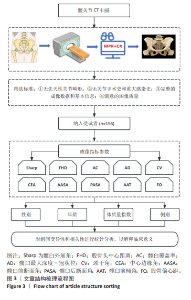
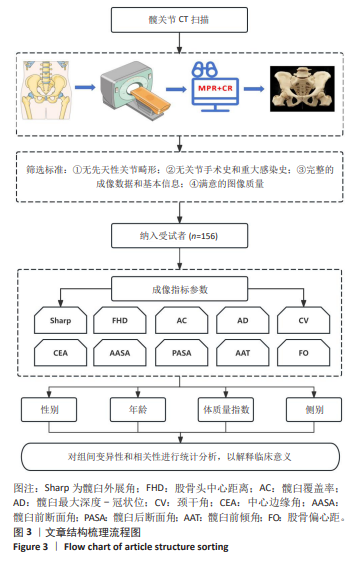
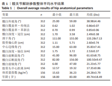
2.1 参与者数量分析 共纳入156例受试者,全部进入结果分析,无脱落。 2.2 试验流程图 见图3。 2.3 基线资料分析 共纳入156例受试者,共312侧,年龄18-83岁,平均(49.74±18.49)岁;男88例(56.4%),女68例(43.6%);组1为31例(19.9%),组2为25例(16%),组3为21例(13.5%),组4为27例(17.3%),组5为25例(16%),组6为27例(17.3%)。髋关节解剖参数指标结果见表1。经组内相关系数检验,髋臼外展角、髋臼覆盖率-传统法、髋臼覆盖率-革新法、髋臼最大深度-冠状位、颈干角、股骨偏心距、中心边缘角、髋臼最大深度-横轴位、髋臼前断面角、髋臼后断面角、髋臼前倾角、股骨头中心距离的测量者间内组内相关系数值分别为0.919,0.958,0.949,0.971,0.996,0.976,0.965,0.949,0.965,0.963,0.943和0.998。"
"
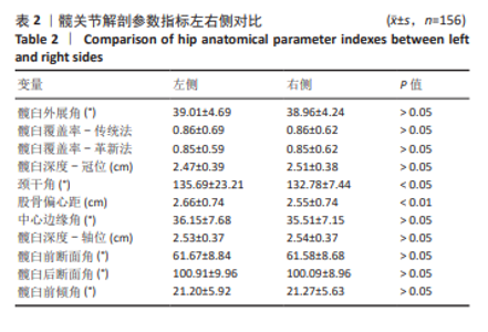
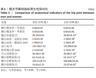
2.4 按左右侧比较髋关节解剖参数指标结果 颈干角和股骨偏心距在左右侧存在显著性差异(P < 0.05,P < 0.01),其余解剖指标左右侧相比无显著性差异,见表2。 2.5 按性别比较髋关节解剖参数指标结果 髋臼外展角、髋臼最大深度-冠状位、髋臼最大深度-横轴位、股骨偏心距、中心边缘角、髋臼前断面角、髋臼前倾角在性别间存在显著性差异 (P < 0.05-0.01),且在髋臼外展角、股骨偏心距、髋臼前倾角中女性大于男性,髋臼最大深度-冠状位、髋臼最大深度-横轴位、中心边缘角、髋臼前断面角指标中男性大于女性,见表3。"
"
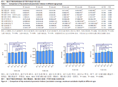
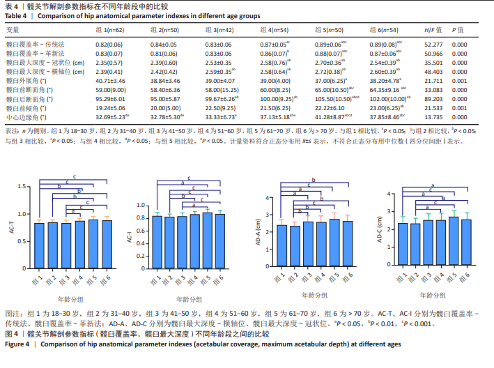
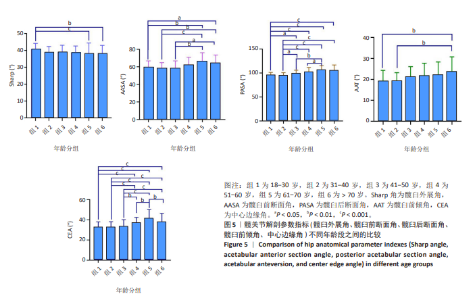
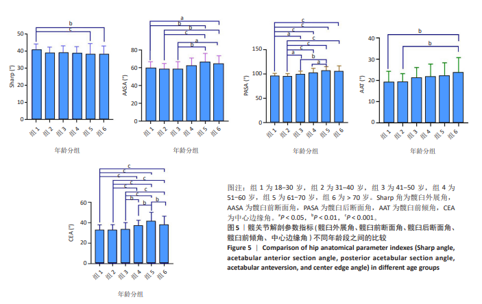
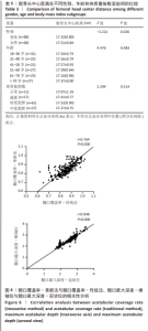
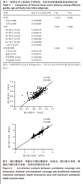
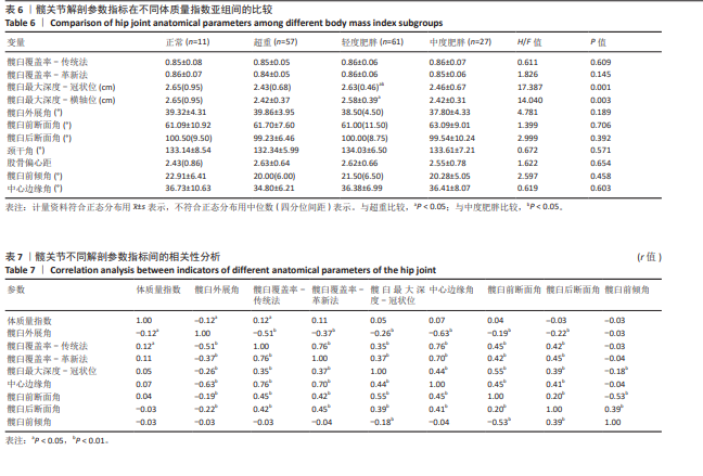
2.6 按不同年龄段、不同体质量指数分组比较髋关节解剖参数指标结果 不同年龄段在髋臼覆盖率、髋臼最大深度、髋臼外展角、髋臼前断面角、髋臼后断面角、中心边缘角、髋臼前倾角均存在显著性差异(P < 0.05)。年龄组1,2与组5,6在髋臼覆盖率、髋臼最大深度、髋臼前断面角、髋臼后断面角、中心边缘角存在显著性差异(P < 0.01);组3与组5,6 的髋臼覆盖率、髋臼前断面角、中心边缘角存在著性差异(P < 0.05)。在髋臼覆盖率、髋臼最大深度、髋臼前断面角、髋臼后断面角、中心边缘角指标上年龄组1,2均值小于组5,6均值(P < 0.05);髋臼覆盖率、髋臼前断面角、中心边缘角指标在组3的均值小于组5,6均值(P < 0.05);而髋臼外展角年龄组1均值大于组5,6均值(P < 0.05)。见表4及图4,5。 股骨头中心距离在不同年龄段、不同体质量指数之间,差异无显著性意义;不同性别之间股骨头中心距离比较,差异有显著性意义(P=0.026),且女性大于男性,见表5。 髋臼最大深度-冠状位、髋臼最大深度-横轴位在不同体质量指数中差异均有显著性意义(H=17.387,P=0.001;H=14.04,P=0.003)。组间比较发现髋臼最大深度-冠状位、髋臼最大深度-横轴位在超重和轻度肥胖分组中差异均有显著性意义(P=0.001,P=0.004);髋臼最大深度-冠状位在轻度肥胖和中度肥胖分组中差异有显著性意义(P=0.022);其余解剖指标在不同体质量指数分组中差异无显著性意义,见表6。"
"
"
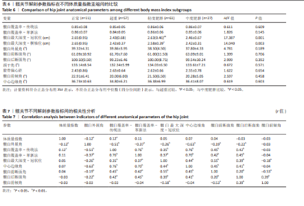
2.7 髋关节解剖参数指标相关性分析结果 髋臼覆盖率-传统法与髋臼覆盖率-革新法比较,差异无显著性意义(P=0.16);髋臼最大深度-冠状位与髋臼最大深度-横轴位比较,差异无显著性意义(P=0.32);髋臼覆盖率-传统法与髋臼覆盖率-革新法存在显著相关性(r=0.76,P < 0.01);髋臼最大深度-冠状位与髋臼最大深度-横轴位存在显著相关性(r=0.95,P < 0.01),见图6。 髋臼覆盖率、髋臼深度、中心边缘角、髋臼前断面角及髋臼后断面角彼此之间呈正相关(P < 0.01),髋臼外展角与体质量指数、髋臼覆盖率、髋臼深度、中心边缘角、髋臼前断面角、髋臼后断面角呈负相关(P < 0.05-0.01),且髋臼覆盖率-传统法与体质量指数呈正相关(P < 0.05);髋臼前倾角与髋臼深度、髋臼前断面角呈负相关(P < 0.01),与髋臼后断面角呈正相关(P < 0.01),见表7。股骨偏心距与颈干角呈负相关(r=-0.51,P < 0.01),髋臼深度与颈干角呈正相关(r=0.11,P < 0.05)。"
| 1] 高波,王丰,王琦, 等. 改良S-P入路不切断股直肌治疗股骨头骨折合并髋关节后脱位[J]. 中国实用医刊,2017,44(22):47-50. [2] LIU J, YE WX, LIANG WG, et al. One case of periprosthetic fracture after total hip arthroplasty. J Clin Orthop. 2018;21(6):754. [3] PIVEC R, JOHNSON AJ, MEARS SC, et al. Hip arthroplasty. Lancet. 2012; 380(9855):1768-1777. [4] AMANATULLAH DF, LANDA J, STRAUSS EJ, et al. Comparison of surgical outcomes and implant wear between ceramic-ceramic and ceramic-polyethylene articulations in total hip arthroplasty. J Arthroplasty. 2011; 26(6 Suppl):72-77. [5] BRAMMING IB, KIERKEGAARD S, LUND B, et al. High relative reliability and responsiveness of the forgotten joint score-12 in patients with femoroacetabular impingement undergoing hip arthroscopic treatment. A prospective survey-based study. J Hip Preserv Surg. 2019;6(2):149-156. [6] 王剑,云文科,李格当, 等. 数字化测量正常成人男女性髋臼前柱解剖学参数的临床意义[J].中国组织工程研究,2018,22(11):1713-1718. [7] 中华人民共和国国家卫生健康委员会医政司.肥胖症中国诊疗指南(2024年版)[J].协和医学杂志,2025,16(1):90-108. [8] ROWE SP, ZIMMERMAN SL, JOHNSON PT, et al. Evaluation of Kawasaki’s disease-associated coronary artery aneurysms with 3D CT cinematic rendering. Emerg Radiol. 2018;25(4):449-453. [9] EID M, DE CECCO CN, NANCE JW JR, et al. Cinematic Rendering in CT: A Novel, Lifelike 3D Visualization Technique. AJR Am J Roentgenol. 2017; 209(2):370-379. [10] 霍佳邦,赵畅,黄广鑫, 等. CT数据三维规划预测全髋关节置换假体型号及截骨的准确性与可复性[J].中国组织工程研究,2021,25(27): 4294-4299. [11] YU TJ, BANGURA A, BODANAPALLY U, et al. Dual-Energy CT and Cinematic Rendering to Improve Assessment of Pelvic Fracture Instability. Radiology. 2022;304(2):353-362. [12] PATTAMAPASPONG N, KANTHAWANG T, SINGSUWAN P, et al. Efficacy of three-dimensional cinematic rendering computed tomography images in visualizing features related to age estimation in pelvic bones. Forensic Sci Int. 2019;294:48-56. [13] LIU Y, LU W, OUYANG K, et al. The imaging evaluation of acetabular labral lesions. J Orthop Traumatol. 2021;22(1):34. [14] RöLING MA, MATHIJSSEN NMC, BLOEM RM. Diagnostic sensitivity and specificity of dynamic three-dimensional CT analysis in detection of cam and pincer type femoroacetabular impingement. BMC Musculoskelet Disord. 2020;21(1):37. [15] 徐亦鹏. 3-D打印个性化髋关节精确制备模板在全髋关节置换术中的应用[D].青岛:青岛大学,2017. [16] RENKAWITZ T, HAIMERL M, DOHMEN L, et al. Minimally invasive computer-navigated total hip arthroplasty, following the concept of femur first and combined anteversion: design of a blinded randomized controlled trial. BMC Musculoskelet Disord. 2011;12:192. [17] ABDEL MP, VON ROTH P, JENNINGS MT, et al. What Safe Zone? The Vast Majority of Dislocated THAs Are Within the Lewinnek Safe Zone for Acetabular Component Position. Clin Orthop Relat Res. 2016;474(2): 386-391. [18] MURPHY WS, YUN HH, HAYDEN B, et al. The Safe Zone Range for Cup Anteversion Is Narrower Than for Inclination in THA. Clin Orthop Relat Res. 2018;476(2):325-335. [19] DELSOLE EM, VIGDORCHIK JM, SCHWARZKOPF R, et al. Total Hip Arthroplasty in the Spinal Deformity Population: Does Degree of Sagittal Deformity Affect Rates of Safe Zone Placement, Instability, or Revision? J Arthroplasty. 2017;32(6):1910-1917. [20] LAUMONERIE P, OLLIVIER M, LIARNO S, et al. Which factors influence proximal femoral asymmetry: a 3D CT analysis of 345 femoral pairs. Bone Joint J. 2018;100-B(7):839-844. [21] DIMITRIOU D, TSAI TY, YUE B, et al. Side-to-side variation in normal femoral morphology: 3D CT analysis of 122 femurs. Orthop Traumatol Surg Res. 2016;102(1):91-97. [22] WANG Q, CHEN D, ChENG SM, et al. Growth and aging of proximal femoral bone: a study with women spanning three generations. J Bone Miner Res. 2015;30(3):528-534. [23] ITO K, LEUNIG M, GANZ R. Histopathologic features of the acetabular labrum in femoroacetabular impingement. Clin Orthop Relat Res. 2004;(429): 262-271. [24] 徐雯倩,蔡振存,梅昱,等. 3D打印技术辅助三维CT分型方法在外伤性髋关节骨折中的临床应用[J]. 中国临床实用医学,2017,8(2):33-37. [25] GANZ R, LEUNIG M, LEUNIG-GANZ K, et al. The etiology of osteoarthritis of the hip: an integrated mechanical concept. Clin Orthop Relat Res. 2008; 466(2):264-272. [26] BEZODIS N, COLYER S, NAGAHARA R, et al. Ratio of forces during sprint acceleration: A comparison of different calculation methods. J Biomech. 2021;127:110685. [27] SPONSELLER PD, JONES KB, AHN NU, et al. Protrusio acetabuli in Marfan syndrome: age-related prevalence and associated hip function. J Bone Joint Surg Am. 2006;88(3):486-495. [28] 王梓鑫.成人双侧髋关节解剖参数的研究[D].大连:大连医科大学, 2021. [29] 周永刚.股骨髋臼撞击综合征的MRI研究[D].天津:天津医科大学, 2012. [30] 张会良,林志炯,韩海宁. 重建股骨偏心距对人工髋关节置换术后髋关节功能的影响[J].中国医药科学,2022,12(20):165-168. [31] LECERF G, FESSY MH, PHILIPPOT R, et al. Femoral offset: anatomical concept, definition, assessment, implications for preoperative templating and hip arthroplasty. Orthop Traumatol Surg Res. 2009;95(3):210-219. [32] 胡项俊.人工髋关节在体生物力学与动态功能评价研究[D].上海:上海交通大学, 2021. [33] HECKMANN N, MCKNIGHT B, STEFL M, et al. Late Dislocation Following Total Hip Arthroplasty: Spinopelvic Imbalance as a Causative Factor. J Bone Joint Surg Am. 2018;100(21):1845-1853. |
| [1] | Liao Guangtao, Feng Ziyu, Fu Xiaoyong, Zhao Qinglan, Chen Chao, Hong Jinsong. Subtalar arthroereisis for treatment of pediatric flexible flatfoot: relationship between radiographic indicators and clinical efficacy [J]. Chinese Journal of Tissue Engineering Research, 2026, 30(3): 661-670. |
| [2] | Zheng Xing, Zhang Qiang, Ye Guomin, Deng Bin, Chang Sha. Correlation of bone mineral density assessed using quantitative CT with obesity indicators, blood pressure, platelet count, and blood lipids in perimenopausal women [J]. Chinese Journal of Tissue Engineering Research, 2026, 30(29): 7548-7554. |
| [3] | Yang Zimeng, Zheng Hongrong, Yu Hao, Gao Peng, Sun Lezhong, Sheng Xiangmei. Correlation between lower limb biomechanics during single-leg landing and hip joint muscle strength in patients with anterior cruciate ligament reconstruction [J]. Chinese Journal of Tissue Engineering Research, 2026, 30(27): 7023-7029. |
| [4] | He Zhuoqun, Wang Haiyan, Li Xiaohe, Zhang Ruofan, Shi Guopeng, Bai Lili, Wang Xiaolu. Correlation between sacro-femoro-pubic angle and spine-pelvic parameters in patients with Lenke types 1, 5, and 6 adolescent idiopathic scoliosis [J]. Chinese Journal of Tissue Engineering Research, 2026, 30(27): 7030-7036. |
| [5] | Peng Guofei, Zhai Yifa, Lu Shuang, Jiang Kundou, Zhang Bin, Zhang Yi, She Rongfeng. Impact of lesser trochanter displacement on hip function following minimally invasive intramedullary nailing for geriatric intertrochanteric fractures [J]. Chinese Journal of Tissue Engineering Research, 2026, 30(27): 7160-7166. |
| [6] | Cao Meng, Li Lun, Tian Simiao, Cao Fang, Song Liqun, Zhao Dewei. Morphometric analysis of principal stress trabeculae in the proximal femur [J]. Chinese Journal of Tissue Engineering Research, 2026, 30(21): 5477-5484. |
| [7] | Zhang Junwei, Chen Lingling, Ma Zhenyuan, Nie Weizhi, Li Chaohui, Wang Haitao, Duan Laibao, Hou Jinyong, Bi Hongzheng. Three-dimensional displacement and risk factors of midshaft clavicle fractures treated with titanium elastic intramedullary nailing [J]. Chinese Journal of Tissue Engineering Research, 2026, 30(2): 269-277. |
| [8] | Zhou Zigui, Liu Jingjing. Association between triglyceride-glucose-body mass index and the risk of hypertension in middle-aged and elderly Chinese population [J]. Chinese Journal of Tissue Engineering Research, 2026, 30(16): 4137-4145. |
| [9] | Liang Zhou, Pan Chengzhen, Chen Feng, Zhang Chi, Yang Bo, Wei Zongbo, Meng Jianhua, Zhou Zhu. Identification of diagnostic biomarkers related to osteoporosis exosomes and preliminary drug screening [J]. Chinese Journal of Tissue Engineering Research, 2026, 30(13): 3458-3473. |
| [10] | Sun Xiaojun, Wang Huaming, Zhang Dehong, Song Xuewen, Huang Jin, Zhang Chen, Pei Shengtai. Effect of finite element method in treatment of developmental dysplasia of the hip in children [J]. Chinese Journal of Tissue Engineering Research, 2025, 29(9): 1897-1904. |
| [11] | Liang Haobo, Wang Zeyu, Ma Wenlong, Liu Hao, Liu Youwen. Hot issues in the field of joint revision: infection, rehabilitation nursing, bone defect, and prosthesis loosening [J]. Chinese Journal of Tissue Engineering Research, 2025, 29(9): 1963-1971. |
| [12] | Han Jie, Pan Chengzhen, Shang Yuzhi, Zhang Chi. Identification of immunodiagnostic biomarkers and drug screening for steroid-induced osteonecrosis of the femoral head [J]. Chinese Journal of Tissue Engineering Research, 2025, 29(35): 7690-7700. |
| [13] | Zhang Jian, Cai Feng, Li Tingwen, Ren Pengbo. Fatigue gait recognition of athletes based on fish swarm algorithm [J]. Chinese Journal of Tissue Engineering Research, 2025, 29(30): 6489-6498. |
| [14] | Chen Jiayi, Li Huijing, Nong Yuxuan, Yin Yunfang, Liu Xiaobo, Chen Yue, Hu Xiaoshen, Zhong Dongling, Li Juan, Liu Tianyu, Jin Rongjiang. Meta-analysis of the correlation between phase angle and sarcopenia and its diagnostic indexes [J]. Chinese Journal of Tissue Engineering Research, 2025, 29(12): 2575-2589. |
| [15] | Guo Sutong, Feng Dehong, Guo Yu, Wang Ling, Ding Yujian, Liu Yi, Qian Zhengying, Li Mingyang. Construction and finite element analysis of normal and osteoporotic hip models [J]. Chinese Journal of Tissue Engineering Research, 2024, 28(9): 1342-1346. |
| Viewed | ||||||
|
Full text |
|
|||||
|
Abstract |
|
|||||