[1] CZABANKA M, THOMÉ C, RINGEL F, et al. Operative Versorgung degenerativer Erkrankungen der Lendenwirbelsäule [Operative treatment of degenerative diseases of the lumbar spine]. Nervenarzt. 2018;89(6):639-647.
[2] SCHNAKE KJ, RAPPERT D, STORZER B, et al. Lumbar fusion-Indications and techniques. Orthopade. 2019;48(1):50-58.
[3] ENO JJ, BOONE CR, BELLINO MJ, et al. The prevalence of sacroiliac joint degeneration in asymptomatic adults. J Bone Joint Surg Am. 2015;97(11):932-936.
[4] HUANG Z, LI G, DENG W, et al. Lumbar Disc Herniation is a Nonnegligible Factor for the Degeneration of Sacroiliac Joints. Pain Physician. 2021;24(3):E357-E365.
[5] GAHLEITNER A, PAMNANI S, HUSCHBECK A, et al. Spontaneous ankylosis of the sacroiliac joint: prevalence and risk factors. Eur J Orthop Surg Traumatol. 2023;33(5):1821-1825.
[6] KIAPOUR A, JOUKAR A, ELGAFY H, et al. Biomechanics of the Sacroiliac Joint: Anatomy, Function, Biomechanics, Sexual Dimorphism, and Causes of Pain. Int J Spine Surg. 2020;14(Suppl 1):3-13.
[7] POLLY DW JR. The Sacroiliac Joint: A Current State-of-the-Art Review. JBJS Rev. 2024;12(2):e23.00151.
[8] TOYOHARA R, KUROSAWA D, HAMMER N, et al. Finite element analysis of load transition on sacroiliac joint during bipedal walking. Sci Rep. 2020;10(1):13683.
[9] VOGLER JB 3RD, BROWN WH, HELMS CA, et al. The normal sacroiliac joint: a CT study of asymptomatic patients. Radiology. 1984;151(2):433-437.
[10] RESNICK D, NIWAYAMA G, GOERGEN TG. Comparison of radiographic abnormalities of the sacroiliac joint in degenerative disease and ankylosing spondylitis. AJR Am J Roentgenol. 1977;128(2):189-196.
[11] SUK SI, LEE CK, KIM WJ, et al. Adding posterior lumbar interbody fusion to pedicle screw fixation and posterolateral fusion after decompression in spondylolytic spondylolisthesis. Spine (Phila Pa 1976). 1997;22(2): 210-220.
[12] ZIEGELER K, KREUTZINGER V, DIEKHOFF T, et al. Impact of age, sex, and joint form on degenerative lesions of the sacroiliac joints on CT in the normal population. Sci Rep. 2021;11(1):5903.
[13] KWON BT, KIM HJ, YANG HJ, et al. Comparison of sacroiliac joint degeneration between patients with sagittal imbalance and lumbar spinal stenosis. Eur Spine J. 2020;29(12):3038-3043.
[14] TELLI H, HÜNER B, KURU Ö. Determination of the Prevalence From Clinical Diagnosis of Sacroiliac Joint Dysfunction in Patients With Lumbar Disc Hernia and an Evaluation of the Effect of This Combination on Pain and Quality of Life. Spine (Phila Pa 1976). 2020; 45(8):549-554.
[15] HA KY, LEE JS, KIM KW. Degeneration of sacroiliac joint after instrumented lumbar or lumbosacral fusion: a prospective cohort study over five-year follow-up. Spine (Phila Pa 1976). 2008;33(11):1192-1198.
[16] NAKASHIMA H, KANEMURA T, SATAKE K, et al. Sacroiliac Joint Degeneration After Lumbopelvic Fixation. Global Spine J. 2022;12(6): 1158-1164.
[17] NAGAMOTO Y, IWASAKI M, SAKAURA H, et al. Sacroiliac joint motion in patients with degenerative lumbar spine disorders. J Neurosurg Spine. 2015;23(2):209-216.
[18] IVANOV AA, KIAPOUR A, EBRAHEIM NA, et al. Lumbar fusion leads to increases in angular motion and stress across sacroiliac joint: a finite element study. Spine (Phila Pa 1976). 2009;34(5):E162-E169.
[19] SATO Y, KASHIWABARA K, TANIGUCHI Y, et al. Associated factors for and progression rate of sacroiliac joint degeneration in subjects undergoing comprehensive medical checkups. Eur Spine J. 2020; 29(3):579-585.
[20] VLEEMING A, SCHUENKE MD, MASI AT, et al. The sacroiliac joint: an overview of its anatomy, function and potential clinical implications. J Anat. 2012;221(6):537-567.
[21] KLANG E, LIDAR M, LIDAR Z, et al. Prevalence and awareness of sacroiliac joint alterations on lumbar spine CT in low back pain patients younger than 40 years. Acta Radiol. 2017;58(4):449-455.
[22] LONGO UG, LOPPINI M, BERTON A, et al. Degenerative changes of the sacroiliac joint after spinal fusion: an evidence-based systematic review. Br Med Bull. 2014;112(1):47-56.
[23] RAIZMAN NM, O’BRIEN JR, POEHLING-MONAGHAN KL, et al. Pseudarthrosis of the spine. J Am Acad Orthop Surg. 2009;17(8): 494-503.
[24] DERMAN PB, SINGH K. Surgical Strategies for the Treatment of Lumbar Pseudarthrosis in Degenerative Spine Surgery: A Literature Review and Case Study. HSS J. 2020;16(2):183-187.
[25] PETERS M, WILLEMS P, WEIJERS R, et al. Pseudarthrosis after lumbar spinal fusion: the role of 18F-fluoride PET/CT. Eur J Nucl Med Mol Imaging. 2015;42(12):1891-1898.
[26] CHUN DS, BAKER KC, HSU WK. Lumbar pseudarthrosis: a review of current diagnosis and treatment. Neurosurg Focus. 2015;39(4):E10.
[27] RAIZMAN NM, O’BRIEN JR, POEHLING-MONAGHAN KL, et al. Pseudarthrosis of the spine. J Am Acad Orthop Surg. 2009;17(8):494-503.
[28] SINGH V, OPPERMANN M, EVANIEW N, et al. L5-S1 Pseudoarthrosis Rate with ALIF Versus TLIF in Adult Spinal Deformity Surgeries: A Retrospective Analysis of 100 Patients. World Neurosurg. 2023;175: e1265-e1276.
[29] LIU J, XIE R, CHIN CT, et al. Comparison of Lumbosacral Fusion Grade in Patients after Transforaminal and Anterior Lumbar Interbody Fusion with Minimum 2-Year Follow-Up. Orthop Surg. 2023;15(9):2334-2341.
[30] NOURIAN AA, HARRINGTON J, PULIDO PA, et al. Fusion Rates of Lateral Lumbar Interbody Fusion Using Recombinant Human Bone Morphogenetic Protein-2. Global Spine J. 2019;9(4):398-402.
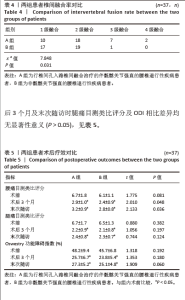
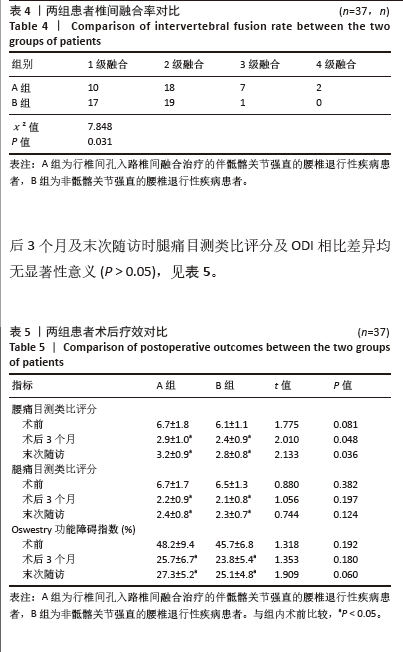
|