Chinese Journal of Tissue Engineering Research ›› 2025, Vol. 29 ›› Issue (22): 4710-4719.doi: 10.12307/2025.439
Previous Articles Next Articles
Injectable hydrogel microspheres that enhance autophagy can improve cartilage microenvironment and resist chondrocyte senescence
Li Wenming, Li Yonghang, Yan Caiping, Wang Xingkuan, Xiang Chao, Zhang Yuan, Jiang Ke, Chen Lu
- Affiliated Hospital of North Sichuan Medical College, Nanchong 637000, Sichuan Province, China
-
Received:2024-03-04Accepted:2024-05-11Online:2025-08-08Published:2024-12-06 -
Contact:Chen Lu, Master’s supervisor, Chief physician, Affiliated Hospital of North Sichuan Medical College, Nanchong 637000, Sichuan Province, China Jiang Ke, PhD, Associate chief physician, Master’s supervisor, Affiliated Hospital of North Sichuan Medical College, Nanchong 637000, Sichuan Province, China -
About author:Li Wenming, Master candidate, Affiliated Hospital of North Sichuan Medical College, Nanchong 637000, Sichuan Province, China Li Yonghang, Master candidate, Affiliated Hospital of North Sichuan Medical College, Nanchong 637000, Sichuan Province, China -
Supported by:Nanchong City 2023 Municipal Science and Technology Plan Special Fund Project, No. 23JCYJPT0036 (to WXK); 2023 University-Level Scientific Research Project of North Sichuan Medical College, No. CBY23-QNA01 (to WXK)
CLC Number:
Cite this article
Li Wenming, Li Yonghang, Yan Caiping, Wang Xingkuan, Xiang Chao, Zhang Yuan, Jiang Ke, Chen Lu. Injectable hydrogel microspheres that enhance autophagy can improve cartilage microenvironment and resist chondrocyte senescence[J]. Chinese Journal of Tissue Engineering Research, 2025, 29(22): 4710-4719.
share this article
Add to citation manager EndNote|Reference Manager|ProCite|BibTeX|RefWorks

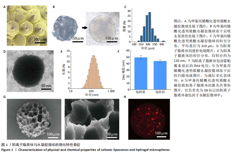
2.1 复合水凝胶微球的制备及其物化特性表征 采用基于pdms芯片的微流控装置制备水凝胶微球[16],水相为甲基丙烯酰化透明质酸单体,油相为95%石蜡油+ 5%Span80混合物,油相对水相施加剪切力,导致混合物分裂形成乳状液滴,在紫外光照射下液滴聚合成水凝胶微球(图1A),使用时将其放入去离子水中可见快速吸水膨胀为均匀球形[17](图1B)。通过调节水相与油相的比值(Qd∶Qc=1/9)可得到平均直径为310 μm的水凝胶微球[18](图1C)。采用旋转蒸发法制备阳离子脂质体(图1D),使用马尔文纳米粒度及Zeta电位分析仪测得其平均粒径约为120 nm(图1E),因(2,3-二油酰基-丙基)-三甲基铵-氯盐为阳离子磷脂,故测得脂质体Zeta电位为+49.5 mV,包封雷帕霉素后正电位略有减少,约+44 mV(图1F)。水凝胶为多孔网状内部结构,可保留大部分水分,冻干后低温放置可保存较长时间,扫描电镜下观察为疏松多孔状球体(图1G)。甲基丙烯酰化透明质酸微凝胶网络内可形成氢键,进一步锚定微凝胶中的脂质体[19],为证明脂质体在甲基丙烯酰化透明质酸微凝胶中的成功固定化,Dil标记脂质体,置于共聚焦激光显微镜下观察,可见Dil标记的脂质体被成功包封于水凝胶微球中(图1H)。"

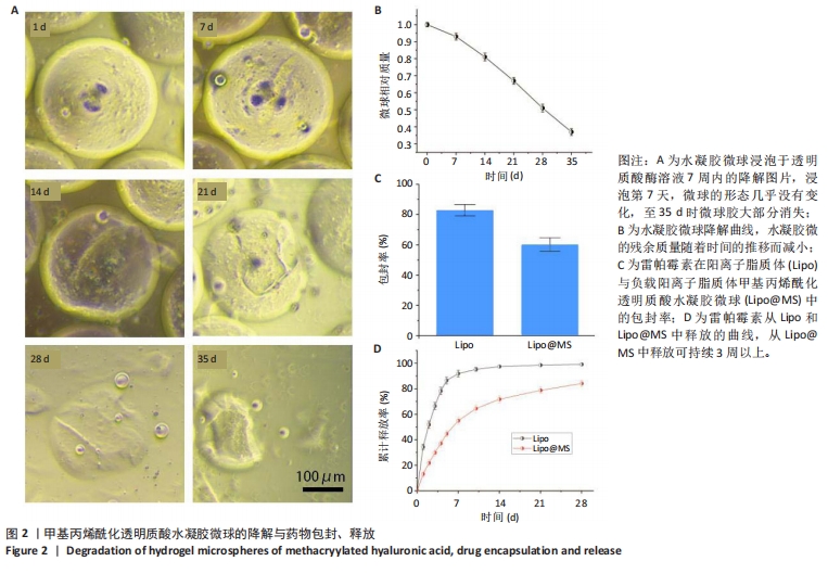
2.2 水凝胶微球降解与药物的包封与释放 为了模拟体内的逐渐降解过程,将甲基丙烯酰化透明质酸水凝胶微球在含有1 000 U/mL透明质酸酶的PBS中,在37 ℃下孵育5周[20]。浸泡第7天,微球的形态几乎没有变化;浸泡第14天,虽然形貌大致完整,但在微球边缘观察到小部分降解缺损;浸泡第21天,微球形态开始变形,难以维持饱满形态,开始塌陷,出现裂缝;浸泡第35天,微球胶大部分消失(图2A)。同时,水凝胶微的残余质量随着时间的推移而减小,这与形貌变化一致(图2B)。 RAPA@Lipo与RAPA@Lipo@MS中雷帕霉素的包封效率分别为(82.85±3.74)%,(60.21±4.54)%(图2C)。在药物释放实验中,RAPA@Lipo与RAPA@Lipo@MS中雷帕霉素的累计释放谱呈双相,初始为快速释放期(约为5 d),随后为缓释期,并且RAPA@Lipo@MS中雷帕霉素的持续释放时间> 3周(图2D)。"
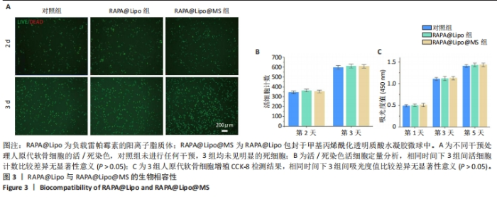
2.3 RAPA@Lipo与RAPA@Lipo@MS的生物相容性 活/死染色结果显示3组均未见明显的死细胞,3组间活细胞数量比较差异无显著性意义(P > 0.05),见图3A,B。CCK-8检测结果显示,相同时间下,3组间细胞增殖吸光度值比较差异无显著性意义(P > 0.05),见图3C。"

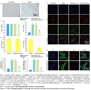
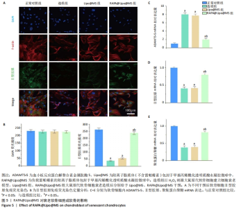
2.4 RAPA@Lipo@MS对衰老软骨细胞的影响 2.4.1 软骨细胞自噬与衰老的相关表征 细胞衰老的表征通过β-半乳糖苷酶检测[21]。如图4A所示,与正常对照组相比,造模组软骨细胞β-半乳糖苷酶染色强度增强。通过免疫荧光观察p62蛋白的表达,造模组、Lipo@MS组p62蛋白表达高于正常对照组(P < 0.05),RAPA@Lipo@MS组p62蛋白表达低于造模组、Lipo@MS组(P < 0.05),造模组与Lipo@MS组p62蛋白表达比较差异无显著性意义(P > 0.05),见图4B,C。同时通过免疫荧光观察p53蛋白的表达,RAPA@Lipo@MS组p53蛋白表达低于造模组(P < 0.05),见图4D,E。 RT-PCR检测结果显示,与正常对照组比较,造模组细胞内白细胞介素6、基质金属蛋白酶13 mRNA表达升高(P < 0.05);与造模组、Lipo@MS组比较,RAPA@Lipo@MS组细胞内白细胞介素6、基质金属蛋白酶13 mRNA表达降低(P < 0.05);造模组与Lipo@MS组细胞内白细胞介素6、基质金属蛋白酶13 mRNA表达比较差异无显著性意义(P > 0.05),见图4F,G。"

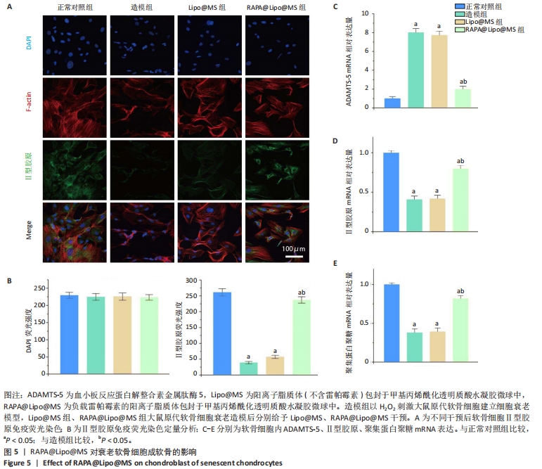
2.4.2 成软骨相关表征 免疫荧光染色结果显示,造模组细胞内Ⅱ型胶原蛋白表达低于正常对照组(P < 0.05),RAPA@Lipo@MS组细胞内Ⅱ型胶原蛋白表达高于造模组、Lipo@MS组(P < 0.05),造模组与Lipo@MS组细胞内Ⅱ型胶原蛋白表达比较差异无显著性意义(P > 0.05),见图5A,B。 RT-PCR检测结果显示,与正常对照组比较,造模组细胞内Ⅱ型胶原、聚集蛋白聚糖mRNA表达降低(P < 0.05),ADAMTS-5 mRNA表达升高(P < 0.05);与造模组、Lipo@MS组比较,RAPA@Lipo@MS组细胞内Ⅱ型胶原、聚集蛋白聚糖mRNA表达升高(P < 0.05),ADAMTS-5 mRNA表达降低(P < 0.05);造模组与Lipo@MS组细胞内Ⅱ型胶原、聚集蛋白聚糖、ADAMTS-5 mRNA表达比较差异均无显著性意义(P > 0.05),见图5C-E[22]。"
| [1] CORYELL PR, DIEKMAN BO, LOESER RF. Mechanisms and therapeutic implications of cellular senescence in osteoarthritis. Nat Rev Rheumatol. 2021;17(1):47-57. [2] ABRAMOFF B, CALDERA FE. Osteoarthritis: Pathology, Diagnosis, and Treatment Options. Med Clin North Am. 2020;104(2):293-311. [3] SWAHN H, LI K, DUFFY T, et al. Senescent cell population with ZEB1 transcription factor as its main regulator promotes osteoarthritis in cartilage and meniscus. Ann Rheum Dis. 2023;82(3):403-415. [4] ZHANG L, PITCHER LE, YOUSEFZADEH MJ, et al. Cellular senescence: a key therapeutic target in aging and diseases. J Clin Invest. 2022; 132(15):e158450. [5] CHILDS BG, GLUSCEVIC M, BAKER DJ, et al. Senescent cells: an emerging target for diseases of ageing. Nat Rev Drug Discov. 2017;16(10):718-735. [6] ROBBINS PD, JURK D, KHOSLA S, et al. Senolytic Drugs: Reducing Senescent Cell Viability to Extend Health Span. Annu Rev Pharmacol Toxicol. 2021;61:779-803. [7] TABIBZADEH S. Role of autophagy in aging: The good, the bad, and the ugly. Aging Cell. 2023;22(1):e13753. [8] SHIRAKABE A, IKEDA Y, SCIARRETTA S, et al. Aging and Autophagy in the Heart. Circ Res. 2016;118(10):1563-1576. [9] ZHANG Y, ZHANG J, WANG S. The Role of Rapamycin in Healthspan Extension via the Delay of Organ Aging. Ageing Res Rev. 2021;70:101376. [10] DOS SANTOS KC, DOS REIS LR, RODERO CF, et al. Bioproperties, Nanostructured System and Analytical and Bioanalytical Methods for Determination of Rapamycin: A Review. Crit Rev Anal Chem. 2023:1-9. doi: 10.1080/10408347.2020.1839737-test. [11] EBADA HM, NASRA MM, NASSRA RA, et al. Cationic nanocarrier of rhein based on hydrophobic ion pairing approach as intra-articular targeted regenerative therapy for osteoarthritis. Colloids Surf B Biointerfaces. 2022;211:112285. [12] AJEESHKUMAR KK, ANEESH PA, RAJU N, et al. Advancements in liposome technology: Preparation techniques and applications in food, functional foods, and bioactive delivery: A review. Compr Rev Food Sci Food Saf. 2021;20(2):1280-1306. [13] ZHU H, ZHENG J, OH XY, et al. Nanoarchitecture-Integrated Hydrogel Systems toward Therapeutic Applications. ACS Nano. 2023;17(9):7953-7978. [14] ZHOU X, LI WK, ZHUANG C, et al. Lei’s formula attenuates osteoarthritis mediated by suppression of chondrocyte senescence via the mTOR axis: in vitro and in vivo experiments. Aging (Albany NY). 2024;16(5): 4250-4269. [15] GAO H, NEPOVIMOVA E, HEGER Z, et al. Role of hypoxia in cellular senescence. Pharmacol Res. 2023;194:106841. [16] SPEARMAN BS, AGRAWAL NK, RUBIANO A, et al. Tunable methacrylated hyaluronic acid-based hydrogels as scaffolds for soft tissue engineering applications. J Biomed Mater Res A. 2020;108(2):279-291. [17] WU J, LI G, YE T, et al. Stem cell-laden injectable hydrogel microspheres for cancellous bone regeneration. Chem Eng J. 2020; 393:124715. [18] ZHAO Z, WANG Z, LI G, et al. Injectable microfluidic hydrogel microspheres for cell and drug delivery. Adv Funct Mater. 2021;31: 2103339. [19] CHANG H, CAI F, ZHANG Y, et al. Silencing Gene-Engineered Injectable Hydrogel Microsphere for Regulation of Extracellular Matrix Metabolism Balance. Small Methods. 2022;6(4): e2101201. [20] GERBER PA, BUHREN BA, BÖLKE E, et al. Time- and Dose-Dependent Effects of Hyaluronidase on the Degradation of Different Hyaluronan-Based Fillers In Vitro. Plast Reconstr Surg. 2023;151(3): 560-567. [21] CHILDS BG, GLUSCEVIC M, BAKER DJ, et al. Senescent cells: an emerging target for diseases of ageing. Nat Rev Drug Discov. 2017;16(10):718-735. [22] PICCA A, FAITG J, AUWERX J, et al. Mitophagy in human health, ageing and disease. Nat Metab. 2023;5(12):2047-2061. [23] JEON OH, KIM C, LABERGE RM, et al. Local clearance of senescent cells attenuates the development of post-traumatic osteoarthritis and creates a pro-regenerative environment. Nat Med. 2017;23(6):775-781. [24] CHAIB S, TCHKONIA T, KIRKLAND JL. Cellular senescence and senolytics: the path to the clinic. Nat Med. 2022;28(8):1556-1568. [25] LEVINE B, KROEMER G. Biological Functions of Autophagy Genes: A Disease Perspective. Cell. 2019;176(1-2):11-42. [26] CHEN X, GONG W, SHAO X, et al. METTL3-mediated m6A modification of ATG7 regulates autophagy-GATA4 axis to promote cellular senescence and osteoarthritis progression. Ann Rheum Dis. 2022;81(1): 87-99. [27] MU W, REZEK V, MARTIN H, et al. Autophagy inducer rapamycin treatment reduces IFN-I-mediated Inflammation and improves anti-HIV-1 T cell response in vivo. JCI Insight. 2022;7(22): e159136. [28] ARRIOLA APELO SI, LAMMING DW. Rapamycin: An InhibiTOR of Aging Emerges From the Soil of Easter Island. J Gerontol A Biol Sci Med Sci. 2016;71(7):841-849. [29] XIE Y, LEI X, ZHAO G, et al. mTOR in programmed cell death and its therapeutic implications. Cytokine Growth Factor Rev. 2023;71-72:66-81. [30] BURKE JA, ZHANG X, BOBBALA S, et al. Subcutaneous nanotherapy repurposes the immunosuppressive mechanism of rapamycin to enhance allogeneic islet graft viability. Nat Nanotechnol. 2022; 17(3):319-330. [31] ZHONG Y, ZHOU Y, DING R, et al. Intra-articular treatment of temporomandibular joint osteoarthritis by injecting actively-loaded meloxicam liposomes with dual-functions of anti-inflammation and lubrication. Mater Today Bio. 2023;19:100573. [32] EBADA HM, NASRA MM, NASSRA RA, et al. Cationic nanocarrier of rhein based on hydrophobic ion pairing approach as intra-articular targeted regenerative therapy for osteoarthritis. Colloids Surf B Biointerfaces. 2022;211:112285. [33] XU XL, XUE Y, DING JY, et al. Nanodevices for deep cartilage penetration. Acta Biomater. 2022;154:23-48. [34] LEI Y, WANG Y, SHEN J, et al. Injectable hydrogel microspheres with self-renewable hydration layers alleviate osteoarthritis. Sci Adv. 2022; 8(5):eabl6449. [35] JÖRGENSEN AM, WIBEL R, BERNKOP-SCHNÜRCH A. Biodegradable Cationic and Ionizable Cationic Lipids: A Roadmap for Safer Pharmaceutical Excipients. Small. 2023;19(17):e2206968. [36] QIAN Y, LIANG X, YANG J, et al. Hyaluronan Reduces Cationic Liposome-Induced Toxicity and Enhances the Antitumor Effect of Targeted Gene Delivery in Mice. ACS Appl Mater Interfaces. 2018;10(38):32006-32016. [37] VARGAS JNS, HAMASAKI M, KAWABATA T, et al. The mechanisms and roles of selective autophagy in mammals. Nat Rev Mol Cell Biol. 2023;24(3):167-185. [38] LAMARK T, SVENNING S, JOHANSEN T. Regulation of selective autophagy: the p62/SQSTM1 paradigm. Essays Biochem. 2017;61(6): 609-624. [39] OU HL, SCHUMACHER B. DNA damage responses and p53 in the aging process. Blood. 2018;131(5):488-495. [40] BIRCH J, GIL J. Senescence and the SASP: many therapeutic avenues. Genes Dev. 2020;34(23-24):1565-1576. [41] WAKALE S, WU X, SONAR Y, et al. How are Aging and Osteoarthritis Related? Aging Dis. 2023;14(3):592-604. [42] TIKU ML, MADHAN B. Preserving the longevity of long-lived type II collagen and its implication for cartilage therapeutics. Ageing Res Rev. 2016:62-71. [43] GONG Y, LI S, WU J, et al. Autophagy in the pathogenesis and therapeutic potential of post-traumatic osteoarthritis. Burns Trauma. 2023;11:tkac060. |
| [1] | Ma Chi, Wang Ning, Chen Yong, Wei Zhihan, Liu Fengji, Piao Chengzhe. Application of 3D-printing patient-specific instruments combined with customized locking plate in opening wedge high tibial osteotomy [J]. Chinese Journal of Tissue Engineering Research, 2025, 29(9): 1863-1869. |
| [2] | Yu Shuai, Liu Jiawei, Zhu Bin, Pan Tan, Li Xinglong, Sun Guangfeng, Yu Haiyang, Ding Ya, Wang Hongliang. Hot issues and application prospects of small molecule drugs in treatment of osteoarthritis [J]. Chinese Journal of Tissue Engineering Research, 2025, 29(9): 1913-1922. |
| [3] | Zhao Jiyu, Wang Shaowei. Forkhead box transcription factor O1 signaling pathway in bone metabolism [J]. Chinese Journal of Tissue Engineering Research, 2025, 29(9): 1923-1930. |
| [4] | Sun Yundi, Cheng Lulu, Wan Haili, Chang Ying, Xiong Wenjuan, Xia Yuan. Effect of neuromuscular exercise for knee osteoarthritis pain and function: a meta-analysis [J]. Chinese Journal of Tissue Engineering Research, 2025, 29(9): 1945-1952. |
| [5] | Deng Keqi, Li Guangdi, Goswami Ashutosh, Liu Xingyu, He Xiaoyong. Screening and validation of Hub genes for iron overload in osteoarthritis based on bioinformatics [J]. Chinese Journal of Tissue Engineering Research, 2025, 29(9): 1972-1980. |
| [6] | Yin Lu, Jiang Chuanfeng, Chen Junjie, Yi Ming, Wang Zihe, Shi Houyin, Wang Guoyou, Shen Huarui. Effect of Complanatoside A on the apoptosis of articular chondrocytes [J]. Chinese Journal of Tissue Engineering Research, 2025, 29(8): 1541-1547. |
| [7] | Wang Peiguang, Zhang Xiaowen, Mai Meisi, Li Luqian, Huang Hao. Generalized equation estimation of the therapeutic effect of floating needle therapy combined with acupoint embedding on different stages of human knee osteoarthritis [J]. Chinese Journal of Tissue Engineering Research, 2025, 29(8): 1565-1571. |
| [8] | Li Huayuan, Li Chun, Liu Junwei, Wang Ting, Li Long, Wu Yongli. Effect of warm acupuncture on PINK1/Parkin pathway in the skeletal muscle of rats with chronic fatigue syndrome [J]. Chinese Journal of Tissue Engineering Research, 2025, 29(8): 1618-1625. |
| [9] | Zhou Panpan, Cui Yinglin, Zhang Wentao, Wang Shurui, Chen Jiahui, Yang Tong . Role of cellular autophagy in cerebral ischemic injury and the regulatory mechanism of traditional Chinese medicine [J]. Chinese Journal of Tissue Engineering Research, 2025, 29(8): 1650-1658. |
| [10] | Wang Qiuyue, Jin Pan, Pu Rui . Exercise intervention and the role of pyroptosis in osteoarthritis [J]. Chinese Journal of Tissue Engineering Research, 2025, 29(8): 1667-1675. |
| [11] | Zhu Hanmin, Wang Song, Xiao Wenlin, Zhang Wenjing, Zhou Xi, He Ye, Li Wei, . Mitophagy regulates bone metabolism [J]. Chinese Journal of Tissue Engineering Research, 2025, 29(8): 1676-1683. |
| [12] | Chen Yueping, Chen Feng, Peng Qinglin, Chen Huiyi, Dong Panfeng . Based on UHPLC-QE-MS, network pharmacology, and molecular dynamics simulation to explore the mechanism of Panax notoginseng in treating osteoarthritis [J]. Chinese Journal of Tissue Engineering Research, 2025, 29(8): 1751-1760. |
| [13] | Yang Zhihang, Sun Zuyan, Huang Wenliang, Wan Yu, Chen Shida, Deng Jiang. Nerve growth factor promotes chondrogenic differentiation and inhibits hypertrophic differentiation of rabbit bone marrow mesenchymal stem cells [J]. Chinese Journal of Tissue Engineering Research, 2025, 29(7): 1336-1342. |
| [14] | Zheng Rongfa, Mo Weibin, Huang Peng, Chen Junji, Liang Ting, Zi Fangyu, Li Guofeng. Effects of electroacupuncture on the expression of metabolic enzymes and autophagy genes in gastrocnemius muscle tissues of exercising rats [J]. Chinese Journal of Tissue Engineering Research, 2025, 29(6): 1127-1136. |
| [15] | Chen Yuning, Jiang Ying, Liao Xiangyu, Chen Qiongjun, Xiong Liang, Liu Yue, Liu Tong. Buqi Huoxue Compounds intervene with the expression of related factors and autophagy related proteins in a rat model of cerebral ischemia/reperfusion [J]. Chinese Journal of Tissue Engineering Research, 2025, 29(6): 1152-1158. |
| Viewed | ||||||
|
Full text |
|
|||||
|
Abstract |
|
|||||