Chinese Journal of Tissue Engineering Research ›› 2025, Vol. 29 ›› Issue (23): 4924-4930.doi: 10.12307/2025.100
Previous Articles Next Articles
Ectomesenchymal stem cells-derived extracellular vesicles promote neuronal axonal elongation
Sun Haitao1, Ren Chunpeng1, Yang Yongtao1, Huang Yonghui2, Qin Rujie1, Li Zhen1
- 1First People’s Hospital of Lianyungang, Lianyungang 222000, Jiangsu Province, China; 2Affiliated Hospital of Jiangsu University, Zhenjiang 212000, Jiangsu Province, China
-
Received:2024-03-25Accepted:2024-06-18Online:2025-08-18Published:2024-09-28 -
Contact:Qin Rujie, Master, Chief physician, First People’s Hospital of Lianyungang, Lianyungang 222000, Jiangsu Province, China; Co-corresponding author: Li Zhen, Master, Physician, First People’s Hospital of Lianyungang, Lianyungang 222000, Jiangsu Province, China -
About author:Sun Haitao, Master, Physician, First People’s Hospital of Lianyungang, Lianyungang 222000, Jiangsu Province, China -
Supported by:Youth Talent Project of First People’s Hospital of Lianyungang, No. QN2205 (to SHT); Scientific Research Project of Zhenjiang City of Jiangsu Province, No. SH2020053 (to HYH)
CLC Number:
Cite this article
Sun Haitao, Ren Chunpeng, Yang Yongtao, Huang Yonghui, Qin Rujie, Li Zhen. Ectomesenchymal stem cells-derived extracellular vesicles promote neuronal axonal elongation[J]. Chinese Journal of Tissue Engineering Research, 2025, 29(23): 4924-4930.
share this article
Add to citation manager EndNote|Reference Manager|ProCite|BibTeX|RefWorks

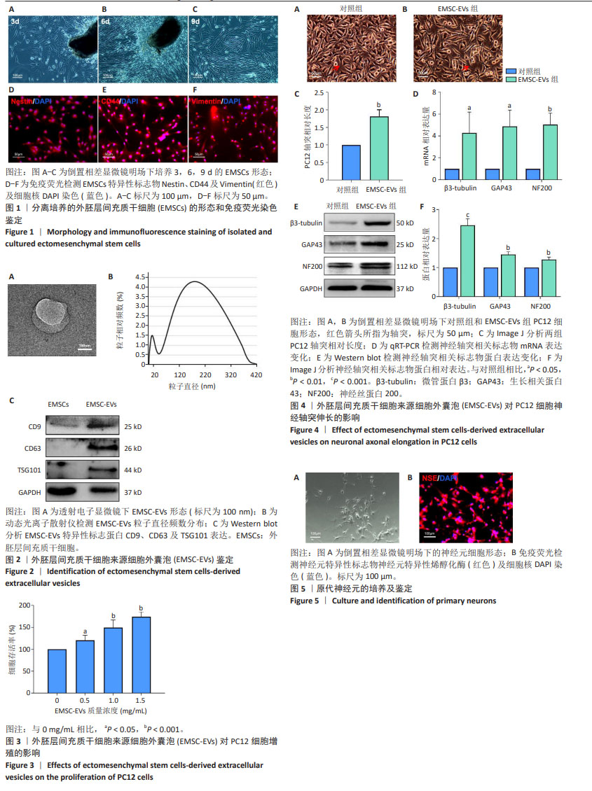
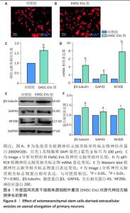
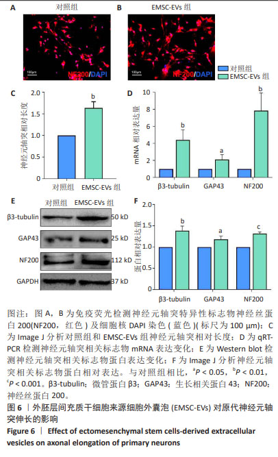
2.1 EMSCs培养及鉴定结果 鼻黏膜组织块静置培养皿中约3 d后,可于镜下观察到少量梭形及不规则形细胞聚集于组织块周围,培养皿中有较多黑色杂质,见图1A;予保留组织块半量换液培养6 d后,可见组织块周围细胞数量增加,呈长梭形,培养皿背景干净,见图1B;予去除组织块培养至9 d后,可见细胞铺满皿底,绝大部分呈长梭形,少数呈不规则形,生长较为迅速,见图1C。取第3代EMSCs进行免疫荧光染色,镜下可见其高表达Nestin、CD44及Vimentin等间充质干细胞标志物,见图1D-F。以上提示成功分离获得EMSCs,可用于后续实验。 2.2 EMSC-EVs形态学表现及鉴定结果 透射电子显微镜观察显示,EMSC-EVs呈凹陷圆球样结构,直径约200 nm,见图2A;动态光粒子散射结果显示,EMSC-EVs粒子直径大小分布为30-400 nm,见图2B;蛋白免疫印迹结果显示,EMSC-EVs相比于EMSCs,高表达细胞外囊泡标记蛋白CD9、CD63以及TSG101,见图2C;以上提示,所提取EMSC-EVs符合细胞外囊泡的生物学标准,可用于后续实验。 2.3 EMSC-EVs促进PC12细胞增殖 CCK-8检测结果显示,EMSC-EVs对PC12细胞无明显的细胞毒性作用,却能够促进PC12细胞增殖,且在已测试蛋白质量浓度0.5-1.5 mg/mL范围内,EMSC-EVs质量浓度越高PC12细胞增殖能力越强,见图3。据此,综合考虑实验效果及减少EMSC-EVs用量,该研究所用EMSC-EVs质量浓度为1 mg/mL。 2.4 EMSC-EVs促进PC12细胞轴突伸长 EMSC-EVs与PC12细胞共孵育72 h后,镜下观察PC12细胞形态,可见EMSC-EVs组PC12细胞轴突(红色箭头所指)普遍长于对照组,见图4A,B。Image J软件测量轴突相对长度,结果显示EMSC-EVs组轴突相对长度为对照组1.5-2.0倍,见图4C。另外,qRT-PCR结果显示,神经轴突相关标志物微管蛋白β3(早期)、生长相关蛋白43(中期)和神经丝蛋白200(成熟)mRNA表达在EMSC-EVs干预后均明显高于对照组,见图4D。Western blot结果也显示,EMSC-EVs干预后微管蛋白β3、生长相关蛋白43和神经丝蛋白200蛋白表达明显高于对照组,见图4E,F。以上结果提示,EMSC-EVs能够促进PC12细胞轴突伸长。 2.5 原代神经元的分离培养及鉴定结果 酶消化法培养6 d的原代神经元镜下观察呈不规则芒星状,见图5A。荧光显微镜观察显示,原代神经元表达神经元特异性标志物——神经元特异性烯醇化酶,见图5B。 2.6 EMSC-EVs促进神经元轴突伸长 图6A,B荧光标记神经丝蛋白200显示,EMSC-EVs与纯化的原代神经元共孵育72 h后,神经元细胞轴突相对长度长于对照组,见图6C。qRT-PCR结果显示,EMSC-EVs促进神经元轴突微管蛋白β3、生长相关蛋白43和神经丝蛋白200的mRNA表达,见图6D。Western blot结果显示,EMSC-EVs促进了神经元微管蛋白β3、生长相关蛋白43和神经丝蛋白200蛋白表达,见图6E,F。以上结果提示,EMSC-EVs能够促进神经元轴突伸长。"
"
| [1] LEMAITRE P, TAREEN SH, PASCIUTO E, et al. Molecular and cognitive signatures of ageing partially restored through synthetic delivery of IL2 to the brain. EMBO Mol Med. 2023;15(5):e16805. [2] SHREE S, SUTRADHAR S, TROTTIER O, et al. Dynamic instability of dendrite tips generates the highly branched morphologies of sensory neurons. Sci Adv. 2022;8(26):eabn0080. [3] HAN Q, XIE Y, ORDAZ JD, et al. Restoring Cellular Energetics Promotes Axonal Regeneration and Functional Recovery after Spinal Cord Injury. Cell Metab. 2020;31(3):623-641.e8. [4] HUTSON TH, KATHE C, PALMISANO I, et al. Cbp-dependent histone acetylation mediates axon regeneration induced by environmental enrichment in rodent spinal cord injury models. Sci Transl Med. 2019; 11(487):eaaw2064. [5] ROCHFORD AE, CARNICER-LOMBARTE A, CURTO VF, et al. When Bio Meets Technology: Biohybrid Neural Interfaces. Adv Mater. 2020; 32(15):e1903182. [6] WEN Q, WENG H, LIU T, et al. Inactivating Celsr2 promotes motor axon fasciculation and regeneration in mouse and human. Brain. 2022;145(2):670-683. [7] GARCÍA-CASTRO J, TRIGUEROS C, MADRENAS J, et al. Mesenchymal stem cells and their use as cell replacement therapy and disease modelling tool. J Cell Mol Med. 2008;12(6B):2552-2565. [8] ZHOU H, HE Y, XIONG W, et al. MSC based gene delivery methods and strategies improve the therapeutic efficacy of neurological diseases. Bioact Mater. 2022;23:409-437. [9] ANDRZEJEWSKA A, DABROWSKA S, LUKOMSKA B, et al. Mesenchymal Stem Cells for Neurological Disorders. Adv Sci (Weinh). 2021;8(7): 2002944. [10] IZQUIERDO-ALTAREJOS P, MORENO-MANZANO V, FELIPO V. Pathological and therapeutic effects of extracellular vesicles in neurological and neurodegenerative diseases. Neural Regen Res. 2024;19(1):55-61 [11] NAKAZAKI M, MORITA T, LANKFORD KL, et al. Small extracellular vesicles released by infused mesenchymal stromal cells target M2 macrophages and promote TGF-β upregulation, microvascular stabilization and functional recovery in a rodent model of severe spinal cord injury. J Extracell Vesicles. 2021;10(11):e12137. [12] HARRELL CR, JOVICIC N, DJONOV V, et al. Mesenchymal Stem Cell-Derived Exosomes and Other Extracellular Vesicles as New Remedies in the Therapy of Inflammatory Diseases. Cells. 2019; 8(12):1605. [13] 贯世豪,黄永辉,龚爱华,等.外胚层间充质干细胞来源细胞外囊泡诱导大鼠星形胶质细胞向神经元的转分化[J].中国组织工程研究,2022,26(30):4840-4846. [14] HU X, LIU Z, ZHOU X, et al. Small extracellular vesicles derived from mesenchymal stem cell facilitate functional recovery in spinal cord injury by activating neural stem cells via the ERK1/2 pathway. Front Cell Neurosci. 2022;16:954597. [15] DENG W, SHAO F, HE Q, et al. EMSCs Build an All-in-One Niche via Cell-Cell Lipid Raft Assembly for Promoted Neuronal but Suppressed Astroglial Differentiation of Neural Stem Cells. Adv Mater. 2019;31(10):e1806861. [16] SASAKI K, DAVIES J, DOLDÁN NG, et al. 3,4,5-Tricaffeoylquinic acid induces adult neurogenesis and improves deficit of learning and memory in aging model senescence-accelerated prone 8 mice. Aging (Albany NY). 2019;11(2):401-422. [17] CAO T, CHEN H, HUANG W, et al. hUC-MSC-mediated recovery of subacute spinal cord injury through enhancing the pivotal subunits β3 and γ2 of the GABAA receptor. Theranostics. 2022;12(7):3057-3078. [18] LIU C, GAO W, ZHAO L, et al. Progesterone attenuates neurological deficits and exerts a protective effect on damaged axons via the PI3K/AKT/mTOR-dependent pathway in a mouse model of intracerebral hemorrhage. Aging (Albany NY). 2022;14(6):2574-2589. [19] QIAN Y, HAN Q, ZHAO X, et al. Asymmetrical 3D Nanoceria Channel for Severe Neurological Defect Regeneration. iScience. 2019;12:216-231. [20] FAN L, LIU C, CHEN X, et al. Exosomes-Loaded Electroconductive Hydrogel Synergistically Promotes Tissue Repair after Spinal Cord Injury via Immunoregulation and Enhancement of Myelinated Axon Growth. Adv Sci (Weinh). 2022;9(13):e2105586. [21] ZHOU W, KE S, LI W, et al. Mapping the Function of Whole-Brain Projection at the Single Neuron Level. Adv Sci (Weinh). 2022;9(33): e2202553. [22] GAUDET AD, POPOVICH PG, RAMER MS. Wallerian degeneration: gaining perspective on inflammatory events after peripheral nerve injury. J Neuroinflammation. 2011;8:110. [23] VAHSEN BF, RIBAS VT, SUNDERMEYER J, et al. Inhibition of the autophagic protein ULK1 attenuates axonal degeneration in vitro and in vivo, enhances translation, and modulates splicing. Cell Death Differ. 2020;27(10):2810-2827. [24] LI F, SAMI A, NORISTANI HN, et al. Glial Metabolic Rewiring Promotes Axon Regeneration and Functional Recovery in the Central Nervous System. Cell Metab. 2020;32(5):767-785.e7. [25] 李森.骨髓间充质干细胞外泌体对PC12细胞氧糖剥夺/复氧后轴突再生的作用及机制研究[D].太原:山西医科大学,2021. [26] LI Z, ZHAO T, DING J, et al. A reactive oxygen species-responsive hydrogel encapsulated with bone marrow derived stem cells promotes repair and regeneration of spinal cord injury. Bioact Mater. 2022;19:550-568. [27] HUANG Y, YANG L. Mesenchymal stem cells and extracellular vesicles in therapy against kidney diseases. Stem Cell Res Ther. 2021;12(1):219. [28] LI JK, YANG C, SU Y, et al. Mesenchymal Stem Cell-Derived Extracellular Vesicles: A Potential Therapeutic Strategy for Acute Kidney Injury. Front Immunol. 2021;12:684496. [29] SACCU G, MENCHISE V, GAI C, et al. Bone Marrow Mesenchymal Stromal/Stem Cell-Derived Extracellular Vesicles Promote Corneal Wound Repair by Regulating Inflammation and Angiogenesis. Cells. 2022;11(23):3892. [30] SHI Y, SHI H, NOMI A, et al. Mesenchymal stem cell-derived extracellular vesicles: a new impetus of promoting angiogenesis in tissue regeneration. Cytotherapy. 2019;21(5):497-508. [31] VONK LA, VAN DOOREMALEN SFJ, LIV N, et al. Mesenchymal Stromal/stem Cell-derived Extracellular Vesicles Promote Human Cartilage Regeneration In Vitro. Theranostics. 2018;8(4):906-920. [32] LI S, LIU J, LIU S, et al. Chitosan oligosaccharides packaged into rat adipose mesenchymal stem cells-derived extracellular vesicles facilitating cartilage injury repair and alleviating osteoarthritis. J Nanobiotechnology. 2021;19(1):343. [33] SUN H, CAO X, GONG A, et al. Extracellular vesicles derived from astrocytes facilitated neurite elongation by activating the Hippo pathway. Exp Cell Res. 2022;411(1):112937. |
| [1] | Hu Taotao, Liu Bing, Chen Cheng, Yin Zongyin, Kan Daohong, Ni Jie, Ye Lingxiao, Zheng Xiangbing, Yan Min, Zou Yong. Human amniotic mesenchymal stem cells overexpressing neuregulin-1 promote skin wound healing in mice [J]. Chinese Journal of Tissue Engineering Research, 2025, 29(7): 1343-1349. |
| [2] | Jin Kai, Tang Ting, Li Meile, Xie Yuan. Effects of conditioned medium and exosomes of human umbilical cord mesenchymal stem cells on proliferation, migration, invasion, and apoptosis of hepatocellular carcinoma cells [J]. Chinese Journal of Tissue Engineering Research, 2025, 29(7): 1350-1355. |
| [3] | Li Dijun, Jiu Jingwei, Liu Haifeng, Yan Lei, Li Songyan, Wang Bin. Three-dimensional gelatin microspheres loaded human umbilical cord mesenchymal stem cells for chronic tendinopathy repair [J]. Chinese Journal of Tissue Engineering Research, 2025, 29(7): 1356-1362. |
| [4] | Liu Qi, Li Linzhen, Li Yusheng, Jiao Hongzhuo, Yang Cheng, Zhang Juntao. Icariin-containing serum promotes chondrocyte proliferation and chondrogenic differentiation of stem cells in the co-culture system of three kinds of cells [J]. Chinese Journal of Tissue Engineering Research, 2025, 29(7): 1371-1379. |
| [5] | He Longcai, Song Wenxue, Ming Jiang, Chen Guangtang, Wang Junhao, Liao Yidong, Cui Junshuan, Xu Kaya. An experimental method for simultaneous extraction and culture of primary cortical neurons and microglial cells from SD rats [J]. Chinese Journal of Tissue Engineering Research, 2025, 29(7): 1395-1400. |
| [6] | Zhao Nannan, Li Yanjie, Qin Hewei, Zhu Bochao, Ding Huimin, Xu Zhenhua. Changes in ferroptosis in hippocampal neurons of vascular dementia model rats treated with Tongmai Kaiqiao Pill [J]. Chinese Journal of Tissue Engineering Research, 2025, 29(7): 1401-1407. |
| [7] | Zhang Zhenyu, Liang Qiujian, Yang Jun, Wei Xiangyu, Jiang Jie, Huang Linke, Tan Zhen. Target of neohesperidin in treatment of osteoporosis and its effect on osteogenic differentiation of bone marrow mesenchymal stem cells [J]. Chinese Journal of Tissue Engineering Research, 2025, 29(7): 1437-1447. |
| [8] | Li Jialin, Zhang Yaodong, Lou Yanru, Yu Yang, Yang Rui. Molecular mechanisms underlying role of mesenchymal stem cell secretome [J]. Chinese Journal of Tissue Engineering Research, 2025, 29(7): 1512-1522. |
| [9] | Cao Yue, Ye Xinjian, Li Biyao, Zhang Yining, Feng Jianying. Effect of extracellular vesicles for diagnosis and therapy of oral squamous cell carcinoma [J]. Chinese Journal of Tissue Engineering Research, 2025, 29(7): 1523-1530. |
| [10] | Yang Zhihang, Sun Zuyan, Huang Wenliang, Wan Yu, Chen Shida, Deng Jiang. Nerve growth factor promotes chondrogenic differentiation and inhibits hypertrophic differentiation of rabbit bone marrow mesenchymal stem cells [J]. Chinese Journal of Tissue Engineering Research, 2025, 29(7): 1336-1342. |
| [11] | Zhang Shuai, Li Zichun, Xu Yihao, Xie Xiaofeng, Guo Zhongsheng, Zhao Qingyang. Effect of transcranial magneto-acousto-electrical stimulation on the plasticity of the prefrontal cortex network in mice [J]. Chinese Journal of Tissue Engineering Research, 2025, 29(6): 1108-1117. |
| [12] | Liu Zhezhe, Yu Meiqing, Wang Tingting, Zhang Min, Li Baiyan. Troxerutin modulates nuclear factor-kappaB signaling pathway to inhibit brain injury and neuronal apoptosis in cerebral infarction rats [J]. Chinese Journal of Tissue Engineering Research, 2025, 29(6): 1137-1143. |
| [13] | He Bo, Chen Wen, Ma Suilu, He Zhijun, Song Yuan, Li Jinpeng, Liu Tao, Wei Xiaotao, Wang Weiwei, Xie Jing . Pathogenesis and treatment progress of flap ischemia-reperfusion injury [J]. Chinese Journal of Tissue Engineering Research, 2025, 29(6): 1230-1238. |
| [14] | Lu Ranran, Zhou Xu, Zhang Lijie, Yang Xinling. Dimethyl fumarate alleviates nerve damage in a mouse model of Parkinson’s disease [J]. Chinese Journal of Tissue Engineering Research, 2025, 29(5): 989-994. |
| [15] | Sun Xianjuan, Wang Qiuhua, Zhang Jinyi, Yang Yangyang, Wang Wenshuang, Zhang Xiaoqing. Adhesion, proliferation, and vascular smooth muscle differentiation of bone marrow mesenchymal stem cells on different electrospinning membranes [J]. Chinese Journal of Tissue Engineering Research, 2025, 29(4): 661-669. |
| Viewed | ||||||
|
Full text |
|
|||||
|
Abstract |
|
|||||