Chinese Journal of Tissue Engineering Research ›› 2024, Vol. 28 ›› Issue (33): 5388-5395.doi: 10.12307/2024.667
Previous Articles Next Articles
Effect of MRI preoperative quantitative assessment of the range of talus osteochondral injury on surgical selection and medium- to long-term follow-up results
Liu Hongda, Yan Rongliang, Gao Yan, Chen Jianghua, Qu Pingyan, Wang Lei, Peng Yi, Cao Lihai, Du Xiaojian, Qu Jiafu
- Second Department of Foot and Ankle Surgery, The Second Hospital of Tangshan, Tangshan 063000, Hebei Province, China
-
Received:2023-08-18Accepted:2023-09-22Online:2024-11-28Published:2024-01-31 -
About author:Liu Hongda, Master, Associate chief physician, Second Department of Foot and Ankle Surgery, The Second Hospital of Tangshan, Tangshan 063000, Hebei Province, China -
Supported by:Hebei Provincial Health and Family Planning Commission Project, No. 20181284 (to LHD)
CLC Number:
Cite this article
Liu Hongda, Yan Rongliang, Gao Yan, Chen Jianghua, Qu Pingyan, Wang Lei, Peng Yi, Cao Lihai, Du Xiaojian, Qu Jiafu. Effect of MRI preoperative quantitative assessment of the range of talus osteochondral injury on surgical selection and medium- to long-term follow-up results[J]. Chinese Journal of Tissue Engineering Research, 2024, 28(33): 5388-5395.
share this article
Add to citation manager EndNote|Reference Manager|ProCite|BibTeX|RefWorks
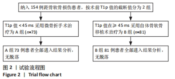
2.1 参与者数量分析 共纳入 154例距骨骨软骨损伤患者,所有患者资料完整,均获得12个月以上随访,全部进入结果分析,无脱落。 2.2 试验流程图 见图2。"
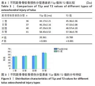
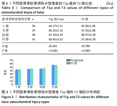
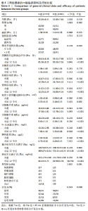
2.3 不同距骨骨软骨损伤型的T1ρ值和T2值比较 154例患者的距骨骨软骨损伤分型如下:Ⅰ型36例,Ⅱ型37例,Ⅲ型40例,Ⅳ型41例,4组分型的T1ρ值及T2值差异均有显著性意义(P < 0.05),两两比较差异亦有显著性意义(P均< 0.05),见表3及图3。"
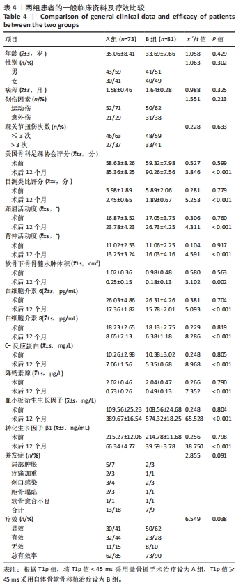
2.4 两组一般临床资料、疗效比较 154例患者治疗后,局部肿胀7例(4.6%),疼痛加重3例(2.0%),创口感染5例(3.3%),软骨愈合不良2例(1.3%)。根据表3的T1ρ值,将T1ρ值< 45 ms 采用微骨折手术治疗设为A组(n=73),T1ρ值≥45 ms 采用自体骨软骨移植治疗设为B组(n=81),比较两组患者的临床特征及疗效结果显示,两组治疗后美国骨科足踝协会评分、目测类比评分、跖屈活动度、背伸活动度、软骨下骨骨髓水肿体积、IL-6、IL-8、CRP、降钙素原、血小板衍生生长因子、TGF-β1、疗效方面比较差异均有显著性意义(P < 0.05),其中B组的疗效总有效率为90%,比A组总有效率(85%)高;两组其余的指标相比差异无显著性意义(P > 0.05)。见表4。"
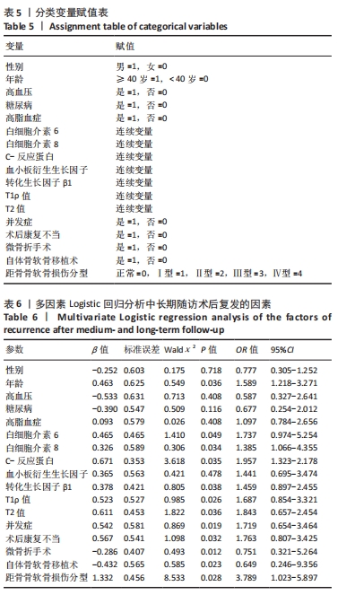
2.5 多因素Logistic回归分析中长期随访术后复发的危险因素分析 以术后是否复发作为因变量(否=0,是=1),自变量赋值见表5,结果显示,年龄(OR=1.589,95%CI:0.305-1.252,P=0.036)、IL-6(OR=1.737,95%CI:0.974-5.254,P=0.049)、IL-8(OR=1.385,95%CI:1.066-4.355,P=0.034)、CRP(OR=1.957,95%CI:1.323-2.178,P=0.035)、TGF-β1(OR=1.459,95%CI:0.897-2.455,P=0.038)、T1ρ值(OR=1.687,95%CI:0.854-3.321,P=0.026)、T2值(OR=1.843,95%CI:0.657-2.454,P=0.036)、并发症(OR=1.719,95%CI:0.654-3.464,P=0.019)、距骨骨软骨损伤分型(OR=3.789,95%CI:1.023-5.897,P=0.028)均是影响患者远期术后复发的独立危险因素;而微骨折手术(OR=0.751,95%CI:0.321-1.264,P=0.012)、自体骨软骨移植(OR=0.649,95%CI:0.246-1.356,P=0.023)是中长期随访术后复发的独立保护因素,见表6。"
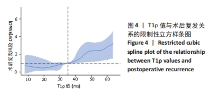
2.6 限制性立方样条图分析术前T1ρ值与术后复发的关系 将多因素分析调整后的T1ρ值、年龄、性别、病程、创伤因素、踝关节扭伤次数等纳入限制性立方样条图,分析T1ρ值与术后复发的关系,结果如图4所示,T1ρ值与术后复发风险间呈明显的非线性效应关系(P=0.028),取OR=1时,T1ρ值为35 ms作为截值点,当T1ρ值≤35 ms时,术后复发的风险迅速降低;当T1ρ值> 35 ms时,术后复发风险迅速升高。"

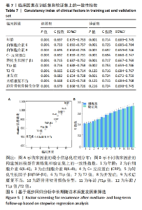
2.7 临床因素在训练集和验证集上的一致性指数 随机抽取124例患者数据作为深度学习训练集,其余30例作为验证集。经多因素Cox风险比例模型计算,显示年龄、IL-6、IL-8、CRP、TGF-β1、T1ρ值、T2值、并发症、术后康复不当预测模型在训练集和校验集上均具有最大的一致性指数(C指数),分别为0.733和0.753,表明年龄、IL-6、IL-8、CRP、TGF-β1、T1ρ值、T2值、并发症及术后康复不当组成的多因素模型对中长期随访术后复发具有较准确的表现,见表7及图5。"
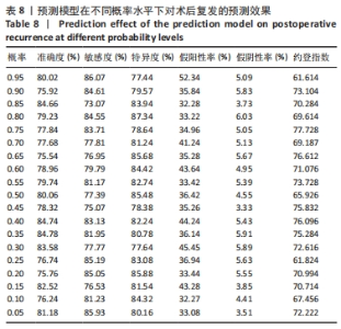
2.8 预测模型构建和验证 利用多因素Logistic回归模型对术后复发的影响因素进行预测,把上述结果放入到回归方程y=1-1/(1+e-z)中,得到预测中长期随访术后复发的公式:P=1-1/(1+e1.106 + 0.463×年龄 + 0.465×IL-6+0.326×IL-8+ 0.671×CRP + 0.378×TGF-β1+0.523×T1ρ+0.611×T2值+0.542×并发症+0.567×术后康复不当+距骨骨软骨损伤分型×1.332),利用回归方程计算术后复发发生的可能性。结果表明,P=0.75时,约登指数数值最大,为77.728,提示模型预测效果较好;当P > 0.75时,表示患者术后复发。模型预测准确度为84.66%,敏感度和特异度分别为73.07%和83.94%。见表8。"

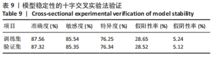
通过十字交叉验证法对上述模型的稳定性进行验证,结果表明上述模型的训练集、验证集参数非常接近,提示模型具有良好的预测性能。利用Nagelkerke R2对模型的拟合程度进行评价,结果显示,Nagelkerke R2=0.456时,模型的拟合程度最优。见表9。"
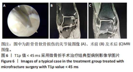
2.9 典型病例 A组:女性患者,36岁,T1ρ值36 ms,美国骨科足踝协会评分57分,目测类比评分6分,跖屈活动度18°,背伸活动度10°,软骨下骨骨髓水肿体积1 cm3,IL-6为27.45 pg/mL,IL-8为16.35 pg/mL,CRP为9.36 mg/L,降钙素原为1.96 μg/L,血小板衍生生长因子为106.32 ng/L,TGF-β1为210.35 ng/mL,疗效为显效。影像学图片见图6。"


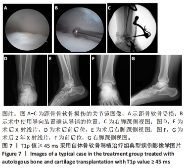
B组:男性患者,38岁,T1ρ值53 ms,美国骨科足踝协会评分62分,目测类比评分8分,跖屈活动度14°,背伸活动度8°,软骨下骨骨髓水肿体积1.5 cm3,IL-6为28.65 pg/mL,IL-8为19.26 pg/mL,CRP为11.45 mg/L,降钙素原为2.13 μg/L,血小板衍生生长因子为115.32 ng/L,TGF-β1为216.89 ng/mL,疗效为有效。影像学图片见图7。"
| [1] BRUNS J, HABERMANN C, WERNER M. Osteochondral Lesions of the Talus: A Review on Talus Osteochondral Injuries, Including Osteochondritis Dissecans. Cartilage. 2021;13(1):1380-1401. [2] BURGER D, FEUCHT M, MUENCH LN, et al. Good clinical outcomes after patellar cartilage repair with no evidence for inferior results in complex cases with the need for additional patellofemoral realignment procedures: a systematic review. Knee Surg Sports Traumatol Arthrosc. 2022;30(5):1752-1768. [3] SHAFFER JJ JR, MANI M, SCHMITZ SL, et al. Proton Exchange Magnetic Resonance Imaging: Current and Future Applications in Psychiatric Research. Front Psychiatry. 2020;11:532606. [4] SHAFSHAK TS, ELNEMR R. The Visual Analogue Scale Versus Numerical Rating Scale in Measuring Pain Severity and Predicting Disability in Low Back Pain. J Clin Rheumatol. 2021;27(7):282-285. [5] BALAJI G, BHUKYA S, NEMA S, et al. Predictors of Functional Outcome in Unstable Ankle Fractures Treated Surgically - A Prospective Cohort Study. Malays Orthop J. 2021;15(1):85-92. [6] 申成春, 黄雷, 张峰, 等. 踝关节镜下微骨折术联合富血小板血浆治疗滑轨样距骨骨软骨损伤疗效分析[J]. 中国骨与关节损伤杂志, 2022,37(6):652-653. [7] PFLÜGER P, ZYSKOWSKI M, WEBER A, et al. Patient reported outcome of 33 operatively treated talar fractures. BMC Musculoskelet Disord. 2021;22(1):698. [8] HU W, CHEN Y, DOU C, et al. Microenvironment in subchondral bone: predominant regulator for the treatment of osteoarthritis. Ann Rheum Dis. 2021;80(4):413-422. [9] ROUGEREAU G, NOAILLES T, KHOURY GE, et al. Is lateral ankle sprain of the child and adolescent a myth or a reality? A systematic review of the literature. Foot Ankle Surg. 2022;28(3):294-299. [10] 赵振拴, 李军, 于晓光, 等. 骨软骨移植术对距骨骨软骨损伤术后凝血功能步态参数下肢力线的影响[J]. 河北医学,2023,29(2):323-328. [11] ANASTASIO AT, BAGHERI K, PEAIRS EM, et al. Juvenile Osteochondral Lesions of the Talus: Current Concepts Review and an Update on the Literature. Children (Basel). 2023;10(5):884. [12] 邱元洲, 高彦军, 王士波, 等. 关节镜下微骨折联合自体富血小板血浆治疗Hepple Ⅲ-Ⅳ型距骨骨软骨损伤[J]. 实用骨科杂志, 2020,26(2):182-184. [13] 高丽香, 袁慧书. T1ρ技术定量评估踝关节距骨骨软骨损伤[J]. 中国医学影像技术,2020,36(3):444-447. [14] YANG Z, XIE C, OU S, et al. Cutoff points of T1 rho/T2 mapping relaxation times distinguishing early-stage and advanced osteoarthritis. Arch Med Sci. 2021;18(4):1004-1015. [15] 保超宇, 潘斯学, 何光雄, 等. MRI测量正常成人距骨关节面软骨的厚度[J]. 昆明医科大学学报,2022,43(5):140-143. [16] 丰凡翔, 王勇, 张鑫, 等. 踝关节富血小板血浆注射结合冲击波治疗距骨软骨损伤微骨折术后患者的疗效及对踝关节功能恢复的影响[J]. 中国临床医生杂志,2023,51(6):715-719. [17] BUERKE M, SHERIFF A, GARLICHS CD. CRP-Apherese bei akutem Myokardinfarkt bzw. COVID-19 CRP apheresis in acute myocardial infarction and COVID-19. Med Klin Intensivmed Notfmed. 2022; 117(3):191-199. [18] LODYGA M, HINZ B. TGF-β1 - A truly transforming growth factor in fibrosis and immunity. Semin Cell Dev Biol. 2020;101(1):123-139. [19] LU L, ZHANG H, DAUPHARS DJ, et al. A Potential Role of Interleukin 10 in COVID-19 Pathogenesis. Trends Immunol. 2021;42(1):3-5. [20] YE Z, HU Y. TGF‑β1: Gentlemanly orchestrator in idiopathic pulmonary fibrosis (Review). Int J Mol Med. 2021;48(1):132. [21] 王梅, 张晓东. 化学交换饱和转移成像技术在肌肉骨骼系统的研究进展[J]. 磁共振成像,2021,12(9):116-120. [22] MARQUEZ-LARA A, STUBBS AJ. Editorial Commentary: Microfracture Remains a Foundational Tool for Cartilage Restoration During Hip-Preservation Procedures: Defying the Odds. Arthroscopy. 2023;39(5): 1195-1197. [23] DI MARTINO A, SILVA S, ANDRIOLO L, et al. Osteochondral autograft transplantation versus autologous bone-cartilage paste grafting for the treatment of knee osteochondritis dissecans. Int Orthop. 2021;45(2):453-461. [24] BAJURI MY, SABRI S, MAZLI N, et al. Osteochondral Injury of the Talus Treated With Cell-Free Hyaluronic Acid-Based Scaffold (Hyalofast®) - A Reliable Solution. Cureus. 2021;13(9):e17928. [25] CHOI SW, LEE GW, LEE KB. Arthroscopic Microfracture for Osteochondral Lesions of the Talus: Functional Outcomes at a Mean of 6.7 Years in 165 Consecutive Ankles. Am J Sports Med. 2020;48(1): 153-158. [26] DILLEY JE, EVERHART JS, KLITZMAN RG. Hyaluronic acid as an adjunct to microfracture in the treatment of osteochondral lesions of the talus: a systematic review of randomized controlled trials. BMC Musculoskelet Disord. 2022;23(1):313. [27] BERNSTEIN EM, KELSEY TJ, COCHRAN GK, et al. Femoral Neck Stress Fractures: An Updated Review. J Am Acad Orthop Surg. 2022;30(7): 302-311. [28] HIGEUCHI M, NAMAKI S, FURUKAWA A, et al. Radiological and histochemical study of bone regeneration using the costal cartilage in rats. J Oral Sci. 2023;65(2):90-95. [29] PASHA S, RAJAPASKE CR, REDDY R, et al. Quantitative imaging of the spine in adolescent idiopathic scoliosis: shifting the paradigm from diagnostic to comprehensive prognostic evaluation. Eur J Orthop Surg Traumatol. 2021;31(7):1273-1285. [30] YEUNG KH, MAN GCW, LAM TP, et al. Accuracy on the preoperative assessment of patients with adolescent idiopathic scoliosis using biplanar low-dose stereoradiography: a comparison with computed tomography. BMC Musculoskelet Disord. 2020;21(1):558. [31] SHARAFI A, BABOLI R, CHANG G, et al. 3D-T1ρ prepared zero echo time-based PETRA sequence for in vivo biexponential relaxation mapping of semisolid short-T2 tissues at 3 T. J Magn Reson Imaging. 2019;50(4):1207-1218. |
| [1] | Shan Jiaxin, Zhang Yilong, Wu Hongtao, Zhang Jiayuan, Li Anan, Liu Wengang, Xu Xuemeng, Zhao Chuanxi. Changes in muscle strength and pain in patients receiving Jianpi Yiqi Huoxue Formula after total knee arthroplasty [J]. Chinese Journal of Tissue Engineering Research, 2024, 28(9): 1378-1382. |
| [2] | Tian Jiaqing, He Mincong, Wei Yurou, He Xianshun, Zhong Yuan, Jiang Yulai, Zhan Zhiwei, Wei Tengfei, He Xiaoming, Wei Qiushi. Distribution law and pathological characteristics of cystic degeneration in steroid-induced femoral head necrosis [J]. Chinese Journal of Tissue Engineering Research, 2023, 27(31): 4996-5001. |
| [3] | Ning Ziwen, Wang Xu, Shi Zhengliang, Qin Yihua, Wang Guoliang, Jia Di, Wang Yang, Li Yanlin. Meniscal injury repair methods for non-blood supply area [J]. Chinese Journal of Tissue Engineering Research, 2023, 27(3): 420-426. |
| [4] | Zhou Weibo, Zhang Yi, Liang Wenwei, Zhu Chunhui, Zhou Fulin. Limited-incision knotless repair for acute closed Achilles tendon ruptures [J]. Chinese Journal of Tissue Engineering Research, 2023, 27(17): 2687-2691. |
| [5] | Xin Daqi, Wang Guoqiang, Han Di, Xing Wenhua, Fu Yu, Zhu Yong, Zhou Yang, Bai Xianming, He Chenyang, Zhao Yan. Finite element simulation surgical modeling of Lenke 3 adult idiopathic scoliosis: modeling evaluation twice in 5 years [J]. Chinese Journal of Tissue Engineering Research, 2022, 26(36): 5755-5763. |
| [6] | Ma Qiaoqiao, Wu Zerui, Guo Zhuotao, Zhang Kai, Zha Guochun, Guo Kaijin. Absence of a tourniquet during total knee arthroplasty: a prospective randomized controlled trial [J]. Chinese Journal of Tissue Engineering Research, 2022, 26(36): 5831-5836. |
| [7] | Ma Ning, Wang Cong, Liu Ningqiang, Xie Tong, Yang Chaojian, Guo Chongjun, Niu Dongsheng, Liang Yuqi. Stability of 3D-printed titanium trabecular metal socket cups during total hip arthroplasty [J]. Chinese Journal of Tissue Engineering Research, 2022, 26(33): 5278-5282. |
| [8] | Sheng Xiaolei, Liu Su, Wang Jin, Zhao Lei, Zhu Yi, Zhang Wei, Gu Qi, Yuan Feng, Tian Shoujin, Ge Jianfei. Femoral neck fractures in middle-aged and young adults using femoral neck system assisted by 3D printed guide plate [J]. Chinese Journal of Tissue Engineering Research, 2022, 26(33): 5290-5296. |
| [9] | Liao Xinyu, He Lu, Li Yanlin, Wang Fuke, Zhou Xiaoxiang, Wang Xu, Zhong Ruiying, Wang Guoliang. Single-bundle anatomical reconstruction of the anterior cruciate ligament with residual ligament stump is beneficial to the recovery of proprioception [J]. Chinese Journal of Tissue Engineering Research, 2022, 26(17): 2631-2635. |
| [10] | He Xiangzhong, Chen Haiyun, Liu Jun, Lü Yang, Pan Jianke, Yang Wenbin, He Jingwen, Huang Junhan. Platelet-rich plasma combined with microfracture versus microfracture in the treatment of knee cartilage lesions: a meta-analysis [J]. Chinese Journal of Tissue Engineering Research, 2021, 25(6): 964-969. |
| [11] | Zhang Tao, Liang Chunyu, Zhang Dingding, Bai Xiaosong, Shen Xiaoyang. Clinical application and progress of computer assisted navigation system in total knee arthroplasty [J]. Chinese Journal of Tissue Engineering Research, 2021, 25(33): 5388-5394. |
| [12] | Zou Shouping, Lu Daoyun, Ye Li. Minimally invasive percutaneous pedicle screw technique for thoracolumbar fractures: biomechanical changes of the spine during 6-month follow-up [J]. Chinese Journal of Tissue Engineering Research, 2021, 25(24): 3865-3869. |
| [13] | Ma Long, Tan Xiaoxin, Sun Guoshao. A 5-year follow-up on sagittal alignment and radiological outcomes of consecutive three-level anterior cervical discectomy and fusion and hybrid surgery [J]. Chinese Journal of Tissue Engineering Research, 2021, 25(12): 1879-1885. |
| [14] | Jing Yucheng, Wang Le, Wang Xianyun, Wei Mei, Li Min, Ji Lishuang, Ma Fangfang, Liu Gang , Zheng Mingqi. Umbilical cord mesenchymal stem cell transplantation in the treatment of ischemic heart disease: a 3-year follow-up [J]. Chinese Journal of Tissue Engineering Research, 2021, 25(1): 6-12. |
| [15] | Zhang Yaochun, Zheng Lifang, Liu Rong. Prospects for 3D printing in the treatment of cerebral aneurysm [J]. Chinese Journal of Tissue Engineering Research, 2020, 24(32): 5243-5248. |
| Viewed | ||||||
|
Full text |
|
|||||
|
Abstract |
|
|||||