Chinese Journal of Tissue Engineering Research ›› 2024, Vol. 28 ›› Issue (10): 1490-1496.doi: 10.12307/2024.370
Previous Articles Next Articles
Hemostatic effect of oxidized regenerated cellulose hemostatic material on minipig liver hemorrhage models
Huo Yun, Sun Xiaoqing
- Hangzhou Singclean Medical Products Co., Ltd., Hangzhou 310018, Zhejiang Province, China
-
Received:2023-01-10Accepted:2023-05-27Online:2024-04-08Published:2023-08-17 -
About author:Huo Yun, Master, Intermediate engineer, Hangzhou Singclean Medical Products Co., Ltd., Hangzhou 310018, Zhejiang Province, China
CLC Number:
Cite this article
Huo Yun, Sun Xiaoqing. Hemostatic effect of oxidized regenerated cellulose hemostatic material on minipig liver hemorrhage models[J]. Chinese Journal of Tissue Engineering Research, 2024, 28(10): 1490-1496.
share this article
Add to citation manager EndNote|Reference Manager|ProCite|BibTeX|RefWorks
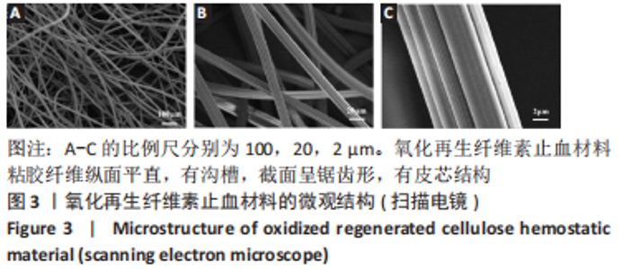
2.1 氧化再生纤维素止血材料微观结构 扫描电镜显示,氧化再生纤维素止血材料粘胶纤维纵面平直,有沟槽,截面呈锯齿形,有皮芯结构,如图3所示。"

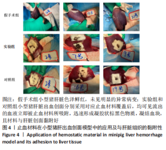
2.2 实验动物数量分析 24只巴马小型猪全部进入结果分析。 2.3 止血材料在小型猪肝脏出血创面模型中的应用及与组织黏附性 如图4所示,术中可见假手术组小型猪肝脏色泽鲜红,未见明显的异常病变;实验组和对照组小型猪肝脏出血创面分别采用对应止血材料覆盖后,均可见流出的血液立即被止血材料所吸附,迅速形成凝胶状棕黑色物质,凝结血块,且材料与肝脏创面黏附好,未见脱落、滑移。结果表明,氧化再生纤维素止血材料具有良好的组织黏附性。"

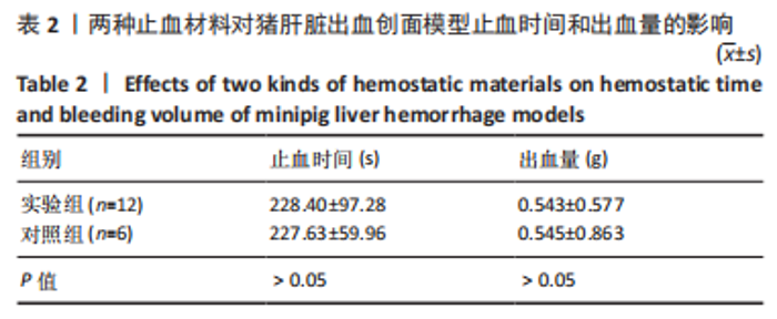
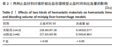
2.4 止血材料对小型猪肝脏出血创面模型止血时间与出血量的影响 表2结果显示,实验组与对照组小型猪肝脏出血创面模型止血时间与出血量比较差异均无显著性意义(P > 0.05),说明在猪肝脏出血程度相似的情况下,使用相同数量的两组止血材料有效性差异不大,这是因为两组产品均是氧化再生纤维素材质,其止血机制相同[13]。"

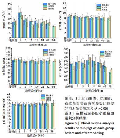
2.5 各组小型猪血常规分析结果 如图5所示,3组间白细胞、红细胞、血红蛋白等血液学参数比较差异均无显著性意义 (P > 0.05)。造模后第1天,3组白细胞含量大幅增加,推断由急性失血造成,血小板含量增加也考虑急性应激反应,这些指标变化符合急性失血的临床表现,因整个过程出血量不多,故红细胞、血红蛋白和平均血红蛋白水平波动不大。造模后第3天,3组血小板和白细胞含量较第1天有所降低,说明机体发挥造血功能作用,并且这些指标在术后1周时已基本上恢复到造模前正常生理水平,故这些指标的波动可能与手术因素和术后创伤愈合有关,术后1周后这些影响因素基本消失,进一步表明氧化再生纤维素止血材料对小型猪的血液系统无产生明显毒副作用,其与对照品速即纱的反应类似,提示氧化再生纤维素止血材料具有较好的生物相容性。"
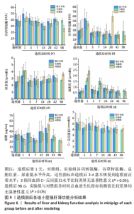
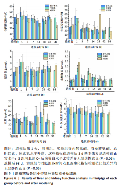
2.6 各组小型猪肝肾功能分析结果 如图6所示,造模后第1天,对照组、实验组谷丙转氨酶、谷草转氨酶、总胆红素、尿素氮水平升高,这可能与术中对肝脏的牵拉与制作肝出血创面有关,导致对肝脏有一定的损伤作用,这些指标在造模后72 h基本恢复到造模前正常水平。C-反应蛋白是重要的炎症指标,研究结果显示,3组间血清C-反应蛋白水平比较差异无显著性意义(P > 0.05),表明氧化再生纤维素止血材料不会引起机体全身剧烈的炎性反应。造模后98 d,实验组和对照组小型猪的生化指标、炎症因子等与造模前基本相同,说明实验动物状态稳定,两种止血材料均未对实验动物的肝肾功能造成损害,并且实验组与对照组各时间点血液生化指标检测值比较差异均无显著性意义(P > 0.05)。"
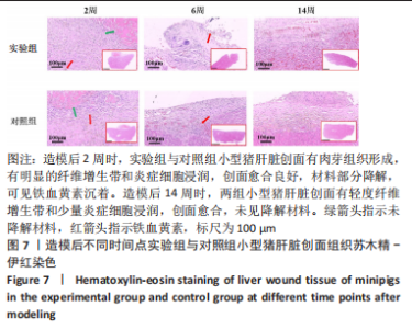
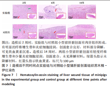
2.7 各组小型猪肝脏创面组织病理学观察结果 各组小型猪肝脏创面苏木精-伊红染色结果,如图7图注所示。造模后2周时,实验组与对照组小型猪肝脏创面有肉芽组织形成,有明显的纤维增生带和炎症细胞浸润,创面愈合良好,与材料接触处的肝细胞形态正常、结构清晰,降解材料被炎症细胞包围,处于吸收阶段,并含有铁血黄素沉着。造模后6周时,两组小型猪的肝脏创面肉芽组织老化,且纤维增生和炎症细胞浸润均较造模后2周时减少,并有含铁血黄素沉着(这可能由于止血材料中酸性的羧基可与血红蛋白中的Fe2+相结合,快速吸附血小板、红血球等凝血因子形成血栓,进而起到止血的作用有关),创面基本愈合,未见降解材料。造模后14周时,两组小型猪肝脏创面有轻度纤维增生带和少量炎症细胞浸润,创面愈合,未见降解材料。结果表明,实验组小型猪肝脏创面的病理反应与对照组类似。"
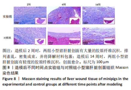
各组小型猪肝脏创面Masson染色结果,如图8所示。造模后2周时,两组小型猪肝脏创面有大量的胶原纤维沉积,排列紊乱,密集成束,并将降解材料包裹,这可能与肉芽组织产生有关;造模后6周时,两组肝脏创面的胶原纤维沉积较造模后2周时有所减少,这与肉芽组织老化有关;造模后14周时,两组小型猪肝脏创面有轻度的胶原纤维沉积,创面愈合。"


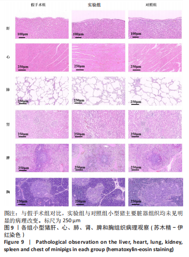
2.8 止血材料的生物相容性 各组小型猪主要脏器组织病理学观察结果,见图9所示,与假手术组对比,实验组与对照组小型猪主要脏器组织均未见明显的病理改变。由血常规、肝肾功能、创面及脏器组织病理学观察结果可知,氧化再生纤维素止血材料具有良好的组织相容性。"
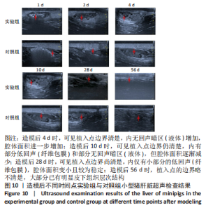
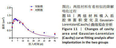
2.9 各组小型猪肝脏超声检查结果 造模后不同时间点实验组与对照组小型猪肝脏超声检测结果,见图10。两组造模后即刻均可见局部点状回声增强(植入物),且有一定的夹层厚度;造模后2 d时,可见植入点的边界逐渐清晰,局部伴有点状回声增强(残留物)且部分无回声暗区(液体),使得腔体面积增加;造模后4 d时,可见植入点边界清楚,内无回声暗区(液体)增加,腔体面积进一步增加,提示材料降解变化较大;造模后10 d时,可见植入点边界仍清楚,内有部分低回声(纤维包膜)和部分无回声暗区(液体),但腔体面积逐渐减少,提示材料降解吸收明显;造模后28 d时,可见植入点边界尚清楚,内仅有小部分的低回声(纤维包膜),腔体面积变小且较为稳定;造模后56 d时,植入点的边界略不清楚,大部分已有明显皮下组织层次结构。"

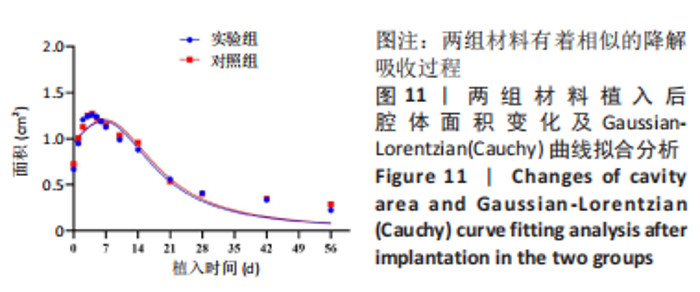
另外,将超声检测获得的液腔面积进行Gaussian-Lorentzian(Cauchy)曲线拟合,结果如图11显示,两组材料也有着相似的降解吸收过程。"
| [1] BEHRENS AM, SIKORSKI MJ, KOFINAS P. Hemostatic strategies for traumatic and surgical bleeding. Biomed Mater Res Part A. 2014; 102(11):4182-4194. [2] WANG J, ZHANG HL, WANG JK, et al. Efficacy of New Zeolite-based Hemostatic Gauze in a Gunshot Model of Junctional Femoral Artery Hemorrhage in Swine. J Surg Res. 2021;(263):176-185. [3] CHEN H, SHANG XQ, YU LS, et al. Safety evaluation of a low-heat producing zeolite granular hemostatic dressing in a rabbit femoral artery hemorrhage model. J Biomater Appl. 2020;34(7):988-997. [4] CHENG Y, HU Z, ZHAO Y, et al. Sponges of carboxymethyl chitosan grafted with collagen peptides for wound healing. Int J Mol Sci. 2019; 20(16):3890. [5] MAEVSKAIA EN, DRESVYANINA EN, SHABUNIN AS, et al. Preparation and Study of Hemostatic Materials Based on Chitosan and Chitin Nanofibrils. Nanotechnol Russ. 2020;15(7-8):466-475. [6] JALAL R, MOJTABA K, HONG C, et al. Novel chitosan/gelatin/oxidized cellulose sponges as absorbable hemostatic agents. Cellulose. 2021; 28:3663-3675. [7] AHMAD M, JAFARSADEGH M, SEYEDHOSEIN J, et al. Biodegradable cellulose-based superabsorbent as potent hemostatic agent. Chem Eng J. 2021;418(1):129252. [8] 胡鸣,毕大鹏,汪颖峰,等. 医用可吸收止血材料的临床应用与研究进展[J]. 踝外科电子杂志,2020,7(3):58-62. [9] 王璐璐.局部可吸收止血材料的应用现状及其研究进展[J].医学研究生学报,2018,31(1):109-112. [10] LIU L, HU WL, YU K, et al. Recent advances in materials for hemostatic management. Biomater Sci-Uk. 2021;9(22):7343-7378. [11] 冯靖祎,黄进,吴航.医疗机构止血材料管理专家共识[J].中国医院建筑与装备,2021,22(7):19-28. [12] 张爽,叶红,朱玉,等.纤维素类可吸收止血材料体外效果评价方法[J].中国医药生物技术,2022,17(2):176-182. [13] 喻炎,马凤森,陆佳骏,等.纤维素类可吸收止血材料的体外细胞毒性评价[J]. 材料导报,2018,32(3):874-880. [14] 李伟,王玲爽,孙伟庆.纤维素类止血材料的临床应用及作用机理的研究[J].当代医药论丛,2019,17(22):13-15. [15] 张爽,徐庆华,童琳,等.可吸收止血材料的研究现状与应用[J].中国组织工程研究,2021,25(10):1628-1634. [16] GABRIELA J, NADA P, VLADIMIR S, et al. Microdispersed Oxidized Cellulose as a novel potential substancewith hypolipidemic properties. Nutrition. 2008;24(11-12):1174-1181. [17] MOMIN M, MISHRA V, GHARAT S, et al. Recent advancements in cellulose-based biomaterials for management of infected wounds. Expert Opin Drug Deliv. 2021;18(11):1741-1760. [18] 俞文峰,徐金明,盛宏旭,等.可吸收再生氧化纤维素在肺癌手术中的临床效果评价[J].中国肺癌杂志,2020,23(6):492-495. [19] PATERNÒ VA, BISIN A, ADDIS A. Comparison of the efficacy of five standard topical hemostats: a study in porcine liver and spleen models of surgical bleeding. BMC Surg. 2020;20(1):215. [20] 崔国栋,陶宇,顾强,等.液态可吸收生物止血材料的安全性及有效性研究[J].中国微侵袭神经外科杂志,2020,25(11):512-516. [21] CHENG WL, HE JM, WU YD, et al. Preparation and characterization of oxidized regenerated cellulose film for hemostasis and the effect of blood on its surface. Cellulose. 2013;20(5):2547-2558. [22] 唐新雯,简祖琴.氧化再生纤维素纱布的止血作用[J].母婴世界, 2015(10):417-418. [23] 仲光勇,郭佩利,宋延平,等.吸收性氧化纤维素止血胶囊的止血作用研究[J].西北药学杂志,2016,31(3):274-276. [24] PALM MD, ALTMAN JS. Topical hemostatic agents: a review. Dermatol Surg. 2008;34(4):431-445. [25] ASHTON WH. Oxidized cellulose product and method for preparing the same. US Paten. 3364200,1968. [26] 王玲爽,童健星,赵哲哲,等.一种可吸收止血纤丝的有效性和安全性评价[J].浙江大学学报(医学版),2021,50(5):633-641. [27] GALGUT PN. Oxide cellulose mesh.I.Biodegradable membrane in periodontal surgery. Biomaterials. 1990;11(8):561-564. [28] 郭鑫.不同材质及形态的纤维素类可吸收止血材料的止血性能评价[D].石家庄:河北医科大学,2021. [29] 崔智慧,张雅菲,田田,等.国产氧化纤维素可吸收止血纱布对大鼠皮肤伤口愈合影响的机制研究[J]. 河北医科大学学报,2019, 40(10):1193-1196. [30] KRÍZOVÁ P, MÁSOVÁ L, SUTTNAR J, et al. The influence of intrinsic coagulation pathway on blood platelets activation by oxidized cellulose. J Biomed Mater Res A. 2007;82(2):274-280. [31] 喻炎.氧化再生纤维素可降解材料及其降解产物的生物学评价[D].杭州:浙江工业大学,2017. [32] 林媛媛.氧化再生纤维素钠盐可吸收止血纱的降解机制研究[D].哈尔滨:哈尔滨工业大学,2013. |
| [1] | Li Rui, Zhang Guihong, Wang Tao, Fan Ping. Effect of ginseng polysaccharide on the expression of prostaglandin E2/6-keto-prostaglandin 1alpha in traumatic osteoarthritis model rats [J]. Chinese Journal of Tissue Engineering Research, 2024, 28(8): 1235-1240. |
| [2] | Hu Zhixing, Li Qun, Yang Chao, Wang Xiaoxiao, Fang Luochangting, Hou Wuqiong, Lin Na, Chen Weiheng, Liu Chunfang, Lin Ya. Network meta-analysis of the modeling effects of different factors on rabbit models of steroid-induced osteonecrosis of femoral head [J]. Chinese Journal of Tissue Engineering Research, 2024, 28(6): 976-984. |
| [3] | Zhou Shibo, Guan Jianbin, Yu Xing, Zhao He, Yang Yongdong, Liu Tao. Animal models of femoral bone defects: preparation status and characteristics [J]. Chinese Journal of Tissue Engineering Research, 2024, 28(4): 633-638. |
| [4] | Ran Lei, Han Haihui, Xu Bo, Wang Jianye, Shen Jun, Xiao Lianbo, Shi Qi. Molecular docking analysis of the anti-inflammatory mechanism of Cibotium barometz and Epimedium for rheumatoid arthritis: animal experiment validation [J]. Chinese Journal of Tissue Engineering Research, 2024, 28(2): 208-215. |
| [5] | Chen Simin, Hu Yingjun, Yan Wenrui, Ji Le, Shao Mengli, Sun Ze, Zheng Hongxing, Qi Shanshan. Establishment and evaluation of a streptozotocin-induced diabetic encephalopathy rat model [J]. Chinese Journal of Tissue Engineering Research, 2024, 28(2): 237-241. |
| [6] | Wu Xiaodan, Wang Zhiguo, Zhan Ying, Zhang Guoxu. Establishment of an acute radioactive skin injury model induced by 32P-beta ray radiation and the mechanism of injury [J]. Chinese Journal of Tissue Engineering Research, 2024, 28(14): 2173-2179. |
| [7] | Xiong Bohan, Lu Xiaojun, Xue Wenqiang, Liu Jinrui, Gao Xianling, Yu Hong, Li Yajuan, Liu Haolong, Li Yanlin. Protective effect of anterior cruciate ligament reconstruction assisted by internal tension-reduction technique on the articular cartilage of southern Yunnan small-ear pigs [J]. Chinese Journal of Tissue Engineering Research, 2024, 28(14): 2221-2226. |
| [8] | Zhou Ziying, Ou Shangkun, Huang Chao, Jiang Hao, Zhang Liying, Gu Hao. Changes of the meibomian gland in a mouse model of aqueous deficient dry eye [J]. Chinese Journal of Tissue Engineering Research, 2024, 28(11): 1666-1671. |
| [9] | Han Zhongxiao, Ou Yaying, Zhuang Xinqing, Zhang Xiang, Li Biaoping, Jiang Zhirui, Zhang Jingyi, Yang Jiashun, Tang Ling, Xiao Wei. Mechanical puncture combined with tumor necrosis factor alpha and complete Freund’s adjuvant to construct a rat discogenic low back pain model [J]. Chinese Journal of Tissue Engineering Research, 2024, 28(11): 1672-1677. |
| [10] | Wang Zhongqing, Xiong Xianmei, Zhang Yan, Li Shijie, Ma Liqiong, Lu Zesheng, Gao Yijia. Panax notoginseng saponin promotes fracture healing by upregulating concentrated growth factors in rats [J]. Chinese Journal of Tissue Engineering Research, 2024, 28(11): 1678-1683. |
| [11] | Ruan Ling, Wang Guanghua, Wu Rongping, Jin Zhan, Lyu Zhenqing, Zhang Nan, Li Shoubang. Correlation between exercise intensity and lipid metabolism disorder and oxidative stress in a high-diet rat model [J]. Chinese Journal of Tissue Engineering Research, 2023, 27(8): 1149-1155. |
| [12] | Li Xiaoyin, Yang Xiaoqing, Chen Shulian, Li Zhengchao, Wang Ziqi, Song Zhen, Zhu Daren, Chen Xuyi. Collagen/silk fibroin scaffold combined with neural stem cells in the treatment of traumatic spinal cord injury [J]. Chinese Journal of Tissue Engineering Research, 2023, 27(6): 890-896. |
| [13] | Huang Guijiang, Ji Yuwei, Zhao Xin, Yang Yi, Zhao Yulan, Wang Peijin, Tang Wei, Jiao Jianlin. Effect and mechanism of different administration routes of placenta-derived mesenchymal stem cells in the treatment of tree shrews with osteoporotic fracture [J]. Chinese Journal of Tissue Engineering Research, 2023, 27(6): 909-914. |
| [14] | Song Jian, Zhao Lei, Liu Aishi. Construction and application of myocardial ischemia model in miniature pigs [J]. Chinese Journal of Tissue Engineering Research, 2023, 27(5): 772-778. |
| [15] | Wang Xiaoge, Liu Jiwen, Yang Shuai, Bao Jinyu, Li Cui. Effects of exercise on depression-like behaviors in chronic unpredictable mild stress rodent models: a systematic review and Meta-analysis [J]. Chinese Journal of Tissue Engineering Research, 2023, 27(5): 813-820. |
| Viewed | ||||||
|
Full text |
|
|||||
|
Abstract |
|
|||||