Chinese Journal of Tissue Engineering Research ›› 2024, Vol. 28 ›› Issue (26): 4137-4144.doi: 10.12307/2024.360
Previous Articles Next Articles
Effect and mechanism of angiotensin (1-7) supplementation combined with exercise therapy on cardiac remodeling in rats with renal hypertension
Xu Wenjie1, Xie Xudong1, He Ruibo2, Ma Gang2, Peng peng2
- 1School of Physical Education and Health Management, Changzhou Vocational Institute of Engineering, Changzhou 213164, Jiangsu Province, China; 2Department of Health Service, Logistics University of Chinese People’s Armed Police Forces, Tianjin 300309, China
-
Received:2023-03-24Accepted:2023-05-15Online:2024-09-18Published:2023-09-28 -
Contact:Peng Peng, PhD, Lecturer, Department of Health Service, Logistics University of Chinese People’s Armed Police Forces, Tianjin 300309, China -
About author:Xu Wenjie, Master, Associate professor, School of Physical Education and Health Management, Changzhou Vocational Institute of Engineering, Changzhou 213164, Jiangsu Province, China -
Supported by:Tianjin Natural Science Foundation, No. 17JCYBJC27400 (to PP); Basic Research Project of Logistics University of Chinese People’s Armed Police Forces, No. WHJ202302 (to PP)
CLC Number:
Cite this article
Xu Wenjie, Xie Xudong, He Ruibo, Ma Gang, Peng peng. Effect and mechanism of angiotensin (1-7) supplementation combined with exercise therapy on cardiac remodeling in rats with renal hypertension[J]. Chinese Journal of Tissue Engineering Research, 2024, 28(26): 4137-4144.
share this article
Add to citation manager EndNote|Reference Manager|ProCite|BibTeX|RefWorks

2.1 实验动物数量分析 在实验过程中,由于拒跑、意外死亡等原因共剔除8只动物,最终纳入统计的样本量为n=52,分别为正常血压组(n=12)、高血压对照组(n=10)、运动组(n=10)、Ang-(1-7)组(n=11)、联合治疗组(n=9)。 2.2 各组大鼠尾动脉血压比较 干预前,组间与正常血压组比较,各模型组收缩压和舒张压均升高(P < 0.05)。干预后,组内与干预前比较,高血压对照组和Ang-(1-7)组收缩压和舒张压升高(P < 0.05),联合治疗组收缩压和舒张压降低(P < 0.05),运动组收缩压和舒张压变化无显著性意义(P > 0.05);组间与正常血压组比较,高血压对照组、运动组和Ang-(1-7)组收缩压和舒张压升高(P < 0.05),联合治疗组收缩压和舒张压变化无显著性意义(P > 0.05);组间与高血压对照组比较,运动组和联合治疗组收缩压和舒张压下降(P < 0.05),Ang-(1-7)组血压变化无显著性意义(P > 0.05);组间与运动组和Ang-(1-7)组比较,联合治疗组收缩压和舒张压下降(P < 0.05)。见图1。"


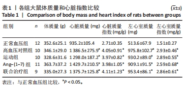
2.3 各组大鼠体质量和心脏指数比较 各组间体质量比较差异无显著性意义(P > 0.05)。与正常血压组比较,高血压对照组、运动组、Ang-(1-7)组和联合治疗组心脏质量、心脏质量指数、左心室质量、左心室质量指数均升高(P < 0.05),然而各模型组间心脏指数参数相比差异无显著性意义(P > 0.05)。见表1。"
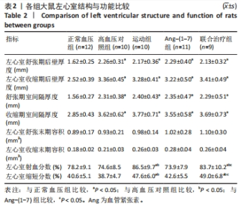
2.4 各组大鼠左心室结构与功能比较 各组间左心室舒张末期容积、左心室收缩末期容积比较差异无显著性意义(P > 0.05)。与正常血压组比较,高血压对照组、运动组、Ang-(1-7)组和联合治疗组左心室舒张期后壁厚度、左心室收缩期后壁厚度、舒张期室间隔厚度、收缩期室间隔厚度增加(P < 0.05),然而各模型组间比较上述参数差异均无显著性意义(P > 0.05)。与高血压对照组比较,运动组和联合治疗组左心室射血分数、左心室缩短分数增加(P < 0.05),Ang-(1-7)组差异无显著性意义(P > 0.05);与运动组比较,联合治疗组左心室射血分数、左心室缩短分数无显著变化(P > 0.05);与Ang-(1-7)组比较,联合治疗组左心室射血分数、左心室缩短分数升高(P < 0.05)。见表2。"

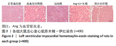
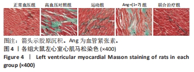
2.5 各组大鼠左心室心肌组织病理学观察 左心室心肌苏木精-伊红染色显示:胞浆呈粉红色,胞核呈蓝色。正常血压组心肌细胞结构完整、排列规则;高血压对照组和Ang-(1-7)组心肌细胞肿胀肥大、排列紊乱;运动组和联合治疗组仍存在心肌细胞肥大,但排列较规整。见图2。"

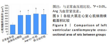
各组左心室心肌细胞横截面积结果显示:与正常血压组比较,高血压对照组、运动组、Ang-(1-7)组和联合治疗组心肌细胞横截面积均增加(P < 0.05),然而各模型组间比较则差异无显著性意义(P > 0.05)。见图3。"

左心室心肌马松染色显示:心肌细胞呈现红色,胶原纤维为蓝色。正常血压组心肌细胞间有极少量胶原纤维;高血压对照组和Ang-(1-7)组细胞间质中出现大量胶原纤维;运动组和联合治疗组心肌纤维化程度显著降低。见图4。"

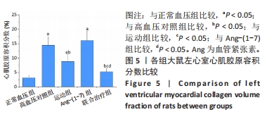
各组大鼠左心室心肌胶原容积分数比较显示:与正常血压组比较,高血压对照组、运动组和Ang-(1-7)组心肌胶原容积分数升高(P < 0.05);与高血压对照组比较,运动组和联合治疗组心肌胶原容积分数降低(P < 0.05),Ang-(1-7)组无显著变化(P > 0.05);与运动组和Ang-(1-7)组比较,联合治疗组心肌胶原容积分数下降(P < 0.05)。见图5。"

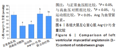
2.6 各组大鼠左心室心肌Ang-(1-7)含量比较 与正常血压组比较,高血压对照组和运动组心肌Ang-(1-7)含量降低(P < 0.05);与高血压对照组比较,Ang-(1-7)组和联合治疗组心肌Ang-(1-7)含量升高(P < 0.05),运动组无显著变化(P > 0.05);与运动组比较,联合治疗组心肌Ang-(1-7)含量升高(P < 0.05);与Ang-(1-7)组比较,联合治疗组心肌Ang-(1-7)含量无显著变化(P > 0.05)。见图6。"

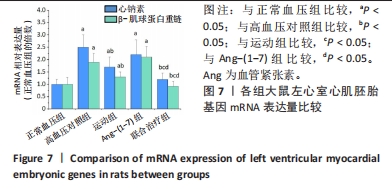
2.7 各组大鼠左心室心肌胚胎基因mRNA表达量比较 与正常血压组比较,高血压对照组、运动组和Ang-(1-7)组心钠素、β-肌球蛋白重链mRNA表达量上调(P < 0.05);与高血压对照组比较,运动组和联合治疗组心钠素、β-肌球蛋白重链mRNA表达量下调(P < 0.05),Ang-(1-7)组无显著变化(P > 0.05);与运动组和Ang-(1-7)组比较,联合治疗组心钠素、β-肌球蛋白重链mRNA表达量下调(P < 0.05)。见图7。"

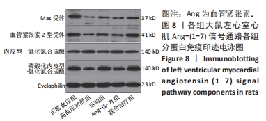
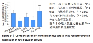
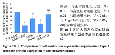
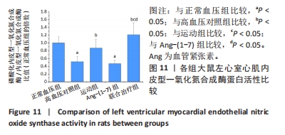
2.8 各组大鼠左心室心肌Ang-(1-7)信号通路蛋白表达量比较 与正常血压组比较,高血压对照组Mas受体、血管紧张素2型受体和内皮型一氧化氮合成酶蛋白表达量下调(P < 0.05);与高血压对照组比较,运动组和联合治疗组Mas受体、血管紧张素2型受体和内皮型一氧化氮合成酶蛋白表达量上调(P < 0.05),Ang-(1-7)组各蛋白表达量差异无显著性意义(P > 0.05);与运动组和Ang-(1-7)组比较,联合治疗组Mas受体、血管紧张素2型受体和内皮型一氧化氮合成酶蛋白表达量上调(P < 0.05)。见图8-11。"

"

"

"
| [1] JEPSON A, DANFORD D, CRAMER JW, et al. Assessment of hypertension, guideline-directed counseling, and outcomes in the ACHD population. Pediatr Cardiol. 2022;43(7):1615-1623. [2] GONZáLEZ A, RAVASSA S, LóPEZ B, et al. Myocardial remodeling in hypertension. Hypertension. 2018;72(3):549-558. [3] PRIETO MC, GONZALEZ AA, VISNIAUSKAS B, et al. The evolving complexity of the collecting duct renin-angiotensin system in hypertension. Nat Rev Nephrol. 2021;17(7):481-492. [4] VARAGIC J, FROHLICH ED. Local cardiac renin-angiotensin system: hypertension and cardiac failure. J Mol Cell Cardiol. 2002;34(11):1435-1442. [5] SANTOS R, OUDIT GY, VERANO-BRAGA T, et al. The renin-angiotensin system: going beyond the classical paradigms. Am J Physiol Heart Circ Physiol. 2019; 316(5):H958-H970. [6] SANTOS R, SAMPAIO WO, ALZAMORA AC, et al. The ACE2/angiotensin-(1-7)/Mas axis of the renin-angiotensin system: focus on angiotensin-(1-7). Physiol Rev. 2018;98(1):505-553. [7] O’NEILL J, HEALY V, JOHNS EJ. Intrarenal Mas and AT1 receptors play a role in mediating the excretory actions of renal interstitial angiotensin-(1-7) infusion in anaesthetized rats. Exp Physiol. 2017;102(12):1700-1715. [8] PEI Z, MENG R, LI G, et al. Angiotensin-(1-7) ameliorates myocardial remodeling and interstitial fibrosis in spontaneous hypertension: role of MMPs/TIMPs. Toxicol Lett. 2010;199(2):173-181. [9] GIANI JF, MAYER MA, MUñOZ MC, et al. Chronic infusion of angiotensin-(1-7) improves insulin resistance and hypertension induced by a high-fructose diet in rats. Am J Physiol Endocrinol Metab. 2009;296(2):E262-E271. [10] BüRGELOVá M, VANOURKOVá Z, THUMOVá M, et al. Impairment of the angiotensin-converting enzyme 2-angiotensin-(1-7)-Mas axis contributes to the acceleration of two-kidney, one-clip Goldblatt hypertension. J Hypertens. 2009;27(10):1988-2000. [11] COUTINHO D, JOVIANO-SANTOS JV, SANTOS-MIRANDA A, et al. Altered heart cytokine profile and action potential modulation in cardiomyocytes from Mas-deficient mice. Biochem Biophys Res Commun. 2022;619:90-96. [12] PATEL VB, MORI J, MCLEAN BA, et al. ACE2 deficiency worsens epicardial adipose tissue inflammation and cardiac dysfunction in response to diet-induced obesity. Diabetes. 2016;65(1):85-95. [13] COLLISTER JP, NAHEY DB. Simultaneous administration of Ang(1-7) or A-779 does not affect the chronic hypertensive effects of angiotensin II in normal rats. J Renin Angiotensin Aldosterone Syst. 2010;11(2):99-102. [14] STUIJ M, ELLING A, ABMA T. Negotiating exercise as medicine: Narratives from people with type 2 diabetes. Health (London). 2021;25(1):86-102. [15] GRONEK P, WIELINSKI D, CYGANSKI P, et al. A Review of Exercise as Medicine in Cardiovascular Disease: Pathology and Mechanism. Aging Dis. 2020;11(2):327-340. [16] PATTI A, MERLO L, AMBROSETTI M, et al. Exercise-based cardiac rehabilitation programs in heart failure patients. Heart Fail Clin. 2021;17(2):263-271. [17] 朱政, 付常喜, 马文超, 等. 有氧运动调控自发性高血压模型大鼠心脏重塑的机制[J]. 中国组织工程研究,2022,26(14):2231-2237. [18] FILHO AG, FERREIRA AJ, SANTOS SH, et al. Selective increase of angiotensin(1-7) and its receptor in hearts of spontaneously hypertensive rats subjected to physical training. Exp Physiol. 2008;93(5):589-598. [19] FRANCHI F, KNUDSEN BE, OEHLER E, et al. Non-invasive assessment of cardiac function in a mouse model of renovascular hypertension. Hypertens Res. 2013; 36(9):770-775. [20] ROSA RM, COLUCCI JA, YOKOTA R, et al. Alternative pathways for angiotensin II production as an important determinant of kidney damage in endotoxemia. Am J Physiol Renal Physiol. 2016;311(3):F496-F504. [21] 黄红梅, 胡宗祥, 刘昭强, 等. 长期高强度间歇训练加重自发性高血压大鼠心肌纤维化[J]. 中国运动医学杂志,2020,39(8):615-625. [22] 袁国强, 秦永生, 彭朋. 高强度间歇运动对自发性高血压模型大鼠病理性心脏肥大的影响及机制[J]. 中国组织工程研究,2020, 24(23):3708-3715. [23] 袁国强, 秦永生, 彭朋. 有氧运动对自发性高血压大鼠心肌纤维化的影响及机制[J]. 天津医药,2020,48(2):100-104. [24] 范朋琦, 秦永生, 彭朋. 不同运动方式对自发性高血压大鼠心脏重塑和运动能力的影响[J]. 现代预防医学,2018,45(23):4341-4345,4351. [25] PEDDI NC, MARASANDRA RAMESH H, GUDE SS, et al. Intrauterine fetal gene therapy: is that the future and is that future now. Cureus. 2022;14(2):e22521. [26] FRANGOGIANNIS NG. Cardiac fibrosis. Cardiovasc Res. 2021;117(6):1450-1488. [27] XI Y, HAO M, LIANG Q, et al. Dynamic resistance exercise increases skeletal muscle-derived FSTL1 inducing cardiac angiogenesis via DIP2A-Smad2/3 in rats following myocardial infarction. J Sport Health Sci. 2021;10(5):594-603. [28] GATI S, MALHOTRA A, SHARMA S. Exercise recommendations in patients with valvular heart disease. Heart. 2019;105(2):106-110. [29] HEIDARI B, ZOLFAGHARI MR, KHADEMVATANI K, et al. Interrelation among exercise training, cardiac hypertrophy, and tissue kallikrein-kinin system in athlete and non-athlete women. J Cardiovasc Thorac Res. 2022;14(3):159-165. [30] WANG J, HE W, GUO L, et al. The ACE2-Ang (1-7)-Mas receptor axis attenuates cardiac remodeling and fibrosis in post-myocardial infarction. Mol Med Rep. 2017;16(2):1973-1981. [31] KANGUSSU LM, GUIMARAES PS, NADU AP, et al. Activation of angiotensin-(1-7)/Mas axis in the brain lowers blood pressure and attenuates cardiac remodeling in hypertensive transgenic (mRen2)27 rats. Neuropharmacology. 2015;97:58-66. [32] 杨吕, 黄煜, 何庆. 调控血管紧张素转化酶2-血管紧张素(1-7)-Mas轴是心脏重构和心力衰竭治疗的新靶点[J]. 中华危重病急救医学,2019,31(11):1425-1428. [33] BOISSIERE J, EDER V, MACHET MC, et al. Moderate exercise training does not worsen left ventricle remodeling and function in untreated severe hypertensive rats. J Appl Physiol (1985). 2008;104(2):321-327. [34] AENGEVAEREN VL, BAGGISH AL, CHUNG EH, et al. Exercise-induced cardiac troponin elevations: from underlying mechanisms to clinical relevance. Circulation. 2021;144(24):1955-1972. [35] PANDORF CE, HADDAD F, OWERKOWICZ T, et al. Regulation of myosin heavy chain antisense long noncoding RNA in human vastus lateralis in response to exercise training. Am J Physiol Cell Physiol. 2020;318(5):C931-C942. [36] COSTA MA, LOPEZ VERRILLI MA, GOMEZ KA, et al. Angiotensin-(1-7) upregulates cardiac nitric oxide synthase in spontaneously hypertensive rats. Am J Physiol Heart Circ Physiol. 2010;299(4): H1205-H1211. [37] LOUGHLIN JM, BROWNE L, HINCHION J. The impact of exogenous nitric oxide during cardiopulmonary bypass for cardiac surgery. Perfusion. 2022;37(7):656-667. [38] TIWARI P, TIWARI V, GUPTA S, et al. Activation of Angiotensin-converting enzyme 2 protects against lipopolysaccharide-induced glial activation by modulating angiotensin-converting enzyme 2/angiotensin (1-7)/Mas receptor axis. Mol Neurobiol. 2023;60(1):203-227. |
| [1] | Wu Jing, Yao Yingce, Yang Xiaowei, Xue Boshi, Zhao Jianbin, Yang Chen, Luan Tianfeng, Zhou Zhipeng. Intervention of muscle strength training combined with neuromuscular electrical stimulation on lower limb function and biomechanical changes in patients with patellofemoral pain [J]. Chinese Journal of Tissue Engineering Research, 2024, 28(9): 1365-1371. |
| [2] | Wei Juan, Li Ting, Huan Mengting, Xie Ying, Xie Zhouyu, Wei Qingbo, Wu Yunchuan. Mechanism by which static exercise improves insulin resistance in skeletal muscle of type 2 diabetes [J]. Chinese Journal of Tissue Engineering Research, 2024, 28(8): 1271-1276. |
| [3] | Lou Guo, Zhang Yan, Fu Changxi. Role of endothelial nitric oxide synthase in exercise preconditioning-induced improvement of myocardial ischemia-reperfusion injury [J]. Chinese Journal of Tissue Engineering Research, 2024, 28(8): 1283-1288. |
| [4] | Cheng Jie, Wang Jihong, Zhang Pei. Functional exercise for tendon adhesion in a model of deep flexor tendon II injury of the third toe [J]. Chinese Journal of Tissue Engineering Research, 2024, 28(8): 1161-1167. |
| [5] | Wang Ji, Zhang Min, Li Wenbo, Yang Zhongya, Zhang Long. Effect of aerobic exercise on glycolipid metabolism, skeletal muscle inflammation and autophagy in type 2 diabetic rats [J]. Chinese Journal of Tissue Engineering Research, 2024, 28(8): 1200-1205. |
| [6] | Ruan Rong, Lou Xujia, Jin Qiguan, Zhang Libing, Xu Shang, Hu Yulong. Effect of resveratrol on gluconeogenesis in exercise-induced fatigue rats [J]. Chinese Journal of Tissue Engineering Research, 2024, 28(8): 1229-1234. |
| [7] | Liu Zhiyang, Fu Zeting, Xia Yu, Ding Haili. The role of BMAL1 and MyoD in exercise-induced skeletal muscle damage [J]. Chinese Journal of Tissue Engineering Research, 2024, 28(4): 510-515. |
| [8] | Zhu Xiaofeng, Chen Weiwei, Huang Jian. Effects of maternal high-fat diet and exercise intervention on insulin sensitivity and the hypothalamic arcuate nucleus in male offspring mice [J]. Chinese Journal of Tissue Engineering Research, 2024, 28(4): 556-561. |
| [9] | Wang Shijie, Wen Dengtai, Wang Jingfeng, Gao Yinghui. Mammalian target of rapamycin in relation to exercise, high fat/high salt diet, and aging [J]. Chinese Journal of Tissue Engineering Research, 2024, 28(4): 574-580. |
| [10] | Yang Qihang, Pu Rui, Chen Ziyang, Leng Siyi, Song Yongjing, Liu Hui, Du Guangyou. Intestinal flora and osteoporosis and exercise intervention [J]. Chinese Journal of Tissue Engineering Research, 2024, 28(26): 4250-4256. |
| [11] | Yang Liyuan, Zhang Yeting, Li Chuikun, Wei Cuilan. Effects of aerobic exercise on the expression of Notch1 and Caspase-3 in the hippocampus of Alzheimer’s disease mice [J]. Chinese Journal of Tissue Engineering Research, 2024, 28(26): 4113-4120. |
| [12] | Zhang Min, Lou Guo, Fu Changxi. Aerobic exercise preconditioning improves therapeutic effect of bone marrow mesenchymal stem cells on acute myocardial infarction [J]. Chinese Journal of Tissue Engineering Research, 2024, 28(25): 3988-3993. |
| [13] | Kong Jianda, Xie Yingao, Chen Shijuan, Zhu Lei. Blood flow restriction training interventions for sarcopenia in older adults: biological mechanisms and proposed application protocols [J]. Chinese Journal of Tissue Engineering Research, 2024, 28(23): 3743-3750. |
| [14] | Qi Yuqing, Liu Xiaoran. Mechanisms of exercise-regulated telomere length and health promotion [J]. Chinese Journal of Tissue Engineering Research, 2024, 28(23): 3759-3765. |
| [15] | Yin Gonghua, Xu Ruoyao, Zhang Lijuan, Zhang Yifan, Qi Jie, Zhang Jun. Regulation of N6-methyladenosine on non-coding RNAs in pathological cardiac remodeling [J]. Chinese Journal of Tissue Engineering Research, 2024, 28(20): 3252-2358. |
| Viewed | ||||||
|
Full text |
|
|||||
|
Abstract |
|
|||||