[1] 李获, 王鹏, 高健明, 等. 股骨头坏死血运重建与内部微观结构改变的关系[J]. 中国组织工程研究,2022,26(9):1323-1328.
[2] CUENOD CA, BALVAY D. Perfusion and vascular permeability: basic concepts and measurement in DCE-CT and DCE-MRI. Diagn Interv Imaging. 2013;94(12):1187-1204.
[3] GRIFFITH JF, YEUNG DKW, ANTONIO GE, et al. Vertebral bone mineral density, marrow perfusion, and fat content in healthy men and men with osteoporosis dynamic contrast-enhanced MR imaging and MR spectroscopy. Radiology. 2005; 236(3):945-951.
[4] NOSAS-GARCIA S, MOEHLER T, WASSER K, et al. Dynamic contrast-enhanced MRI for assessing the disease activity of multiple myeloma: a comparative study with histology and clinical markers. J Magn Reson Imaging. 2005;22(1):154-162.
[5] PS T. Modeling tracer kinetics in dynamic Gd-DTPA MR imaging. J Magn Reson Imaging. 1997;1(7):91-101.
[6] THOMASSIN-NAGGARA I, SOUALHI N, BALVAY D, et al. Quantifying tumor vascular heterogeneity with DCE-MRI in complex adnexal masses: A preliminary study. J Magn Reson Imaging. 2017;46(6):1776-1785.
[7] THOMASSIN-NAGGARA I, DARAI E, CUENOD CA, et al. Dynamic contrast-enhanced magnetic resonance imaging: a useful tool for characterizing ovarian epithelial tumors. J Magn Reson Imaging. 2008;28(1):111-120.
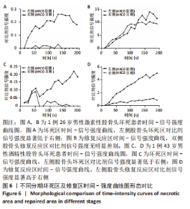
[8] YOON BH, MONT MA, KOO KH, et al. The 2019 Revised Version of Association Research Circulation Osseous Staging System of Osteonecrosis of the Femoral Head. J Arthroplasty. 2020;35(4):933-940.
[9] TT S, HC L, CJ C, et al. Correlation of MR lumbar spine bone marrow perfusion with bone mineral density in female subjects. Radiology. 2004;1(233):121-128.
[10] JS T, WE R. Evolution from empirical dynamic contrast enhanced magnetic resonance imaging to pharmacokinetic MRI. Adv Drug Deliv Rev. 2000;1(41):91-110.
[11] TURKBEY B, THOMASSON D, PANG Y, et al. The role of dynamic contrast-enhanced MRI in cancer diagnosis and treatment. Diagn Interv Radiol. 2010;16(3):186-192.
[12] 胡长根, 陈君长, 刘强, 等. 激素对股骨头微血管及组织细胞的影响[J]. 中华骨科杂志,2004,24(6):359-363.
[13] DOR Y, PORAT R, KESHET E. Vascular endothelial growth factor and vascular adjustments to perturbations in oxygen homeostasis. Am J Physiol Cell Physiol. 2001;280(6):1367-1374.
[14] MALIZOS KN, ZIBIS AH, DAILIANA Z, et al. MR imaging findings in transient osteoporosis of the hip. Eur J Radiol. 2004;50(3):238-244.
[15] GUILLERMAN RP. Marrow: red, yellow and bad. Pediatr Radiol. 2013;43 Suppl 1: S181-S192.
[16] SHENG H, ZHANG G, WANG YX, et al. Functional perfusion MRI predicts later occurrence of steroid-associated osteonecrosis: an experimental study in rabbits. J Orthop Res. 2009;27(6):742-747.
[17] CHAN WP, LIU YJ, HUANG GS, et al. Relationship of idiopathic osteonecrosis of the femoral head to perfusion changes in the proximal femur by dynamic contrast-enhanced MRI. AJR Am J Roentgenol. 2011;196(3):637-643.
[18] SAKAI T, SUGANO N, NISHII T, et al. MR findings of necrotic lesions and the extralesional area of osteonecrosis of the femoral head. Skeletal Radiol. 2000; 29(3):133-141.
[19] SIMMONS DJ, DAUM WJ, TOTTY W, et al. Correlation of MRI images with histology in avascular necrosis in the hip. A preliminary study. J Arthroplasty. 1989;4(1): 7-14.
[20] LANG P, JERGESEN HE, MOSELEY ME, et al. Avascular necrosis of the femoral head high-field-strength MR imaging with histologic correlation. Radiology. 1988; 169(2):517-524.
[21] AARON RK, DYKE JP, CIOMBOR DM, et al. Perfusion abnormalities in subchondral bone associated with marrow edema, osteoarthritis, and avascular necrosis. Ann N Y Acad Sci. 2007;1117: 124-137.
[22] LEE JH, DYKE JP, BALLON D, et al. Assessment of bone perfusion with contrast-enhanced magnetic resonance imaging. Orthop Clin North Am. 2009;40(2):249-257.
[23] MIN HW, LI ZR, CHENG LM, et al. Measuring blood flow change of osteonecrosis of femoral head with laser doppler flowmetry. Zhonghua Wai Ke Za Zhi. 2008; 46(15):1171-1173.
[24] LARRIBE M, GAY A, FREIRE V, et al. Usefulness of dynamic contrast-enhanced MRI in the evaluation of the viability of acute scaphoid fracture. Skeletal Radiol. 2014;43(12):1697-1703.
[25] SCHOIERER O, BLOESS K, BENDER D, et al. Dynamic contrast-enhanced magnetic resonance imaging can assess vascularity within fracture non-unions and predicts good outcome. Eur Radiol. 2014;24(2):449-459.
[26] 赵德伟. 加强对股骨头缺血性坏死病理生理的认识[J]. 中华关节外科杂志(电子版),2014,8(5):560-562.
|