Chinese Journal of Tissue Engineering Research ›› 2023, Vol. 27 ›› Issue (23): 3646-3652.doi: 10.12307/2023.579
Previous Articles Next Articles
Preparation of rabbit models of surgically induced knee osteoarthritis
Zhang Chuancheng1, Shen Meihua1, Chen Lifeng2, Wang Huasong2, Xiang Yang1, Tan Zhangkui2
- 1Wuhan University of Science and Technology, Wuhan 430065, Hubei Province, China; 2Central Theater Command General Hospital of the Chinese PLA, Wuhan 430070, Hubei Province, China
-
Received:2022-06-28Accepted:2022-10-24Online:2023-08-18Published:2023-01-16 -
Contact:Chen Lifeng, MD, Associate chief physician, Central Theater Command General Hospital of the Chinese PLA, Wuhan 430070, Hubei Province, China -
About author:Zhang Chuancheng, Master candidate, Wuhan University of Science and Technology, Wuhan 430065, Hubei Province, China Shen Meihua, Master, Wuhan University of Science and Technology, Wuhan 430065, Hubei Province, China -
Supported by:Young Top-notch Medical Talents of Hubei Province (the First Level), No. 1371 (to CLF)
CLC Number:
Cite this article
Zhang Chuancheng, Shen Meihua, Chen Lifeng, Wang Huasong, Xiang Yang, Tan Zhangkui. Preparation of rabbit models of surgically induced knee osteoarthritis[J]. Chinese Journal of Tissue Engineering Research, 2023, 27(23): 3646-3652.
share this article
Add to citation manager EndNote|Reference Manager|ProCite|BibTeX|RefWorks
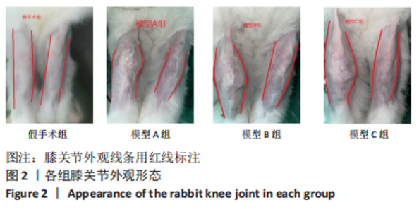
2.1 实验动物实验分析 此次实验共使用雄性新西兰大白兔16只,术前健康状况良好,建模过程中模型B组、模型C组各死亡1只(死亡后模型B、C组样本量满足n≥3,对实验结果影响较小,暂不考虑补充),剩余14只生命体征均稳定。 2.2 动物一般情况 各组实验动物手术前后体质量无明显变化。各模型组术后当天饮食减少,右膝出现明显跛行、皮肤红肿,并有弓背、呼吸急促、流涎、暴躁等现象;7 d后跛行加重,右侧膝关节内出现骨性隆起,质地坚硬。其中模型A、B组患膝可有关节活动;模型C组患膝疼痛明显,活动时屈曲状态。见图2。"
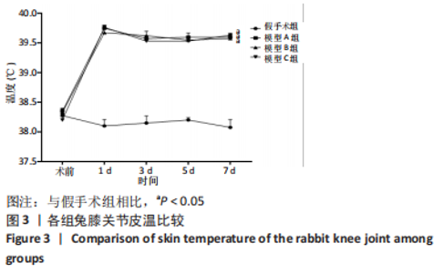
2.3 各组兔右侧膝关节皮温测量 术前各组兔皮温无明显差异(P > 0.05),术后1,3,5,7 d模型组皮温相比于假手术组明显升高,差异有显著性意义(P < 0.05)。各模型组皮温升高差异无显著性意义(P > 0.05)。见图3。"
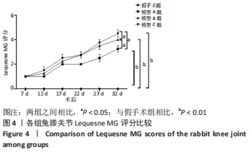
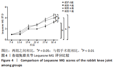
2.4 各组兔Lequesne MG 评分 各组兔Lequesne MG评分显示,模型组评分明显高于假手术组,差异有显著性意义(P < 0.01);各模型组之间相比差异有显著性意义(P < 0.05),见图4。"
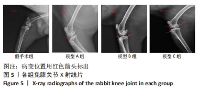
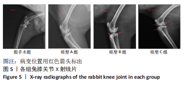
2.5 膝关节X射线片 假手术组X射线片显示关节面平整、光滑,无骨赘、关节间隙狭窄等异常改变;模型A组关节边缘出现轻度骨质增生,关节面轻度粗糙,关节间隙无明显改变;模型B组关节边缘骨质增生明显,关节面粗糙;相比于模型A、B组,模型C组有骨赘形成,关节面破坏,关节间隙明显狭窄。见图5。"
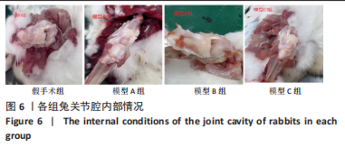
2.6 关节腔内部情况 打开关节囊后观察各组兔膝关节变化情况,假手术组关节面光滑平整,滑膜光泽清亮,均匀覆盖于脂肪垫上,无充血;模型A、B组关节情况大致相仿,表面呈现暗黄色,滑膜充血,关节内脂肪相较于假手术组增多;模型C组脂肪垫明显增生,且多于模型A、B组,滑膜液浑浊,滑膜表面出现裂隙,可见淤血。见图6。"
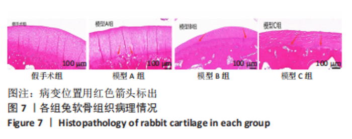
2.7 软骨组织形态 股骨内髁软骨苏木精-伊红染色结果显示,假手术组关节软骨表面平整光滑,表面区、移行区、辐射区和矿化区4层结构清晰,排列规律,细胞分布及染色均匀,潮线清晰完整;模型A组软骨细胞数量减少,潮线部分消失,层次不清,可见少量血管结构;模型B组潮线崎岖不平,软骨细胞数量减少;模型C组软骨结构损坏严重,层次紊乱,潮线消失,软骨细胞大量减少,可见血管生成。见图7。"

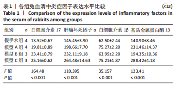
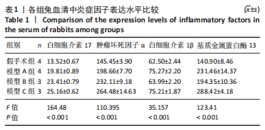
2.8 炎症因子水平 2.8.1 血清炎症因子水平 与假手术组相比,各模型组血清中肿瘤坏死因子α、白细胞介素1β、白细胞介素17、MMP-13水平明显升高,差异有显著性意义(P < 0.001),见表1。"
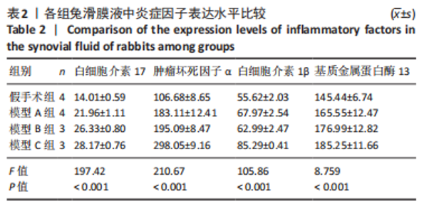
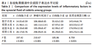
2.8.2 滑膜液炎症因子水平 与假手术组相比,各模型组滑膜液中肿瘤坏死因子α、白细胞介素1β、白细胞介素17、MMP-13水平明显升高,差异有显著性意义(P < 0.001),见表2。"
| [1] KAN HS,CHAN PK,CHIU KY, et al. Non-surgical treatment of knee osteoarthritis. Hong Kong Med J. 2019;25:127-133. [2] 中华医学会骨科分会关节外科学组,吴阶平医学基金会骨科学专家委员会. 膝骨关节炎阶梯治疗专家共识(2018年版)[J]. 中华关节外科杂志(电子版),2019,13(1):124-130. [3] NAJAFI S, SANATI E, KHADEMI M, et al. Intra-articular botulinum toxin type A for treatment of knee osteoarthritis: Clinical trial. Toxicon. 2019; 165:69-77. [4] JIN H, ZUO H, Xu R, et al. A case report of ultrasound-guided knee nerve pulse radiofrequency combined with platelet-rich plasma in the treatment of knee osteoarthritis. Medicine (Baltimore). 2021; 100(51):e27878. [5] GONG J, LI Q, WEI M, et al. Effect of Tongluozhitong Prescription-Assisted Intra-Articular Injection of Sodium Hyaluronate on VAS Score and Knee Lysholm Score in Patients with Knee Osteoarthritis. Evid Based Complement Alternat Med. 2021;2021:3210494. [6] ROEMER FW, GUERMAZI A, ZHANG Y, et al. Hoffa’s Fat Pad: Evaluation on Unenhanced MR Images as a Measure of Patellofemoral Synovitis in Osteoarthritis. AJR Am J Roentgenol. 2009;192(6):1696-700. [7] KLOPPENBURG M, BERENBAUM F. Osteoarthritis year in review 2019: epidemiology and therapy. Osteoarthritis Cartilage. 2020;28(3): 242-248. [8] MCCOY AM. Animal models of osteoarthritis:Comparisons and key considerations. Vet Pathol. 2015;52(5):803-818. [9] DE LANGE-BROKAAR BJ, IOAN-FACSINAY A, YUSUF E, et al. Evolution of synovitis in osteoarthritic knees and its association with clinical features. Osteoarthritis Cartilage. 2016;24:1867-1874. [10] TSAI HC, CHEN TL, CHEN YP, et al. Traumatic osteoarthritis-induced persistent mechanical hyperalgesia in a rat model of anterior cruciate ligament transection plus a medial meniscectomy. J Pain Res. 2018;11: 41-50. [11] BURKE CJ, ALIZAI H, BELTRAN LS, et al. MRI of synovitis and joint fluid. J Magn Reson Imaging. 2019;49:1512-1527. [12] 苏剑清,孙波,丁韵蓉,等.基于“损伤-修复-再损伤”方法制备兔膝骨关节炎滑膜炎模型[J].中国组织工程研究,2022,26(23): 3738-3743. [13] 熊元,赵振国,李传郡,等.兔膝骨关节炎模型的制备及鉴定[J].实验动物科学,2013,30(3):31-34+63. [14] PAS HI, WINTERS M, HAISMA HJ, et al. Stem cell injections in knee osteoarthritis: a systematic review of the literature. Br J Sports Med. 2017;51(15):1125-1133. [15] RODRIGUEZ-MERCHAN EC, DE LA CORTE-RODRIGUEZ H. The role of orthoses in knee osteoarthritis. Hosp Pract(1995). 2019;47:1-5. [16] 曹向昱,刘雨曦,曹永平.老年退行性骨关节炎治疗进展[J].中国临床保健杂志,2022,25(1):25-29. [17] TEICHTAHL AJ, WLUKA AE, WIJETHILAKE P, et al. Wolff’s law in action: a mechanism for early knee osteoarthritis. Arthritis Res Ther. 2015;17: 207. [18] LIU L, ZHAO C, ZHAO S, et al. Evaluation of the effectiveness and safety of icariin in the treatment of knee osteoarthritis: A protocol for a systematic review and meta-analysis. Medicine (Baltimore). 2021;100: e28277. [19] 王欣,罗文,黄文泽,等.单侧膝骨关节炎临床分期与双足足底压力的相关性[J].中国组织工程研究,2021,25(27):4312-4317. [20] YUN SJ, LIM Y, JIN W, et al. Validity of Radiograph-Based Infrapatellar Fat Pad Opacity Grading for Assessing Knee Synovitis: Correlation With Contrast-Enhanced MRI. Am J Roentgenol. 2017;209(6):1321-1330. [21] STEINBRUCK A,WOICZINSKI M,WEBER P, et al. Posterior cruciate ligament balancing in total knee arthroplasty:a numerical study with a dynamic force controlled knee model. Biomed Eng Online. 2014;13:91. [22] HULTH A, LINDBERG L, TELHAG H. Experimental osteoarthritis in rabbits. Acta Orthop Scand. 1970;41:522-530. [23] ZAPATA-LINARES N, EYMARD F, BERENBAUM F, et al. Role of adipose tissues in osteoarthritis. Curr Opin Rheumatol. 2021;33(1):84-93. [24] JARRAYA M,DIAZ LE,ROEMER FW,et al.MRI findings consistent with peripatellar fat pad impingement:how much related to patellofemoral maltracking? Magn Reson Med Sci. 2018;17(3):195-202. [25] BELLUZZI E,STOCCO E,POZZUOLI A,et al. Contribution of infrapatellar fat pad and synovial membrane to knee osteoarthritis pain. Biomed Res Int. 2019;2019:6390182. [26] 陈墅,刘宁,周义钦,等. 髌下脂肪垫在膝骨关节炎发病和进展中的作用[J]. 中华骨科杂志,2018,38(15):953-960. [27] 王勃,李英俊,邱丽莎.比较分析膝骨关节炎患者肌骨超声与X线片的表现特点[J].现代医用影像学,2021,30(12):2307-2309. [28] STUTZ G, KUSTER MS, KLEINSTUK F, et al Arthroscopic management of septic arthritis:stages of infection and results. Knee Surg Sports Traum Arthrosc. 2008;8(5):270-274. [29] 谢平金,柴生颋.膝骨性关节炎X线片观测指标及其应用进展[J].中国中医骨伤科杂志,2017,25(3):72-76. [30] SELLAM J, BERENBAUM F. The role of synovitis in pathophysiology and clinical symptoms of osteoarthritis. Nat Rev Rheumatol. 2010; 6(11):625-635. [31] HAYASHI D, ROEMER FW, KATUR A, et al. Imaging of Synovitis in Osteoarthritis:Current Status and Outlook. Semin Arthritis Rheum. 2011;41(2):116-130. [32] 熊颖,顾武斌.X线、超声及MRI影像诊断膝骨关节炎的价值对比分析[J].影像研究与医学应用,2019,3(6):30-32. [33] MATHIESSEN A, SLATKOWSKY-CHRISTENSEN B, KVIEN TK, et al. Ultrasound-detected inflammation predicts radiographic progression in hand osteoarthritis atier 5 years. Ann Rheum Dis. 2016;75(5):825-830. [34] Hayashi D, Roemer FW, Katur A, et al. Imaging of Synovitis in Osteoarthritis:Current Status and Outlook. Semin Arthritis Rheum. 2010;41(2):116-130. [35] KANDAHARI AM, YANG X, DIGHE AS, et al.Recognition of Immune Response for the Early Diagnosis and Treatment of Osteoarthritis. J Immunol Res. 2015;20(15):1-13. [36] KAPOOR M, MARTEL-PELLETIER J, LAJEUNESSE D, et al. Role of proinflammatory cytokines in the pathophysiology of osteoarthritis. Nat Rev Rheumatol. 2011;7(1):33-42. [37] LIAO CR, WANG SN, ZHU SY, et al. Advanced oxidation protein products increase TNF-α and IL-1β expression in chondrocytes via NADPH oxidase 4 and accelerate cartilage degeneration in osteoarthritis progression. Redox Biol. 2020;28:101306. [38] FEI J, LIANG B, JIANG C, et al. Luteolin inhibits IL-1β-induced inflammation in rat chondrocytes and attenuates osteoarthritis progression in a rat model. Biomed Pharmacother. 2019;109:1586-1592. [39] ZHANG C, ZHU M, WANG H, et al. LOXL2 attenuates osteoarthritis through inactivating Integrin/FAK signaling. Sci Rep. 2021;11(1):17020. [40] HOSSEINZADEH A, KAMRAVA SK, JOGHATAEI MT, et al. Apoptosis signaling pathways in osteoarthritis and possible protective role of melatonin. J Pineal Res. 2016;61(4):411-425. [41] ZHAO Y, LI Y, QU R, et al. Cortistatin binds to TNF-α receptors and protects against osteoarthritis. EBioMedicine. 2019;41:556-570. [42] XU Z, KE T, ZHANG Y, et al. Danshensu inhibits the IL-1β-induced inflammatory response in chondrocytes and osteoarthritis possibly via suppressing NF-κB signaling pathway. Mol Med. 2021;27(1):80. [43] CHENG Y, LI F, ZHANG WS, et al. Silencing BLNK protects against interleukin-1β-induced chondrocyte injury through the NF-κB signaling pathway. Cytokine. 2021;148:155686. [44] OZLER K, AKTAS E, ATAY C, et al.Serum and knee synovial fluid matrix metalloproteinase-13 and tumor necrosis factor-alpha levels in patients with late-stage osteoarthritis. Acta OrthopTraumatol Turc. 2016;50(3): 356-361. |
| [1] | Guo Shuhui, Yang Ye, Jiang Yangyang, Xu Jianwen. Screening and validation of neurogenic bladder miRNA-mRNA regulatory network [J]. Chinese Journal of Tissue Engineering Research, 2023, 27(在线): 1-8. |
| [2] | Sun Kexin, Zeng Jinshi, Li Jia, Jiang Haiyue, Liu Xia. Mechanical stimulation enhances matrix formation of three-dimensional bioprinted cartilage constructs [J]. Chinese Journal of Tissue Engineering Research, 2023, 27(在线): 1-7. |
| [3] | Fang Xingyan, Tian Zhenli, Zhao Zheyi, Wen Ping, Xie Tingting. Effects of sodium arsenite on human umbilical vein endothelial cell injury and sphingosine kinases 1/sphingosine 1-phosphate signaling axis [J]. Chinese Journal of Tissue Engineering Research, 2023, 27(在线): 1-7. |
| [4] | Li Xiaomin, Tian Xiangdong, Tan Yetong, Zhu Guangyu, Wang Rongtian, Wang Jian, Xue Zhipeng, Ma Sheng, Hu Yuanyi, Huang Ye, Ding Tiansong. Changes of lower limb force line and knee function after high tibial osteotomy in osteoporotic medial ventricular knee osteoarthritis [J]. Chinese Journal of Tissue Engineering Research, 2023, 27(9): 1325-1329. |
| [5] | Liang Jiaqi, Liu Hengxu, Yang Jinxin, Yang Yi, Deng Xuhui, Tan Mingjian, Luo Jiong. Health benefit relationship between exercise and intestinal bacteria [J]. Chinese Journal of Tissue Engineering Research, 2023, 27(8): 1292-1299. |
| [6] | Gao Yu, Han Jiahui, Ge Xin. Immunoinflammatory microenvironment after spinal cord ischemia-reperfusion injury [J]. Chinese Journal of Tissue Engineering Research, 2023, 27(8): 1300-1305. |
| [7] | Tang Liang, Li Xiheng, Niu Ruijuan, Li Xinyue, Zou Xinying, Mao Tianjiao, Li Jiang. Naringin regulates the function of RAW264.7 macrophages to affect the osteogenic differentiation of MC-3T3-E1 cells [J]. Chinese Journal of Tissue Engineering Research, 2023, 27(8): 1205-1210. |
| [8] | Huang Linke, Wei Linhua, Jiang Jie, Liu Qian, Chen Weiwei. Effects of estrogen combined with treadmill exercise on bone mass and articular cartilage in ovariectomized mice [J]. Chinese Journal of Tissue Engineering Research, 2023, 27(8): 1166-1171. |
| [9] | Ruan Ling, Wang Guanghua, Wu Rongping, Jin Zhan, Lyu Zhenqing, Zhang Nan, Li Shoubang. Correlation between exercise intensity and lipid metabolism disorder and oxidative stress in a high-diet rat model [J]. Chinese Journal of Tissue Engineering Research, 2023, 27(8): 1149-1155. |
| [10] | Li Cheng, Zheng Guoshuang, Kuai Xiandong, Yu Weiting. Alginate scaffold in articular cartilage repair [J]. Chinese Journal of Tissue Engineering Research, 2023, 27(7): 1080-1088. |
| [11] | Wang Min, Yin Xiushan, Wang Yingxi, Zhang Yan, Zhao Long, Xia Shuyue. Inhalation of bone marrow mesenchymal stem cells-derived exosomes alleviates inflammatory injury in chronic obstructive pulmonary disease [J]. Chinese Journal of Tissue Engineering Research, 2023, 27(6): 827-834. |
| [12] | Li Xiaoyin, Yang Xiaoqing, Chen Shulian, Li Zhengchao, Wang Ziqi, Song Zhen, Zhu Daren, Chen Xuyi. Collagen/silk fibroin scaffold combined with neural stem cells in the treatment of traumatic spinal cord injury [J]. Chinese Journal of Tissue Engineering Research, 2023, 27(6): 890-896. |
| [13] | Huang Guijiang, Ji Yuwei, Zhao Xin, Yang Yi, Zhao Yulan, Wang Peijin, Tang Wei, Jiao Jianlin. Effect and mechanism of different administration routes of placenta-derived mesenchymal stem cells in the treatment of tree shrews with osteoporotic fracture [J]. Chinese Journal of Tissue Engineering Research, 2023, 27(6): 909-914. |
| [14] | Li Zhichao, Tan Guoqing, Su Hui, Xu Zhanwang, Xue Haipeng. Regulatory role of non-coding RNAs as potential therapeutic targets in spinal cord injury [J]. Chinese Journal of Tissue Engineering Research, 2023, 27(5): 758-764. |
| [15] | Song Jian, Zhao Lei, Liu Aishi. Construction and application of myocardial ischemia model in miniature pigs [J]. Chinese Journal of Tissue Engineering Research, 2023, 27(5): 772-778. |
| Viewed | ||||||
|
Full text |
|
|||||
|
Abstract |
|
|||||