Chinese Journal of Tissue Engineering Research ›› 2023, Vol. 27 ›› Issue (11): 1750-1757.doi: 10.12307/2023.125
Previous Articles Next Articles
piRNA-5938 can regulate cardiomyocyte apoptosis and mitochondrial fission
Wu Xiaolei, Han Yu, Li Jialei, Wang Shuang, Cao Jimin, Sun Teng
- Key Laboratory of Cell Physiology of Ministry of Education, Shanxi Provincial Key Laboratory of Cell Physiology, Department of Physiology, Shanxi Medical University, Taiyuan 030001, Shanxi Province, China
-
Received:2022-02-10Accepted:2022-04-24Online:2023-04-18Published:2022-09-27 -
Contact:Sun Teng, MD, Associate professor, Master’s supervisor, Key Laboratory of Cell Physiology of Ministry of Education, Shanxi Provincial Key Laboratory of Cell Physiology, Department of Physiology, Shanxi Medical University, Taiyuan 030001, Shanxi Province, China Cao Jimin, MD, Professor, Doctoral supervisor, Key Laboratory of Cell Physiology of Ministry of Education, Shanxi Provincial Key Laboratory of Cell Physiology, Department of Physiology, Shanxi Medical University, Taiyuan 030001, Shanxi Province, China -
About author:Wu Xiaolei, Master candidate, Key Laboratory of Cell Physiology of Ministry of Education, Shanxi Provincial Key Laboratory of Cell Physiology, Department of Physiology, Shanxi Medical University, Taiyuan 030001, Shanxi Province, China -
Supported by:the National Natural Science Foundation of China, Nos. 82170294 and 81800268 (to ST) and 82170523 (to CJM); Shanxi Province “1331 Project” for Basic Medical Key Discipline Construction, No. XK201708 (to CJM and ST [project participant])
CLC Number:
Cite this article
Wu Xiaolei, Han Yu, Li Jialei, Wang Shuang, Cao Jimin, Sun Teng. piRNA-5938 can regulate cardiomyocyte apoptosis and mitochondrial fission[J]. Chinese Journal of Tissue Engineering Research, 2023, 27(11): 1750-1757.
share this article
Add to citation manager EndNote|Reference Manager|ProCite|BibTeX|RefWorks
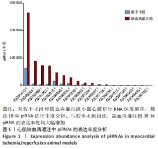
2.1 实验动物数量分析 ①小鼠模型:实验选用小鼠45只,分别作为假手术组和缺血再灌注组,其中缺血再灌注组造模成功的小鼠有21只,成功率为70%。②大鼠模型:实验选用大鼠15只,分别作为假手术组和缺血再灌注组,其中缺血再灌注组造模成功的小鼠有8只,成功率为80%。 2.2 深度测序鉴定心肌缺血再灌注模型中大量piRNA表达水平上调 缺血再灌注模型通常用于临床再灌注损伤的研究。在C57小鼠心脏中构建了缺血再灌注模型及假手术模型,进行RNA深度测序。测序结果显示,小鼠心脏中约表达1 200种piRNA,其中高表达的piRNA有10%左右。筛选出正常和缺血再灌注损伤小鼠心脏中差异表达的422种piRNA,其中约有289条piRNA在缺血再灌注模型中表达水平显著上调,133条piRNA表达水平显著下调。选取了其中18条表达水平显著上调的piRNA,对其丰度进行统计分析,见图1。经过筛选,最终以表达水平显著上调的AB350089.1(piRNA-5938)作为研究对象。"


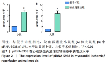
2.3 心肌缺血再灌注动物模型中piRNA-5938表达水平均显著上调 为验证测序结果,分别在C57小鼠和SD大鼠心脏中构建缺血再灌注模型及其假手术模型,实时荧光定量PCR实验检测piRNA-5938表达水平。结果显示,相对于假手术组,缺血再灌注组小鼠心脏组织中piRNA-5938的表达水平上调约3.5倍(P < 0.01),见图2A。相对于假手术组,缺血再灌注组大鼠心脏组织中piRNA-5938的表达水平上调超过6倍(P < 0.01),见图2B。"
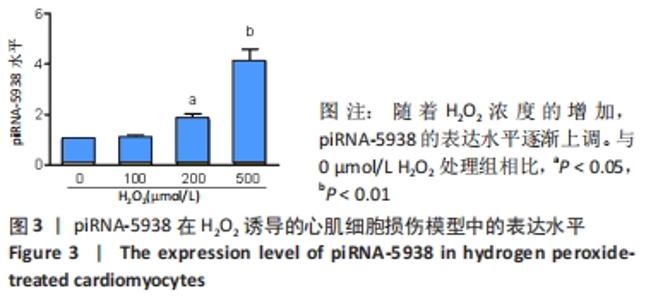
2.4 双氧水诱导心肌细胞凋亡模型中piRNA-5938表达水平显著上调 缺血再灌注损伤发生后,损伤部位会有活性氧自由基释放,引起细胞凋亡。为模拟再灌注损伤中氧化自由基引起的细胞凋亡,使用双氧水诱导SD大鼠乳鼠原代心肌细胞凋亡,实时荧光定量PCR实验结果表明,相较于空白对照组,100 μmol/L双氧水处理后,piRNA-5938的表达水平无显著变化。加大双氧水浓度后,200 μmol/L双氧水处理组中,piRNA的表达水平上调约1.8倍(P < 0.05),500 μmol/L双氧水处理组中,piRNA-5938上调超过4倍(P < 0.01),表明原代心肌细胞中piRNA-5938的上调程度呈与双氧水浓度呈正相关,即具有浓度依赖性,见图3。"

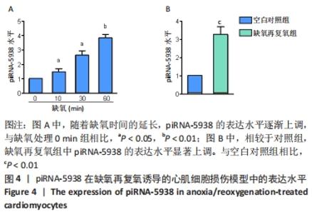
2.5 缺氧再复氧诱导心肌细胞凋亡模型中piRNA-5938表达水平显著上调 缺血再灌注时心肌细胞缺氧再复氧,往往发生氧反常,心肌细胞发生凋亡。因此构建缺氧再复氧诱导心肌细胞凋亡模型,以探究piRNA-5938的表达情况。结果显示,随着缺氧时间的增加,原代心肌细胞中piRNA-5938的表达水平逐渐上调(10 min组上调约1.5倍,30 min组约2.5倍,60 min组3 倍多),见图4A。相较于空白对照组,缺氧再复氧心肌细胞中piRNA-5938的表达水平上调超过3倍(P < 0.01),见图4B。"

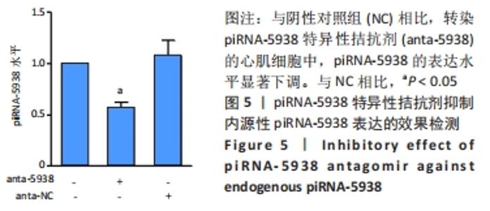
2.6 piRNA-5938拮抗剂显著抑制内源性piRNA-5938的表达 为研究piRNA-5938在心肌损伤中的作用,合成了piRNA-5938特异性拮抗剂(antagomir)及其阴性对照(antagomir NC),转染原代心肌细胞后,实时荧光定量PCR法检测抑制效果。实验结果显示,转染了piRNA-5938拮抗剂的心肌细胞中,piRNA-5938的表达水平较阴性对照组下调约50%(P < 0.05),证实piRNA-5938拮抗剂对内源性piRNA-5938的抑制作用,见图5。"

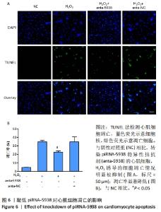
2.7 piRNA-5938调控心肌细胞凋亡 文献报道双氧水可诱导细胞凋亡,TUNEL实验结果也证实,200 μmol/L双氧水处理组后,细胞凋亡率相对空白对照组显著提高,即低浓度的双氧水诱导了原代心肌细胞凋亡,见图6。之前的结果显示,双氧水处理心肌细胞后piRNA-5938的表达水平显著上调,因此猜测piRNA-5938可能调控了双氧水诱导的心肌细胞凋亡。TUNEL法检测心肌细胞凋亡结果显示,与阴性对照组相比,转染piRNA-5938拮抗剂的心肌细胞再经双氧水处理,发生凋亡的细胞数目显著减少,凋亡率由约34%降低到约22%,降低了30%左右(P < 0.05)。以上结果表明,抑制内源性的piRNA-5938显著抑制了双氧水诱导的心肌细胞凋亡,起到心肌保护作用。"
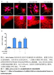
2.8 piRNA-5938调控心肌细胞线粒体分裂 文献报道线粒体分裂参与了细胞凋亡的发生,那么piRNA-5938是否调控线粒体分裂。MitoTracker线粒体探针染色结果显示,相对于空白对照组,双氧水处理后,线粒体分裂率显著提高,说明双氧水诱导了心肌细胞线粒体分裂。而抑制内源性piRNA-5938表达后,相对于阴性对照组,线粒体分裂率降低了约34%,线粒体分裂情况显著减少(P < 0.05),见图7。以上结果表明piRNA-5938调控了双氧水诱导的线粒体分裂,敲低piRNA-5938抑制了线粒体分裂。"

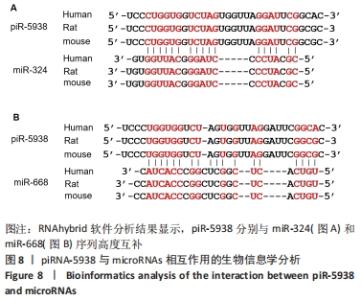
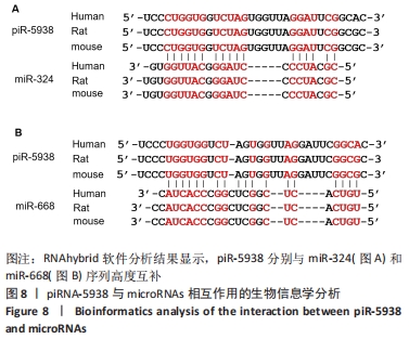
2.9 生物信息学方法预测piRNA-5938靶向线粒体网络调控分子miR-324和miR-668 进一步寻找piRNA-5938调控线粒体分裂的下游靶标。RNAhybrid软件分析了piRNA和microRNA序列,结果显示,piRNA-5938分别与miR-324和miR-668序列能够很好地互补配对,提示piRNA-5938在体内可能与miR-324和miR-668相互结合。已有研究表明miR-324和miR-668是线粒体动态网络调控的重要分子,miR-324通过抑制线粒体分裂调控因子Mtfr1进而抑制了线粒体分裂、心肌细胞凋亡和心肌梗死。miR-668通过靶向线粒体分裂蛋白Mtp18进而抑制了缺氧和缺血引起的线粒体分裂。因此,大胆推测piRNA-5938可能通过靶向结合miR-324或miR-668进而参与miR-324/Mtfr或miR-668/Mtp18线粒体分裂调控途径,见图8。"
| [1] RAMACHANDRA CJA, HERNANDEZ-RESENDIZ S, CRESPO-AVILAN GE, et al. Mitochondria in acute myocardial infarction and cardioprotection. EBioMedicine. 2020;57:102884. [2] DEL RE DP, AMGALAN D, LINKERMANN A, et al. Fundamental Mechanisms of Regulated Cell Death and Implications for Heart Disease. Physiol Rev. 2019;99(4):1765-1817. [3] POHJOISMäKI JL, GOFFART S. The role of mitochondria in cardiac development and protection. Free Radic Biol Med. 2017;106:345-354. [4] SABOUNY R, SHUTT TE. Reciprocal Regulation of Mitochondrial Fission and Fusion . Trends Biochem Sci. 2020;45(7):564-577. [5] VAN DER BLIEK AM, SHEN Q, KAWAJIRI S. Mechanisms of mitochondrial fission and fusion. Cold Spring Harb Perspect Biol. 2013;5(6):a011072. [6] HUANG P, WANG L, LI Q, et al. Atorvastatin enhances the therapeutic efficacy of mesenchymal stem cells-derived exosomes in acute myocardial infarction via up-regulating long non-coding RNA H19. Cardiovasc Res. 2020;116(2):353-367. [7] MAO Q, LIANG XL, ZHANG CL, et al. LncRNA KLF3-AS1 in human mesenchymal stem cell-derived exosomes ameliorates pyroptosis of cardiomyocytes and myocardial infarction through miR-138-5p/Sirt1 axis. Stem Cell Res Ther. 2019;10(1):393. [8] LI A, YU Y, DING X, et al. MiR-135b protects cardiomyocytes from infarction through restraining the NLRP3/caspase-1/IL-1β pathway. Int J Cardiol. 2020;307:137-145. [9] YANG D, YU J, LIU HB, et al. The long non-coding RNA TUG1-miR-9a-5p axis contributes to ischemic injuries by promoting cardiomyocyte apoptosis via targeting KLF5. Cell Death Dis. 2019;10(12):908. [10] OZATA DM, GAINETDINOV I, ZOCH A, et al. PIWI-interacting RNAs: small RNAs with big functions. Nat Rev Genet. 2019;20(2):89-108. [11] DONG J, WANG X, CAO C, et al. UHRF1 suppresses retrotransposons and cooperates with PRMT5 and PIWI proteins in male germ cells. Nat Commun. 2019;10(1):4705. [12] NISHIMURA T, NAGAMORI I, NAKATANI T, et al. PNLDC1, mouse pre-piRNA Trimmer, is required for meiotic and post-meiotic male germ cell development. EMBO Rep. 2018;19(3):e44957. [13] GUAN Y, KEENEY S, JAIN D, et al. yama, a mutant allele of Mov10l1, disrupts retrotransposon silencing and piRNA biogenesis. PLoS Genet. 2021;17(2):e1009265. [14] CHEN P, KOTOV AA, GODNEEVA BK, et al. piRNA-mediated gene regulation and adaptation to sex-specific transposon expression in D. melanogaster male germline. Genes Dev. 2021;35(11-12):914-935. [15] SELLITTO A, GELES K, D’AGOSTINO Y, et al. Molecular and Functional Characterization of the Somatic PIWIL1/piRNA Pathway in Colorectal Cancer Cells. Cells. 2019;8(11):1390. [16] JAIN G, STUENDL A, RAO P, et al. A combined miRNA-piRNA signature to detect Alzheimer’s disease. Transl Psychiatry. 2019;9(1):250. [17] SHEN L, WANG C, CHEN L, et al. Dysregulation of MicroRNAs and PIWI-Interacting RNAs in a Caenorhabditis elegans Parkinson’s Disease Model Overexpressing Human α-Synuclein and Influence of tdp-1. Front Neurosci. 2021;15:600462. [18] TAN L, MAI D, ZHANG B, et al. PIWI-interacting RNA-36712 restrains breast cancer progression and chemoresistance by interaction with SEPW1 pseudogene SEPW1P RNA. Mol Cancer. 2019;18(1):9. [19] GAO XQ, ZHANG YH, LIU F, et al. The piRNA CHAPIR regulates cardiac hypertrophy by controlling METTL3-dependent N(6)-methyladenosine methylation of Parp10 mRNA. Nat Cell Biol. 2020;22(11):1319-1331. [20] YANG J, XUE FT, LI YY, et al. Exosomal piRNA sequencing reveals differences between heart failure and healthy patients. Eur Rev Med Pharmacol Sci. 2018;22(22):7952-7961. [21] ZHANG BF, JIANG H, CHEN J, et al. LncRNA H19 ameliorates myocardial infarction-induced myocardial injury and maladaptive cardiac remodelling by regulating KDM3A. J Cell Mol Med. 2020;24(1):1099-1115. [22] CHEN L, LI S, ZHU J, et al. Mangiferin prevents myocardial infarction-induced apoptosis and heart failure in mice by activating the Sirt1/FoxO3a pathway. J Cell Mol Med. 2021;25(6):2944-2955. [23] VELLA S, GALLO A, LO NIGRO A, et al. PIWI-interacting RNA (piRNA) signatures in human cardiac progenitor cells. Int J Biochem Cell Biol. 2016;76:1-11. [24] LIPPS C, NORTHE P, FIGUEIREDO R, et al. Non-Invasive Approach for Evaluation of Pulmonary Hypertension Using Extracellular Vesicle-Associated Small Non-Coding RNA. Biomolecules. 2019;9(11):666. [25] TAKAHASHI T, HEATON SM, PARRISH NF. Mammalian antiviral systems directed by small RNA. PLoS Pathog. 2021;17(12):e1010091. [26] YAMASHIRO H, SIOMI MC. PIWI-Interacting RNA in Drosophila: Biogenesis, Transposon Regulation, and Beyond. Chem Rev. 2018; 118(8):4404-4421. [27] ZOCH A, AUCHYNNIKAVA T, BERRENS RV, et al. SPOCD1 is an essential executor of piRNA-directed de novo DNA methylation. Nature. 2020; 584(7822):635-639. [28] SHEN S, YU H, LIU X, et al. PIWIL1/piRNA-DQ593109 Regulates the Permeability of the Blood-Tumor Barrier via the MEG3/miR-330-5p/RUNX3 Axis. Mol Ther Nucleic Acids. 2018;10:412-425. [29] LIU X, ZHENG J, XUE Y, et al. PIWIL3/OIP5-AS1/miR-367-3p/CEBPA feedback loop regulates the biological behavior of glioma cells. Theranostics. 2018;8(4):1084-1105. [30] LENG X, MA J, LIU Y, et al. Mechanism of piR-DQ590027/MIR17HG regulating the permeability of glioma conditioned normal BBB. J Exp Clin Cancer Res. 2018;37(1):246. [31] HU YH, SUN J, ZHANG J, et al. Long non-coding RNA ROR sponges miR-138 to aggravate hypoxia/reoxygenation-induced cardiomyocyte apoptosis via upregulating Mst1. Exp Mol Pathol. 2020;114:104430. [32] JIN Y, NI S. miR-496 remedies hypoxia reoxygenation-induced H9c2 cardiomyocyte apoptosis via Hook3-targeted PI3k/Akt/mTOR signaling pathway activation. J Cell Biochem. 2020;121(1):698-712. [33] WANG Y, JIANG Y, SUN X, et al. Downregulation of miR-200a protects cardiomyocyte against apoptosis. Biomed Pharmacother. 2020;123: 109303. [34] YANG J, HUANG X, HU F, et al. LncRNA ANRIL knockdown relieves myocardial cell apoptosis in acute myocardial infarction by regulating IL-33/ST2. Cell Cycle. 2019;18(23):3393-3403. [35] ZHANG X, WANG Q, WANG X, et al. Tanshinone IIA protects against heart failure post-myocardial infarction via AMPKs/mTOR-dependent autophagy pathway. Biomed Pharmacother. 2019;112:108599. [36] ZHANG J, HUANG L, SHI X, et al. Metformin protects against myocardial ischemia-reperfusion injury and cell pyroptosis via AMPK/NLRP3 inflammasome pathway. Aging (Albany NY). 2020;12(23):24270-24287. [37] WU Y, PAN N, AN Y, et al. Diagnostic and Prognostic Biomarkers for Myocardial Infarction. Front Cardiovasc Med. 2020;7:617277. [38] ZHU X, LU X. MiR-423-5p inhibition alleviates cardiomyocyte apoptosis and mitochondrial dysfunction caused by hypoxia/reoxygenation through activation of the wnt/β-catenin signaling pathway via targeting MYBL2. J Cell Physiol. 2019;234(12):22034-22043. [39] ZHU Y, ZHAO P, SUN L, et al. Overexpression of circRNA SNRK targets miR-103-3p to reduce apoptosis and promote cardiac repair through GSK3β/β-catenin pathway in rats with myocardial infarction. Cell Death Discov. 2021;7(1):84. [40] SU Q, LIU Y, LV XW, et al. Inhibition of lncRNA TUG1 upregulates miR-142-3p to ameliorate myocardial injury during ischemia and reperfusion via targeting HMGB1- and Rac1-induced autophagy. J Mol Cell Cardiol. 2019;133:12-25. [41] YANG M, KONG DY, CHEN JC. Inhibition of miR-148b ameliorates myocardial ischemia/reperfusion injury via regulation of Wnt/β-catenin signaling pathway. J Cell Physiol. 2019;234(10):17757-17766. [42] ZHOU R, HUANG W, FAN X, et al. miR-499 released during myocardial infarction causes endothelial injury by targeting α7-nAchR. J Cell Mol Med. 2019;23(9):6085-6097. |
| [1] | Wang Xuejiao, Shi Wenjuan, Zhang Yan, Xing Dehai, Li Dongxue, Jiao Xiangying. Role of thioredoxin-interacting protein-mediated inflammatory response and apoptosis in myocardial infarction [J]. Chinese Journal of Tissue Engineering Research, 2023, 27(11): 1715-1721. |
| [2] | Wang Shuo, Liu Wenying, Lü Chaofan, Li Jiacong, Geng Yi, Zhao Yungang. Cardioprotective effect of 3-nitro-N-methyl salicylamide on the isolated rat heart under cold ischemia preservation [J]. Chinese Journal of Tissue Engineering Research, 2022, 26(8): 1194-1201. |
| [3] | Dong Miaomiao, Lai Han, Li Manling, Xu Xiuhong, Luo Meng, Wang Wenhao, Zhou Guoping. Effect of electroacupuncture on expression of nucleotide binding oligomerization domain-like receptor protein 3/cysteinyl aspartate specific proteinase 1 in rats with cerebral ischemia/reperfusion injury [J]. Chinese Journal of Tissue Engineering Research, 2022, 26(5): 749-755. |
| [4] | Li Yan, Zhang Yan, Zhang Ningkun, Chen Yu. Effect and mechanism of conditioned medium from hypoxia preconditioned human Wharton’s Jelly derived mesenchymal stem cells on myocardial ischemia/reperfusion injury [J]. Chinese Journal of Tissue Engineering Research, 2022, 26(31): 5008-5013. |
| [5] | Cao Shuxing, Song Yongzhou, Li Ming, Zhang Hongliang, Zhao Lihui, Ma Wei. Regulatory role of curcumin in hydrogen peroxide-induced autophagy in chondrocytes [J]. Chinese Journal of Tissue Engineering Research, 2022, 26(14): 2161-2166. |
| [6] | Zhu Chunhui, Zhang Yi, Song Huanghe, Liang Wenwei. Protective effect of astaxanthin on tert-butyl hydrogen peroxide-induced chondrocyte damage [J]. Chinese Journal of Tissue Engineering Research, 2022, 26(11): 1648-1655. |
| [7] | Dong Yi, Deng Jiupeng, Fan Xinhao, Xi Guangwei. Effect of high-concentration H2O2 immersion and ultrasonic treatment with different durations on interfacial bonding strength of fiber post and resin [J]. Chinese Journal of Tissue Engineering Research, 2021, 25(34): 5453-5458. |
| [8] |
Shang Zhizhong, Jiang Yanbiao, Yao Lan, Wang Anan, Wang Hongxia, Tian Yuanxin, Liu Dengrui, Ma Bin .
Feasibility of stem cell therapy for renal ischemia/reperfusion injury: a systematic review based on animal experiments [J]. Chinese Journal of Tissue Engineering Research, 2020, 24(25): 4094-4100. |
| [9] | Long Yanfang1, 2, Wang Xinlei2, Wang Mingpu3, Tang Xingjiang2. Effects of androgen on the expression of Bcl-2, Bax and Cyt-C in brain tissue of adult rat models of middle cerebral artery occlusion [J]. Chinese Journal of Tissue Engineering Research, 2019, 23(27): 4344-4349. |
| [10] | Xu Wen, Qian Yu, Yin Jin . Changes of cardiac function and myocardial apoptosis regulatory factors in rat models of excessive fatigue [J]. Chinese Journal of Tissue Engineering Research, 2019, 23(19): 3074-3079. |
| [11] | Zhou Han, Hui Min, Miao Detian, Wang Le, Dong Xiling, Zhang Xiaoming. Effect of surface treatment reagents and treatment time on bond strength of glass fiber posts to resin cement [J]. Chinese Journal of Tissue Engineering Research, 2019, 23(18): 2852-2857. |
| [12] | Li Manglai, Yang Xuejun. Effects and influencing factors of neurophysiological monitoring on reducing the risk of nerve injury in spine surgery [J]. Chinese Journal of Tissue Engineering Research, 2019, 23(16): 2560-2565. |
| [13] | Yuan Dajiang, Peng Wuxun, Zhang Fei, Wang Zhenwen, Zheng Yinggang. Preincubation with low-concentration hydrogen peroxide enhances anti-oxidative stress ability of bone marrow mesenchymal stem cells [J]. Chinese Journal of Tissue Engineering Research, 2019, 23(13): 1982-1988. |
| [14] | Jiang Wen-jie, Liang Xue-mei. Ginsenoside Rg1 protects against hydrogen peroxide induced damage to bone marrow-derived endothelial progenitor cells [J]. Chinese Journal of Tissue Engineering Research, 2018, 22(33): 5338-5343. |
| [15] | Wang Zhi-qing, Chen Xiong-sheng, Zhou Sheng-yuan, Xu Guo-feng. Role and significance of hydrogen peroxide-induced transforming growth factor beta1 expression in ligamentum flavum hypertrophy [J]. Chinese Journal of Tissue Engineering Research, 2017, 21(12): 1867-1871. |
| Viewed | ||||||
|
Full text |
|
|||||
|
Abstract |
|
|||||