| [1] 丁凡,邵增务.椎间盘组织工程种子细胞研究进展[J].国际骨科学杂志,2010,31(6):376-377,380.
[2] Wang F, Wu X, Wang Y, et al. Research situation of stem cells transplantation for intervertebral disc degeneration. Zhongguo Xiu Fu Chong Jian Wai Ke Za Zhi. 2013;27(5):575-579.
[3] Zhang L, Ning B, Jia T, et al. Microcarrier bioreactor culture system promotes propagation of human intervertebral disc cells. Ir J Med Sci. 2010;179(4):529-534.
[4] 丁树伟,徐学振. 椎间盘组织工程种子细胞的选取、鉴定及培养难点[J].中国医药指南,2013,11:470-472.
[5] 陈国仙,王万明,王国荣,等. 骨髓间充质干细胞透明质酸钠溶液复合体移植修复兔退变椎间盘的实验研究[J].中华临床医师杂志(电子版),2011, 5(6):1547-1553.
[6] 王青,贾长青.干细胞生物技术治疗椎间盘退行性病变的现状[J].中国组织工程研究与临床康复,2007,11(20):4001-4004.
[7] Jin ES, Min J, Jeon SR, et al. Analysis of molecular expression in adipose tissue-derived mesenchymal stem cells : prospects for use in the treatment of intervertebral disc degeneration. J Korean Neurosurg Soc. 2013;53(4):207-212.
[8] Stoyanov JV, Gantenbein-Ritter B, Bertolo A, et al. Role of hypoxia and growth and differentiation factor-5 on differentiation of human mesenchymal stem cells towards intervertebral nucleus pulposus-like cells. Eur Cell Mater. 2011;21:533-547.
[9] Korecki CL, Taboas JM, Tuan RS, et al. Notochordal cell conditioned medium stimulates mesenchymal stem cell differentiation toward a young nucleus pulposus phenotype. Stem Cell Res Ther. 2010;1(2):18.
[10] 张彦男,邵增务,吴永超,等. 非接触共培养脊索细胞诱导骨髓间充质干细胞向类软骨细胞分化的实验研究[J].中国脊柱脊髓杂志,2012,22(8):737-742.
[11] 韩成龙,姜超.骨髓间充质干细胞向髓核细胞的转化[J].中国矫形外科杂志,2009,17(19):1476-1478.
[12] 李全修,陈伯华,刘勇,等.构建兔髓核细胞诱导人骨髓间充质干细胞向类髓核细胞分化的体外模型[J].中国组织工程研究与临床康复,2009,13(45):8961-8965.
[13] 韩成龙,姜超.模拟微重力下转化生长因子β1诱导骨髓间充质干细胞向髓核样细胞分化[J].中国脊柱脊髓杂志,2011,21(5): 358-364.
[14] 李华壮,周跃.椎间盘组织工程研究进展[J].中华骨科杂志,2005, 25(5):316-318.
[15] van den Eerenbeemt KD, Ostelo RW, van Royen BJ, et al. Total disc replacement surgery for symptomatic degenerative lumbar disc disease: a systematic review of the literature. Eur Spine J. 2010;19(8):1262-1280.
[16] Miller LE, Block JE. Safety and effectiveness of bone allografts in anterior cervical discectomy and fusion surgery. Spine (Phila Pa 1976). 2011;36(24):2045-2050.
[17] Meisel HJ, Siodla V, Ganey T, et al. Clinical experience in cell-based therapeutics: disc chondrocyte transplantation A treatment for degenerated or damaged intervertebral disc. Biomol Eng. 2007;24(1):5-21.
[18] Watanabe K, Mochida J, Nomura T, et al. Effect of reinsertion of activated nucleus pulposus on disc degeneration: an experimental study on various types of collagen in degenerative discs. Connect Tissue Res. 2003;44(2): 104-108.
[19] 李军芳,邝晓聪,韦瑛,等.人骨髓间充质干细胞体内移植安全性评价的初步研究[J].广西医科大学学报,2009,26(6):833-836.
[20] 呼和,韩成龙,姜超,等.转化生长因子β1对骨髓干细胞分化类髓核细胞的影响[J].中国组织工程研究与临床康复,2011,15(36): 6722-6726.
[21] 吴健,杨金华,杨宗华,等.可注射型纤维蛋白凝胶转化生长因子β1复合骨髓间充质干细胞移植治疗椎间盘退变[J].中国组织工程研究与临床康复,2011,15(3):478-482.
[22] Risbud MV, Guttapalli A, Stokes DG, et al. Nucleus pulposus cells express HIF-1 alpha under normoxic culture conditions: a metabolic adaptation to the intervertebral disc microenvironment. J Cell Biochem. 2006;98(1):152-159.
[23] Yamamoto Y, Mochida J, Sakai D, et al. Upregulation of the Viability of Nucleus Pulposus Cells by Bone Marrow-Derived Stromal Cells: Significance of Direct Cell-to-Cell Contact in Coculture System. Spine.2004;29(14):1508-1514.
[24] 刘连江,李放,文天用,等.转化生长因子β1/胰岛素样生长因子1与髓核细胞诱导脂肪间充质干细胞向髓核样软骨细胞分化的比较[J].中国组织工程研究与临床康复,2010,14(23):4217-4221.
[25] 邵建树,吴小涛,王运涛,等.兔骨髓间充质干细胞与髓核细胞共培养后的营养效应和类髓核分化效应[J].中国脊柱脊髓杂志,2009, 19(5):381-387.
[26] 陈亮,李大鹏,黄永辉,等.髓核细胞诱导骨髓间充质干细胞向髓核方向的分化[J].中国组织工程研究,2013,17(6):992-998. |

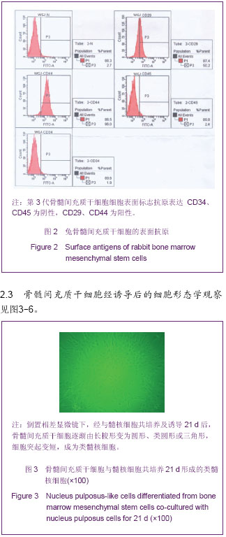


.jpg)