[1] SHI S, SI Y, HAN Y, et al. Recent Progress in Protective Membranes Fabricated via Electrospinning: Adv. Mater., Biomimetic Structures, and Functional Applications. Adv Mater. 2022;34(17):2107938.
[2] SI Y, SHI S, HU J. Applications of electrospinning in human health: From detection, protection, regulation to reconstruction. Nano Today. 2023;48:101723.
[3] SALEHHUDIN HS, MOHAMAD EN, MAHADI WNL, et al. Multiple-jet electrospinning methods for nanofiber processing: A review. Mater Manuf Process. 2018;33(5):479-498.
[4] ZHI C, SHI S, SI Y, et al. Recent Progress of Wearable Piezoelectric Pressure Sensors Based on Nanofibers, Yarns, and Their Fabrics via Electrospinning. Adv Mater Technol. 2023;8(5):2201161.
[5] DOU Y, ZHANG W, KAISER A. Electrospinning of Metal–Organic Frameworks for Energy and Environmental Applications. Adv Sci. 2020;7(3):1902590.
[6] KEIROUZ A, WANG Z, REDDY VS, et al. The History of Electrospinning: Past, Present, and Future Developments. Adv Mater Technol. 2023;8(11):2201723.
[7] LOSCERTALES IG, BARRERO A, GUERRERO I, et al. Micro/Nano Encapsulation via Electrified Coaxial Liquid Jets. Science. 2002;295(5560):1695-1698.
[8] YANG Z, GAO M, LIANG W, et al. One-dimensional electrospinning nanomaterials toward capacitive deionization: Fundamentals, development, and perspectives. Desalination. 2023;567:117010.
[9] ZULKIFLI MZA, NORDIN D, SHAARI N, et al.
Overview of Electrospinning for Tissue Engineering Applications. Polymers. 2023; 15(11):2418.
[10] HAN W, WANG L, SUN J, et al. Dual-Drug-Loaded Core–Shell Electrospun Nanofiber Dressing for Deep Burns. ACS Appl Bio Mater. 2024;7(2):1179-1190.
[11] PIEN N, KRZYSLAK H, SHASTRY KALLAJE S, et al. Tissue engineering of skeletal muscle, tendons and nerves: A review of manufacturing strategies to meet structural and functional requirements. Appl Mater Today. 2023;31:101737.
[12] MACEWAN MR, KOVACS T, OSBUN J, et al. Comparative analysis of a fully-synthetic nanofabricated dura substitute and bovine collagen dura substitute in a large animal model of dural repair. Interdiscip Neurosurg. 2018;13:145-150.
[13] 陈亮,许运.静电纺丝微米/纳米纤维材料在硬膜组织损伤修复中的应用[J].中华实验外科杂志,2016,33(7):1880-1883.
[14] SABATINO G, DELLA PEPA GM, BIANCHI F, et al. Autologous dural substitutes: A prospective study. Clin Neurol Neurosurg. 2014;116:20-23.
[15] BOHOUN CA, GOTO T, MORISAKO H, et al. Skull Base Dural Repair Using Autologous Fat as a Dural Substitute: An Efficient Technique. World Neurosurg. 2019;127:e896-e900.
[16] MIYASHITA K, INUZUKA T, KONDO H, et al.
Creutzfeldt‐Jakob disease in a patient with a cadaveric dural graft. Neurology. 1991;41(6):940.
[17] TURCHAN A, ROCHMAN TF, IBRAHIM A,
et al. Duraplasty using amniotic membrane versus temporal muscle fascia: A clinical comparative study. J Clin Neurosci. 2018; 50:272-276.
[18] KNOPP U, CHRISTMANN F, REUSCHE E, et al. A new collagen biomatrix of equine origin versus a cadaveric dura graft for the repair of dural defects – a comparative animal experimental study. Acta Neurochir. 2005;147(8):877-887.
[19] SEO Y, KIM JW, DHO YS, et al. Evaluation of the safety and effectiveness of an alternative dural substitute using porcine pericardium for duraplasty in a large animal model. J Clin Neurosci. 2018;58:187-191.
[20] WANG W, QIANG A. Research and application progress on dural substitutes. J Neuroresstoratology. 2019;7(4):161-170.
[21] ATTENELLO FJ, MCGIRT MJ, GARCÉS-AMBROSSI GL, et al. Suboccipital decompression for Chiari I malformation: outcome comparison of duraplasty with expanded polytetrafluoroethylene dural substitute versus pericranial autograft. Childs Nerv Syst. 2009;25(2):183-190.
[22] ZHANG Q, LI R, LI J, et al. PDMS/Uhmwpe Dura Mater Substitute Laminated with Conductive Graphene for Intracranial Pressure Sensing. ECS MA. 2019;(44):2097.
[23] SANDOVAL-SÁNCHEZ JH, RAMOS-ZÚÑIGA R, LUQUÍN DE ANDA S, et al. A New Bilayer Chitosan Scaffolding as a Dural Substitute: Experimental Evaluation. World Neurosurgery. 2012;77(3-4):577-582.
[24] MATSUMOTO K, NAKAMURA T, FUKUDA S, et al. A Gelatin Coated Collagen-Polyglycolic Acid Composite Membrane as a Dural Substitute. ASAIO J. 2001;47(6):641-645.
[25] XU C, MA X, CHEN S, et al. Bacterial Cellulose Membranes Used as Artificial Substitutes for Dural Defection in Rabbits. Int J Mol Sci. 2014;15(6):10855-10867.
[26] KIM DW, EUM WS, JANG SH, et al. A transparent artificial dura mater made of silk fibroin as an inhibitor of inflammation in craniotomized rats: Laboratory investigation. J Neurosurg. 2011;114(2):485-490.
[27] NUNAMAKER EA, OTTO KJ, KIPKE DR. Investigation of the material properties of alginate for the development of hydrogel repair of dura mater. J Mech Behav Biomed Mater. 2011;4(1):16-33.
[28] POGORIELOV M, KRAVTSOVA A, REILLY GC, et al. Experimental evaluation of new chitin–chitosan graft for duraplasty. J Mater Sci Mater Med. 2017;28(2):34.
[29] DENG W, TAN Y, RIAZ RAJOKA MS, et al. A new type of bilayer dural substitute candidate made up of modified chitin and bacterial cellulose. Carbohydr Polym. 2021;256:117577.
[30] KINACI A, VAN THOOR S, REDEGELD S, et al. Ex vivo evaluation of a multilayered sealant patch for watertight dural closure: cranial and spinal models. J Mater Sci Mater Med. 2021;32(8):85.
[31] XU Y, CUI W, ZHANG Y, et al. Hierarchical Micro/Nanofibrous Bioscaffolds for Structural Tissue Regeneration. Adv Healthc Mater. 2017;6(13):1601457.
[32] LIU W, THOMOPOULOS S, XIA Y. Electrospun Nanofibers for Regenerative Medicine. Adv Healthc Mater. 2012;1(1): 10-25.
[33] LIU Y, GUO Q, ZHANG X, et al. Progress in Electrospun Fibers for Manipulating Cell Behaviors. Adv Fiber Mater. 2023;5(4):
1241-1272.
[34] POZZOBON LG, SPERLING LE, TEIXEIRA CE, et al. Development of a conduit of PLGA-gelatin aligned nanofibers produced by electrospinning for peripheral nerve regeneration. Chem Biol Interact. 2021;348: 109621.
[35] YIN Z, CHEN X, SONG H, et al. Electrospun scaffolds for multiple tissues regeneration in vivo through topography dependent induction of lineage specific differentiation. Biomaterials. 2015;44:173-185.
[36] KOBAYASHI M, LEI NY, WANG Q, et al.
Orthogonally oriented scaffolds with aligned fibers for engineering intestinal smooth muscle. Biomaterials. 2015;61:75-84.
[37] YAU WWY, LONG H, GAUTHIER NC, et al. The effects of nanofiber diameter and orientation on siRNA uptake and gene silencing. Biomaterials. 2015;37:94-106.
[38] GUO Z, XU J, DING S, et al. In vitro evaluation of random and aligned polycaprolactone/gelatin fibers via electrospinning for bone tissue engineering. J Biomater Sci Polym Ed. 2015;26(15):989-1001.
[39] PROTASONI M, SANGIORGI S, CIVIDINI A, et al. The collagenic architecture of human dura mater: Laboratory investigation. J Neurosurg. 2011;114(6):1723-1730.
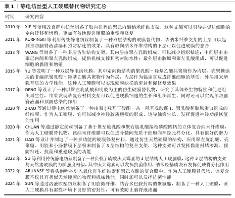
[40] XIE J, MACEWAN MR, RAY WZ, et al. Radially Aligned, Electrospun Nanofibers as Dural Substitutes for Wound Closure and Tissue Regeneration Applications. ACS Nano. 2010; 4(9):5027-5036.
[41] CHUAN D, WANG Y, FAN R, et al. Fabrication and Properties of a Biomimetic Dura Matter Substitute Based on Stereocomplex Poly(lactic Acid) Nanofibers. Int J Nanomed. 2020;15:3729-3740.
[42] 徐敬之,王文博,孙慧雯,等.干细胞工程化双面异性静电纺丝膜促进硬脊膜修复的体外实验[J].中国组织工程研究, 2024,28(10):1540-1546.
[43] YU F, LI Q, YIN S, et al. Reconstructing spinal dura-like tissue using electrospun poly(lactide-co-glycolide) membranes and dermal fibroblasts to seamlessly repair spinal dural defects in goats. J Biomater Appl. 2015;30(3):311-326.
[44] 于凤宾,董波,岑莲,等.体外构建并评价组织工程化复合PLGA人工硬脊膜[J].现代医药卫生,2020,36(5):663-667.
[45] KURPINSKI K, PATEL S. Dura mater regeneration with a novel synthetic, bilayered nanofibrous dural substitute: an experimental study. Nanomedicine. 2011;6(2):325-337.
[46] XU Y, SHI G, TANG J, et al. ECM-inspired micro/nanofibers for modulating cell function and tissue generation. Sci Adv. 2020;6(48):eabc2036.
[47] SU Y, LI Z, ZHU H, et al. Electrohydrodynamic Fabrication of Triple-layered Polycaprolactone Dura Mater Substitute with Antibacterial and Enhanced Osteogenic Capability. CJME:AMF. 2022;1(2):100026.
[48] WANG Y, GUO H, YING D. Multilayer scaffold of electrospun PLA–PCL–collagen nanofibers as a dural substitute. J Biomed Mater Res B Appl Biomater. 2013;101(8):1359-1366.
[49] GHEZZI CE, MARELLI B, MUJA N, et al. Mesenchymal stem cell‐seeded multilayered dense collagen‐silk fibroin hybrid for tissue engineering applications. Biotechnol J. 2011;6(10):1198-1207.
[50] DENG K, YANG Y, KE Y, et al. A novel biomimetic composite substitute of PLLA/gelatin nanofiber membrane for dura repairing. Neurol Res. 2017;39(9):819-829.
[51] LV FY, DONG RH, LI ZJ, et al. In situ precise electrospinning of medical glue fibers as nonsuture dural repair with high sealing capability and flexibility. Int J Nanomed. 2016;11:4213-4220.
[52] MA H, SUN Y, TANG Y, et al. Robust Electrospun Nanofibers from Chemosynthetic Poly(4‐hydroxybutyrate) as Artificial Dural Substitute. Macromol Biosci. 2021;21(7):2100134.
[53] SUN S, LUO H, WANG Y, et al. Artificial spinal dura mater made of gelatin microfibers and bioadhesive for preventing cerebrospinal fluid leakage. Chem Commun. 2024;60(17): 2353-2356.
[54] 王汉斌,苏亦兵,史良,等.一种聚己内酯/明胶膜对兔颅骨缺损模型的防黏连作用[J].中国微侵袭神经外科杂志, 2016,21(11):516-519.
[55] SHI R, XUE J, WANG H, et al. Fabrication and evaluation of a homogeneous electrospun PCL–gelatin hybrid membrane as an anti-adhesion barrier for craniectomy. J Mat Chem B. 2015;3(19):4063-4073.
[56] WANG S, YU P, LI X, et al. Design and fabrication of functional hydrogels with specific surface wettability. Colloid Interface Sci Commun. 2023;52:100697.
[57] LIU S, PAN G, LIU G, et al. Electrospun fibrous membranes featuring sustained release of ibuprofen reduce adhesion and improve neurological function following lumbar laminectomy. J Control Release. 2017;264:1-13.
[58] SHI R, HUANG Y, ZHANG J, et al. Effective delivery of mitomycin‐C and meloxicam by double‐layer electrospun membranes for the prevention of epidural adhesions. J Biomed Mater Res Part B. 2020;108(2):353-366.
[59] JING Y, MA X, XU C, et al. Repair of dural defects with electrospun bacterial cellulose membranes in a rabbit experimental model. Mater Sci Eng C Mater Biol Appl. 2020;117:111246.
[60] WANG J, LI K, XU J, et al. A biomimetic hierarchical small intestinal submucosa-chitosan sponge/chitosan hydrogel scaffold with a micro/nano structure for dural repair. J Mat Chem B. 2021;9(37):7821-7834.
[61] SANPAKITWATTANA A, SUVANNAPRUK W, CHUMNANVEJ S, et al. Cefazolin Loaded Oxidized Regenerated Cellulose/Polycaprolactone Bilayered Composite for Use as Potential Antibacterial Dural Substitute. Polymers. 2022;14(20):4449.
[62] MOHTARAM NK, KO J, AGBAY A, et al. Development of a glial cell-derived neurotrophic factor-releasing artificial dura for neural tissue engineering applications. J Mat Chem B. 2015;3(40):7974-7985.
[63] ZHU Y, WANG A, SHEN W, et al. Nanofibrous Patches for Spinal Cord Regeneration. Adv Funct. Mater. 2010;20(9):1433-1440.
[64] ZHAO T, XU K, WU Q, et al. Duraplasty of PHBV/PLA/Col membranes promotes axonal regeneration by inhibiting NLRP3 complex and M1 macrophage polarization in rats with spinal cord injury. FASEB J. 2020;34(9): 12147-12162. |