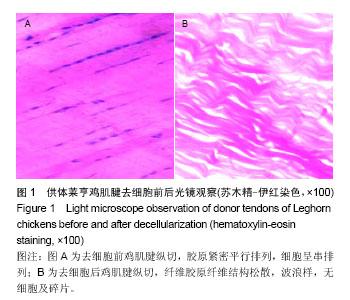
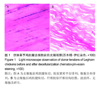
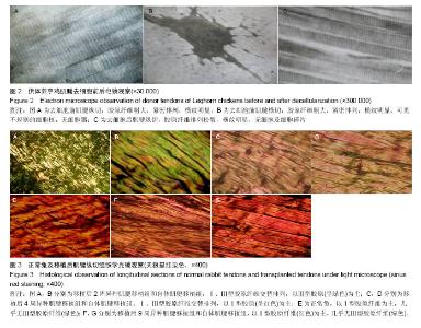
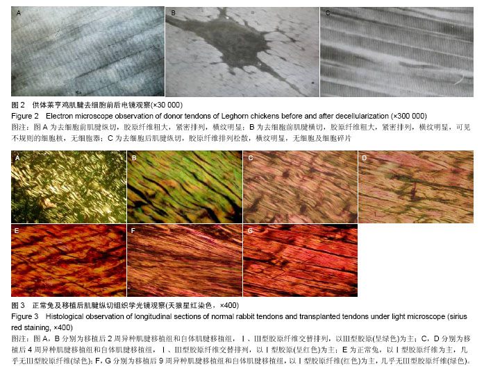
| [1] Cissell DD, Hu JC, Griffiths LG, et al. Antigen removal for the production of biomechanically functional,xenogeneic tissue grafts.J Biomech. 2014;47(9):1987-1996.
[2] Wong ML,Wong JL,Athanasiou KA,Griffiths LG,et al.Stepwise solubilization-based antigen removal for xenogeneic scaffold generation in tissue engineering. Acta Biomater. 2013;9(5): 6492-6501.
[3] Deeken CR, White AK, Bachman SL, et al. Method of preparing a decellularized porcine tendon using tributyl phosphate. J Biomed Mater Res B Appl Biomater. 2011;96(2): 199-206.
[4] Schulze-Tanzil G,Al-Sadi O,Ertel W,et al.Decellularized tendon extracellular matrix-a valuable approach for tendon reconstruction. Cells. 2012;1(4):1010-1028.
[5] 梁黎明,柴家科,杨红明,等.脱细胞肌腱制备的实验研究[J].中国美容医学,2006,(03):239-240.
[6] Omae H, Sun YL, An KN, et al. Engineered tendon with decellularized xenotendon slices and bone marrow stromal cells: an in vivo animal study. J Tissue Eng Regen Med. 2012;6(3):238-244.
[7] 左新成,林子豪,黄昌林,等.不同理化方法处理异种肌腱移植的动物实验[J].中华实验外科杂志,2002,19(6):607.
[8] 陈元良,姜平,夏学颖,等.异种肌腱基质材料生物相容性的体外评价实验.中国美容医学,2011,20(5):787-790.
[9] 邱南海,夏英鹏,组织工程化肌腱的实验研究与临床应用[J]. 中国组织工程研究与临床康复,2008,12(24): 4718-4722.
[10] Tanaka H, Manske PR, Pruitt DL, et al. Effect of cyclic tension on lacerated flexor tendons in vitro. J Hand Surg Am.1995;20(3):467-473.
[11] Chen Z, Hong G, Wan F. [Morphometric study of collagen fibers during healing following partial and complete section of extensor tendons in rats]. Zhongguo Xiu Fu Chong Jian Wai Ke Za Zhi.1997;11(5):276-278.
[12] Forslund C, Aspenberg P. CDMP-2 induces bone or tendon-like tissue depending on mechanical stimulation. J Orthop Res.2002;20(6):1170-1174.
[13] Skutek M, van Griensven M, Zeichen J, et al. Cyclic mechanical stretching modulates secretion pattern of growth factors in human tendon fibroblasts. Eur J Appl Physiol.2001. 86(1):48-52. |
