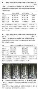
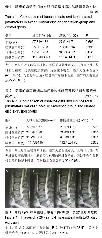
| [1] Kjaer P, Leboeuf-Yde C, Sorensen JS, et al. An epidemiologic study of MRI and low back pain in 13-year-old children. Spine (Phila Pa 1976). 2005; 30(7):798-806.
[2] Hoy D, Brooks P, Blyth F, et al. The Epidemiology of low back pain. Best Pract Res Clin Rheumatol. 2010; 24(6):769-781.
[3] Berg S, Isberg B, Josephson A, et al. The impact of discography on the surgical decision in patients with chronic low back pain. Spine J. 2012;12(4):283-291.
[4] Lu Y, Guzman JZ, Purmessur D, et al. Nonoperative management of discogenic back pain: a systematic review. Spine (Phila Pa 1976). 2014;39(16): 1314-1324.
[5] Battie MC, Videman T, Parent E. Lumbar disc degeneration: epidemiologic and genetic influences. Spine (Phila Pa 1976). 2004;29(23):2679-2690.
[6] Belavý DL, Armbrecht G, Felsenberg D. Evaluation of lumbar disc and spine morphology: long-term repeatability and comparison of methods. Physiol Meas. 2012;33(8):1313-1321.
[7] Yang X, Kong Q, Song Y, et al. The characteristics of spinopelvic sagittal alignment in patients with lumbar disc degenerative diseases. Eur Spine J. 2014;23(3): 569-575.
[8] Endo K, Suzuki H, Tanaka H, et al. Sagittal spinal alignment in patients with lumbar disc herniation. Eur Spine J. 2010;19(3):435-438.
[9] Osterman K, Osterman H. Experimental lumbar spondylolisthesis in growing rabbits. Clin Orthop Relat Res. 1996;(332):274-280.
[10] Dubousset J. Treatment of spondylolysis and spondylolisthesis in children and adolescents. Clin Orthop Relat Res. 1997;(337):77-85.
[11] Ergun T, Lakadamyali H, Sahin MS. The relation be-tween sagittal morphology of the lumbosacral spine and the degree of lumbar intervertebral disc degeneration. Acta Orthop Traumatol Turc. 2010;44(4): 293-299.
[12] Evcik D, Yucel A. Lumbar lordosis in acute and chronic low back pain patients. Rheumatol Int. 2003;23(4): 163-165.
[13] Youdas JW, Garrett TR, Egan KS, et al. Lumbar lordosis and pelvic inclination in adults with chronic low back pain. Phys Ther. 2000;80(3):261-275.
[14] Williams AL, Haughton VM. Daniels DL, et al. Differential diagnosis of extrud nucleus pulposus. Radiology. 1983;148(1):141-148.
[15] Lao L, Daubs MD, Takahashi S, et al. Kinetic magnetic resonance imaging analysis of lumbar segmental motion at levels adjacent to disc herniation. Eur Spine J. 2016;25(1):222-229.
[16] Merot OA, Maugars YM, Berthelot JM. Similar outcome despite slight clinical differences between lumbar radiculopathy induced by lateral versus medial disc herniations in patients without previous foraminal stenosis: a prospective cohort study with 1-year follow-up. Spine J. 2014;14(8):1526-1531
[17] Andreasen ML, Langhoff L, Jensen TS, et al. Reproduction of the lumbar lordosis: a comparison of standing radiographs versus supine magnetic resonance imaging obtained with straightened lower extremities. J Manipulative Physiol Ther. 2007;30(1): 26-30.
[18] Kalichman L, Li L, Hunter DJ, et al. Association between computed tomography-evaluated lumbar lordosis and features of spinal degeneration, evaluated in supine position. Spine J. 2011;11(4):308-315.
[19] Liu H, Li S, Zheng Z, et al. Pelvic retroversion is the key protective mechanism of L4–5 degenerative spondylolisthesis. Eur Spine J. 2015;24(6):1204-1211.
[20] Rajnics P, Templier A, Skalli W, et al. The importance of spinopelvic parameters in patients with lumbar disc lesions.Int Orthop. 2002;26(2):104-108.
[21] Yang X, Kong QQ, Song YM, et al. The characteristics of spinopelvic sagittal alignment in patients with lumbar disc degenerative diseases. Eur Spine J. 2014;23(3): 569-575.
[22] Gelb DE, Lenke LG, Bridwell KH, et al. An analysis of sagittal spinal alignment in 100 asymptomatic middle and older aged volunteers. Spine (Phila Pa 1976). 1995;20(12):1351-1358.
[23] Keorochana G, Taghavi CE, Lee KB, et al. Effect of sagittal alignment on kinematic changes and degree of disc degeneration in the lumbar spine: an analysis using positional MRI. Spine (Phila Pa 1976). 2011; 36(11):893-898.
[24] Wang Z, Parent S, Mac-Thiong JM, et al. Influence of sacral morphology in developmental spondylolisthesis. Spine (Phila Pa 1976). 2008;33(20):2185-2191.
[25] Whitesides TE Jr, Horton WC, Hutton WC, et al. Spondylolytic spondylolisthesis: a study of pelvic and lumbosacral parameters of possible etiologic effect in two genetically and geographically distinct groups with high occurrence. Spine (Phila Pa 1976). 2005;30 (6 Suppl):S12-S21.
[26] Glavas P, Jean-Marc M. Assessment of lumbosacral kyphosis in spondylolisthesis: a computer-assisted reliability study of six measurement techniques. Eur Spine J. 2009;18(2):212-217.
[27] Tanguay F, Labelle H, Wang Z, et al. Clinical significance of lumbosacral kyphosis in adolescent spondylolisthesis. Spine (Phila Pa 1976). 2012;37(4): 304-308.
[28] Oh YM, Eun JP. The relationship between sagittal spinopelvic parameters and the degree of lumbar intervertebral disc degeneration in young adult patients with low-grade spondylolytic spondylolisthesis. Bone Joint J. 2013;95-B(9):1239-1243.
[29] Yang X, Kong Q, Song Y, et al. The characteristics of spinopelvic sagittal alignment in patients with lumbar disc degenerative diseases. Eur Spine J. 2014;23(3): 569-575.
[30] Viester L, Verhagen E, Hengel KM, et al. The relation between body mass index and musculoskeletal symptoms in the working population. BMC Musculoskelet Disord. 2013;14:238. |