Chinese Journal of Tissue Engineering Research ›› 2014, Vol. 18 ›› Issue (40): 6459-6463.doi: 10.3969/j.issn.2095-4344.2014.40.011

Previous Articles     Next Articles

Anterior plate internal fixation and autologous iliac bone graft fusion in the repair of thoracic spinal tuberculosis: bone healing

Huang Jiang1, Li Xiao-feng2, Yang Yuan2   

  1. 1Hepu County People’s Hospital, Hepu 536100, Guangxi Zhuang Autonomous Region, China; 2Guangxi Orthopedics and Traumatology Hospital, Nanning 530012, Guangxi Zhuang Autonomous Region, China
  • Revised:2014-08-04 Online:2014-09-24 Published:2014-09-24
  • Contact: Yang Yuan, Professor, Chief physician, Guangxi Orthopedics and Traumatology Hospital, Nanning 530012, Guangxi Zhuang Autonomous Region, China
  • About author:Huang Jiang, Associate chief physician, Hepu County People’s Hospital, Hepu 536100, Guangxi Zhuang Autonomous Region, China
  • Supported by:

    the Guangxi Zhuang Autonomous Region Health Department Funded Project, No. 200863

Abstract:

BACKGROUND: Thoracic vertebrae connected with the ribs of corresponding segments constitute the thorax. Because of thoracic vertebrae deeply and complicated structure, it is difficult to operate or to expose thoracic vertebrae completely. The traditional anterior ways can thoroughly remove the focus and achieve a good internal fixation, but this treatment needs to cut the rid. The surgeon discovered in years of clinical practice that distraction of intercostal space without cutting off the ribs could completely remove the focus and achieve fixation and fusion.

OBJECTIVE: To explore the feasibility, advantages and disadvantages of anterior plate fixation and autologous bone graft fusion in the repair of thoracic spinal tuberculosis.
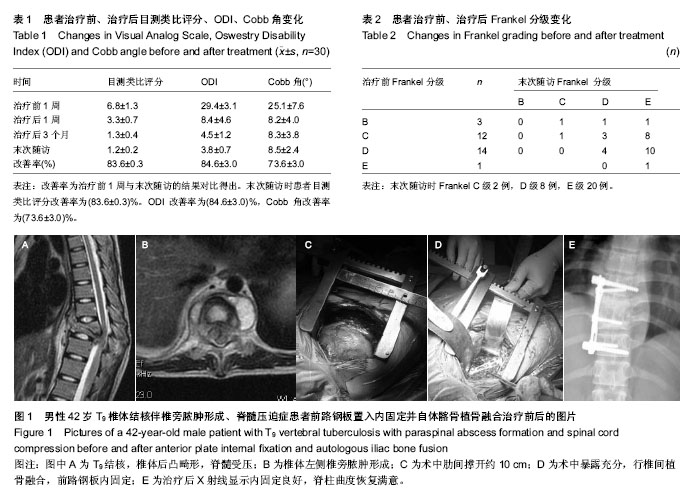
METHODS: A total of 30 cases of thoracic spinal tuberculosis were enrolled. On the base of traditional thoracic spinal anterior surgical approach, the rib was reserved. Anterior distraction device was used to open the ribs for clearance and to move the focus of thoracic spinal tuberculosis. Autologous ilium was obtained and subjected to fusion and anterior plate fixation. Visual Analog Scale, Oswestry Disability Index, Frankel grade and Bridwell bone healing rating were used before and after treatment.

RESULTS AND CONCLUSION: A total of 30 patients were followed up for 3-60 months. The bone healing (class A) was seen, but no fistula formation was visible. Nerve compression symptoms were improved, without aggravating cases. Postoperatively, the average Cobb angle correction was 10.32°. After 6 months, imaging revealed bone fusion of affected vertebral body. After 18 months, the vertebral height was not lost obviously, no recurrence of vertebral tuberculosis. These results indicated that anterior intercostal space without cutting ribs exposed clearly, showing a large space, where can meet the requirement of first-stage debridement fixation and fusion in the repair of thoracic tuberculosis. The pathological changes were visibly clear; the focus was thoroughly removed, and the ribs were reserved. The outcomes are identical to rib resection. Simultaneously, autogenous iliac trilateral cortex has good supporting effects. Loose cancellous bone mesh has good osteogenesis and is helpful to bone healing.


中国组织工程研究杂志出版内容重点:人工关节;骨植入物;脊柱骨折;内固定;数字化骨科;组织工程


全文链接:

Key words:  thoracic vertebrae, tuberculosis, internal fixators, bone transplantation, ilium

CLC Number: