Chinese Journal of Tissue Engineering Research ›› 2013, Vol. 17 ›› Issue (11): 1993-2000.doi: 10.3969/j.issn.2095-4344.2013.11.014
Previous Articles Next Articles
Changes of gliacytes after spinal cord transection in Ambystoma mexicanum
Li Min, Liu Jia, Zhong Yu-hua, Peng Fu-hua
- Department of Neurology, Third Affiliated Hospital of Sun Yat-sen University, Guangzhou 510630, Guangdong Province, China
-
Received:2012-12-10Revised:2013-01-07Online:2013-03-12Published:2013-03-12 -
Contact:Peng Fu-hua, Doctor, Chief physician, Department of Neurology, Third Affiliated Hospital of Sun Yat-sen University, Guangzhou 510630, Guangdong Province, China pfh93@163.com -
About author:Li Min★, Studying for master’s degree, Department of Neurology, Third Affiliated Hospital of Sun Yat-sen University, Guangzhou 510630, Guangdong Province, China plum-min@163.com
CLC Number:
Cite this article
Li Min, Liu Jia, Zhong Yu-hua, Peng Fu-hua. Changes of gliacytes after spinal cord transection in Ambystoma mexicanum[J]. Chinese Journal of Tissue Engineering Research, 2013, 17(11): 1993-2000.
share this article
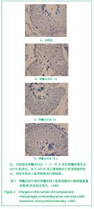
2.1 实验动物数量分析 实验动物分为对照组和手术组,各48条。在实验过程中由于感染等因素有脱失,每组最多不超过3条。在0,1,3及10 d每个时间点每组保证取6条。 2.2 脊髓全切后损伤脊髓周围小胶质细胞和巨噬细胞数量的变化 见图2。"
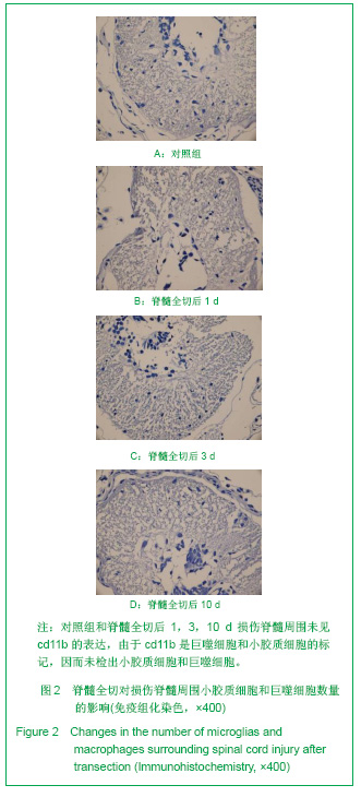
cd11b为小胶质细胞单克隆抗体,可作为小胶质细胞标记[11]。免疫组化结果显示,对照组脊髓不表达cd11b,而脊髓全切后1,3,10 d时,损伤脊髓附近也未见cd11b的表达,说明损伤脊髓周围没有小胶质细胞和巨噬细胞。 2.3 脊髓全切后损伤脊髓周围星形胶质细胞数量的变化 见图3。"
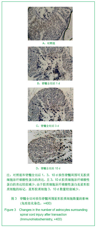
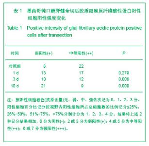
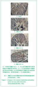
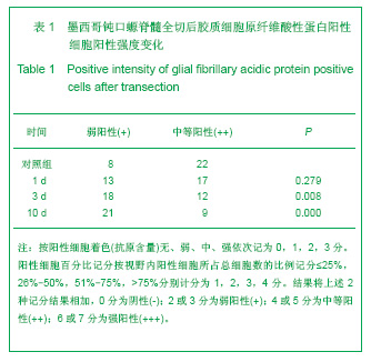
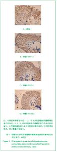
胶质细胞原纤维酸性蛋白被广泛证实仅识别星形胶质细胞,可作为星形胶质细胞的标记[12-19]。免疫组化结果显示,对照组脊髓大量表达胶质细胞原纤维酸性蛋白,脊髓全切后1 d时,损伤脊髓附近胶质细胞原纤维酸性蛋白未发生明显变化(P > 0.05),而3,10 d后,损伤脊髓附近胶质细胞原纤维酸性蛋白表达明显减少(P < 0.05),提示脊髓全切将减少损伤脊髓周围星形胶质细胞数量,见表1。"
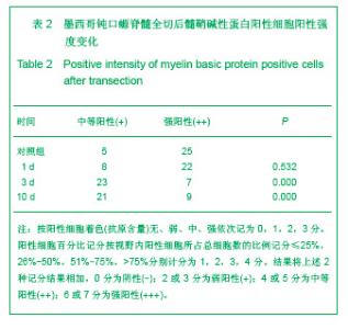
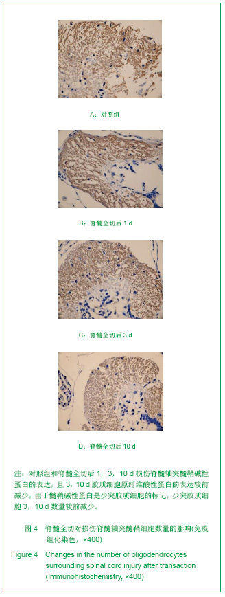
2.4 脊髓全切后损伤脊髓轴突髓鞘细胞的变化 见表2,图4。"
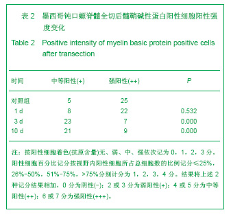
髓鞘碱性蛋白是组成中枢神经系统髓鞘的主要蛋白质单克隆抗体,可特异地显示轴突髓鞘,可作为少突胶质细胞标记。由图4及表2可见,对照组脊髓大量表达髓鞘碱性蛋白,脊髓全切后1 d时,损伤脊髓轴突髓鞘碱性蛋白未发生明显变化(P > 0.05),而3,10 d后,损伤脊髓轴突髓鞘碱性蛋白表达明显减少(P < 0.05),提示脊髓全切将减少损伤脊髓轴突髓鞘细胞数量。"
| [1] Luo QZ. Zhonghua Shenjing Waike Zazhi. 2004(20):88-90. 罗其中.中枢神经损伤后的神经再生与修复研究进展[J].中华神经外科杂志,2004(20):88-90.[2] Ribotta MG, Menet V, Privat A. Glial scar and axonal regeneration in the CNS: lessons from GFAP and vimentin transgenic mice. Acta Neurochir Suppl. 2004;89:87-92.[3] Silver J, Miller JH. Regeneration beyond the glial scar. Nat Rev Neurosci. 2004;5(2):146-156.[4] Davis BM, Ayers JL, Koran L, et al. Time course of salamander spinal cord regeneration and recovery of swimming: HRP retrograde pathway tracing and kinematic analysis. Exp Neurol. 1990;108(3):198-213.[5] Kirsche K, Kirsche W. Experimental study on the influence of olfactory nerve regeneration on forebrain regeneration of Ambystoma mexicanum. J Hirnforsch. 1964;7(3):315-333. [6] Monaghan JR, Walker JA, Page RB, et al. Early gene expression during natural spinal cord regeneration in the salamander Ambystoma mexicanum. J Neurochem. 2007; 101(1):27-40. [7] Scaps P, Bernet F, Gautron J, et al. Activities of acetylcholinesterase, choline acetyltransferase, and catecholamine production in the spinal cord of the axolotl Ambystoma mexicanum during forelimb regeneration. Biochem Cell Biol. 1994;72(5-6):188-194. [8] Vogel S. Regeneration after spinal cord dissection in Ambystoma mexicanum. Z Exp Chir. 1973;6(3):219-224.[9] Davis BM, Duffy MT, Simpson SB Jr. Bulbospinal and intraspinal connections in normal and regenerated salamander spinal cord. Exp Neurol. 1989;103(1):41-51.[10] Zhang MH, Peng L, Cao J. Ningxia Yike Daxue Xuebao. 2009;31(2):261-262. 张鸣号,彭亮,曹军.显微图像分析法与人工计数法在免疫组化结果判读中的应用[J].宁夏医科大学学报,2009,31(2): 261-262.[11] Barrett CP, Guth L, Donati EJ, et al. Astroglial reaction in the gray matter lumbar segments after midthoracic transection of the adult rat spinal cord. Exp Neurol. 1981;73(2):365-377.[12] Chen L, Yang P, Kijlstra A. Distribution, markers, and functions of retinal microglia. Ocul Immunol Inflamm. 2002; 10(1):27-39. [13] Rodnight R, Gonçalves CA, Wofchuk ST, et al. Control of the phosphorylation of the astrocyte marker glial fibrillary acidic protein (GFAP) in the immature rat hippocampus by glutamate and calcium ions: possible key factor in astrocytic plasticity. Braz J Med Biol Res. 1997;30(3):325-338. [14] Gopalan SM, Wilczynska KM, Konik BS, et al. Astrocyte-specific expression of the alpha1- antichymotrypsin and glial fibrillary acidic protein genes requires activator protein-1. J Biol Chem. 2006; 281(4): 1956-1963. [15] Sun Y, Wu S, Bu G, et al. Glial fibrillary acidic protein-apolipoprotein E (apoE) transgenic mice: astrocyte-specific expression and differing biological effects of astrocyte-secreted apoE3 and apoE4 lipoproteins. J Neurosci. 1998;18(9):3261-3272. [16] Ye P, Popken GJ, Kemper A, et al. Astrocyte-specific overexpression of insulin-like growth factor-I promotes brain overgrowth and glial fibrillary acidic protein expression. J Neurosci Res. 2004;78(4):472-484. [17] Besnard F, Brenner M, Nakatani Y, et al. Multiple interacting sites regulate astrocyte-specific transcription of the human gene for glial fibrillary acidic protein. J Biol Chem. 1991;266 (28):18877-18883. [18] Gopalan SM, Wilczynska KM, Konik BS, et al. Nuclear factor-1-X regulates astrocyte-specific expression of the alpha1-antichymotrypsin and glial fibrillary acidic protein genes. J Biol Chem. 2006;281(19):13126-13133. [19] Brock TO, O'Callaghan JP. Quantitative changes in the synaptic vesicle proteins synapsin I and p38 and the astrocyte-specific protein glial fibrillary acidic protein are associated with chemical-induced injury to the rat central nervous system. J Neurosci. 1987;7(4):931-942.[20] Alonso G. NG2 proteoglycan-expressing cells of the adult rat brain: possible involvement in the formation of glial scar astrocytes following stab wound. Glia. 2005;49(3):318-338.[21] West H, Richardson WD, Fruttiger M. Stabilization of the retinal vascular network by reciprocal feedback between blood vessels and astrocytes. Development. 2005;132(8): 1855-1862.[22] Menet V, Prieto M, Privat A, et al. Axonal plasticity and functional recovery after spinal cord injury in mice deficient in both glial fibrillary acidic protein and vimentin genes. Proc Natl Acad Sci U S A. 2003;100(15):8999-9004. [23] Li XS, He XR, Sun JS. Guoji Jianyan Yixue Zazhi. 2008;29(6): 532-534. 李小松,何小蓉,孙建森.少突胶质细胞对中枢神经系统再生抑制作用的研究进展[J].国际检验医学杂志,2008,29(6):532-534.[24] Schwartz M, Lazarov-Spiegler O, Rapalino O, et al. Potential repair of rat spinal cord injuries using stimulated homologous macrophages. Neurosurgery. 1999;44(5):1041-1045. [25] Yin GD. Shanghai: Second Military Medical University, 2009. 尹国栋.表达NgRs的活化巨噬细胞在大鼠脊髓损伤修复中的作用机制[D].上海:第二军医大学,2009. [26] Liu PL. Jinan: Shandong University, 2004. 刘培来.活化巨噬细胞移植治疗脊髓损伤的实验研究[D].济南:山东大学,2004. [27] Liu PL, Li M, Zhang YK, et al. Shandong Daxue Xuebao: Yixue Ban. 2005;43(9):834-838. 刘培来,李明,张元凯,等.活化巨噬细胞移植时间和脊髓损伤后功能恢复的实验研究[J].山东大学学报:医学版,2005,43(9): 834-838. [28] Liu HZ, Zhou LG, He LY, et al. Zhongguo Jiaoxing Waike Zazhi. 2007;15(13):1015-1018. 刘洪智,周路纲,贺兰英,等.周围神经联合活化巨噬细胞移植修复小鼠脊髓损伤[J].中国矫形外科杂志,2007,15(13):1015-1018.[29] Streit WJ, Semple-Rowland SL, Hurley SD, et al. Cytokine mRNA profiles in contused spinal cord and axotomized facial nucleus suggest a beneficial role for inflammation and gliosis. Exp Neurol. 1998;152(1):74-87. [30] Tang XQ, Heron P, Mashburn C, et al. Targeting sensory axon regeneration in adult spinal cord. J Neurosci. 2007;27(22): 6068-6078. [31] Kamei N, Tanaka N, Oishi Y, et al. BDNF, NT-3, and NGF released from transplanted neural progenitor cells promote corticospinal axon growth in organotypic cocultures. Spine (Phila Pa 1976). 2007;32(12):1272-1278. [32] Vidal PM, Lemmens E, Dooley D, et al. The role of "anti-inflammatory" cytokines in axon regeneration. Cytokine Growth Factor Rev. 2013;24(1):1-12. [33] Nadeau S, Filali M, Zhang J, et al. Functional recovery after peripheral nerve injury is dependent on the pro-inflammatory cytokines IL-1β and TNF: implications for neuropathic pain. J Neurosci. 2011;31(35):12533-12542. [34] Cafferty WB, Gardiner NJ, Das P, et al. Conditioning injury-induced spinal axon regeneration fails in interleukin-6 knock-out mice. J Neurosci. 2004;24(18):4432-4443. [35] Dupraz S, Grassi D, Karnas D, et al. The insulin-like growth factor 1 receptor is essential for axonal regeneration in adult central nervous system neurons. PLoS One. 2013;8(1): e54462.[36] Hollis ER 2nd, Lu P, Blesch A, et al. IGF-I gene delivery promotes corticospinal neuronal survival but not regeneration after adult CNS injury. Exp Neurol. 2009;215(1):53-59. [37] Tiangco DA, Papakonstantinou KC, Mullinax KA, et al. IGF-I and end-to-side nerve repair: a dose-response study. J Reconstr Microsurg. 2001;17(4):247-256. [38] Gao Z, Zhao PB, Xu XK, et al. Jilin Yixueyuan Xuebao. 1999 (3):7-9. 高忠,赵蓬波,徐晓琨,等.神经生长因子对感觉轴突再生的作用[J].吉林医学院学报, 1999(3):7-9. [39] Wu YT, Wang YJ. Zhongguo Zuzhi Gongcheng Yanjiu yu Linchuang Kangfu. 2008;12(29):5631-5635. 乌优图,王运杰.神经生长因子对神经干细胞分化及神经元轴突形成的影响[J].中国组织工程研究与临床康复, 2008,12(29): 5631-5635. [40] Sun XD, Li YJ, Dong XJ. Liaoning Yixueyuan Xuebao. 2010; (4):377-380. 孙小单,李羽佳,佟晓杰.神经生长因子促神经再生作用的研究进展[J].辽宁医学院学报, 2010,(4):377-380. [41] Sun JJ, Wang JB, Li GR. Zhonghua Er Bi Yanhou Ke Zazhi. 1996(5):274-276. 孙建军,汪吉宝,李桂榕.生长因子对离体听神经元存活与轴突再生的影响[J].中华耳鼻咽喉科杂志, 1996(5):274-276. [42] Wu YT. Shenyang: China Medical University, 2008. 乌优图.神经生长因子(NGF)对神经干细胞的分化以及对神经元轴突形成的影响[D].沈阳:中国医科大学,2008.[43] Liu W, Zhang XJ. Zhongguo Zuzhong Zazhi. 2008; (8): 597-602. 刘薇,张祥建.胰岛素样生长因子-1神经保护作用的研究进展[J].中国卒中杂志, 2008(8):597-602. [44] Wu LX, Qin Y, Gu LQ, et al. Zhongguo Linchuang kangfu. 2002;6(4):524-525. 吴岚晓,秦煜,顾立强,等.胰岛素样生长因子1在神经特异性再生中的作用机制[J].中国临床康复,2002,6(4):524-525. [45] Zheng LF, Yi XN. Hainan Yixueyuan Xuebao. 2005;11(5): 434-437. 郑林丰,易西南.胰岛素样生长因子与周围神经再生[J].海南医学院学报, 2005,11(5):434-437. [46] Li Y, Liu J, Huang GQ, et al. Zhongguo Zuzhi Gongcheng Yanjiu yu Linchuang kangfu. 2009;13(40):7903-7907. 李云,刘佳,黄桂琴,等.嗅鞘细胞移植对脊髓全横断损伤大鼠大脑运动皮质胰岛素样生长因子1和睫状神经营养因子表达的影响[J].中国组织工程研究与临床康复,2009,13(40):7903-7907.[47] Schonbach C. The neuroglia in the spinal cord of the newt, Triturus viridescens. J Comp Neurol. 1969;135(1):93-120.[48] Zukor KA, Kent DT, Odelberg SJ. Meningeal cells and glia establish a permissive environment for axon regeneration after spinal cord injury in newts. Neural Dev. 2011;6(1):1. [49] Fawcett J. Repair of spinal cord injuries: where are we, where are we going? Spinal Cord. 2002;40(12):615-623. [50] Yan PW, He XJ, Li HP, et al. Zhongguo Linchuang Kangfu. 2006;10(33):27-29. 袁普卫,贺西京,李浩鹏,等.嗅鞘细胞移植后脊髓损伤区髓磷脂相关糖蛋白表达的动态变化[J].中国临床康复, 2006,10(33): 27-29. [51] Liu XW, Shi GD. Zhongguo Jizhu Jisui Zazhi. 2011;21(12): 1030-1033. 刘晓伟,史国栋.髓磷脂相关抑制因子与脊髓损伤后轴突再生抑制的研究进展[J].中国脊柱脊髓杂志,2011,21(12):1030-1033. [52] Qu YM, Feng DX, Xiao BM, et al. Zhongguo Xiandai Yisheng. 2011;19(19):25-27. 屈一鸣,冯大雄,肖百敏,等.脊髓损伤后髓磷脂抑制分子及作用机制的研究进展[J].中国现代医生, 2011,19(19):25-27. |
| [1] | Guan Qian, Luan Zuo, Ye Dou, Yang Yinxiang, Wang Zhaoyan, Wang Qian, Yao Ruiqin. Morphological changes in human oligodendrocyte progenitor cells during passage [J]. Chinese Journal of Tissue Engineering Research, 2021, 25(7): 1045-1049. |
| [2] | Ye Dou, , Ma Xuexia , Guan Qian, , Luan Zuo , Yang Yinxiang , Wang Zhaoyan , Wang Qian , He Ying , Yao Ruiqin. Proportion and morphological characteristics of human oligodendrocyte precursor cells in different cell culture vessels [J]. Chinese Journal of Tissue Engineering Research, 2021, 25(1): 44-49. |
| [3] |
Zhang Cong, Zhao Yan, Du Xiaoyu, Du Xinrui, Pang Tingjuan, Fu Yining, Zhang Hao, Zhang Buzhou, Li Xiaohe, Wang Lidong.
Biomechanical analysis of the lumbar spine and pelvis in adolescent
idiopathic scoliosis with lumbar major curve |
| [4] | Xu Guofeng, Li Xuebin, Tang Yifan, Zhao Yin, Zhou Shengyuan, Chen Xiongsheng, Jia Lianshun. The role of autophagy in ossification of the human ligamentum flavum [J]. Chinese Journal of Tissue Engineering Research, 2020, 24(8): 1174-1181. |
| [5] | He Yujie, Wang Haiyan, Li Zhijun, Li Xiaohe, Cai Yongqiang, Dai Lina, Xu Yangyang, Wang Yidan, Xu Xuebin. Digital measurements of the anatomical parameters of pedicle-rib unit screw fixation in thoracic vertebrae of preschoolers [J]. Chinese Journal of Tissue Engineering Research, 2020, 24(6): 869-876. |
| [6] | Cao Qingjun, Yang Fenghua, Wang Hua. Hippocampal astrocytes in juvenile rats with persistent epilepsy: the role of cannabinoid receptor type 2 in regulating MAPK pathway [J]. Chinese Journal of Tissue Engineering Research, 2020, 24(32): 5179-5185. |
| [7] | Sun Jian, Fang Chao, Gao Fei, Wei Laifu, Qian Jun. Clinical efficacy and complications of short versus long segments of internal fixation for the treatment of degenerative scoliosis: a meta-analysis [J]. Chinese Journal of Tissue Engineering Research, 2020, 24(3): 438-445. |
| [8] | Gao Jianbo, Xia Bing, Li Shengyou, Yang Yujie, Ma Teng, Yu Peng, Luo Zhuojing, Huang Jinghui. Effect of nanoparticles carrying chondroitin sulfate ABC on the migration of Schwann cells in a magnetic field [J]. Chinese Journal of Tissue Engineering Research, 2020, 24(28): 4526-4532. |
| [9] | Xu Yangyang, Zhang Kai, Li Zhijun, Zhang Yunfeng, Su Baoke, Wang Xing, Wang Lidong, Wang Yidan, He Yujie, Li Kun, Wang Haiyan, Li Xiaohe. Morphological analysis of optimal selection of lumbar pedicle screws in adolescents aged 12-15 years [J]. Chinese Journal of Tissue Engineering Research, 2020, 24(21): 3321-3328. |
| [10] | Qin Haikuo, Luo Shixing. Correlation of cortical bone thickness and X-ray gray value in different planes of proximal femur with brittle fracture of female hip [J]. Chinese Journal of Tissue Engineering Research, 2020, 24(18): 2867-2872. |
| [11] | Huang Tianji, Yang Shengdong, Lin Hao, Zhang Chunyang, Deng Zhongqi, Zhong Weiyang, Luo Xiaoji. Mapping knowledge domains of bibliometrics regarding percutaneous vertebroplasty and percutaneous kyphoplasty based on VOSviewer [J]. Chinese Journal of Tissue Engineering Research, 2020, 24(15): 2410-2417. |
| [12] | Qu Renfei, Mai Yuying, Chen Xiaowei, Hu Huanying, Chen Guozhi, Li Dongdong, Liao Hongbing. Relationship between lactic acid concentration and osteoclast differentiation of mouse monocytes [J]. Chinese Journal of Tissue Engineering Research, 2020, 24(14): 2177-2183. |
| [13] | Zhao Hong, Wang Suping, Lian Jianwen. Insight into white matter changes in Alzheimer’s disease: a focus on myelin and oligodendrocyte [J]. Chinese Journal of Tissue Engineering Research, 2020, 24(13): 2120-2125. |
| [14] | Song Xudong, He Yunwu, Li Yonglin, Chen Jing, Hu Junlan. Ultrasound-guided paravertebral nerve block for zoster-associated pain: a Meta-analysis [J]. Chinese Journal of Tissue Engineering Research, 2020, 24(11): 1797-1804. |
| [15] | Liu Yapu, Lin Junyu, Yang Zhou, Wu Xiuhua, Wu Xiaoliang, Zhu Qing’an. Three-dimensional visualization and quantitative analysis of microvessels in rat cervical spinal cord using barium sulfate perfusion combined with micro-CT scanning [J]. Chinese Journal of Tissue Engineering Research, 2019, 23(36): 5836-5840. |
| Viewed | ||||||
|
Full text |
|
|||||
|
Abstract |
|
|||||