Chinese Journal of Tissue Engineering Research
Previous Articles Next Articles
Morphological changes of the smear layer after caries removal using different methods: An observation under scanning electron microscope
Fang Ling, Zhu Yan-li
- Oral Department, Daqing Oilfield General Hospital, Daqing 163000, Heilongjiang Province, China
-
Received:2013-03-04Revised:2013-03-08Online:2013-09-17Published:2013-09-17 -
Contact:Zhu Yan-li, Master, Oral Department, Daqing Oilfield General Hospital, Daqing 163000, Heilongjiang Province, China zhuyanli840220@163.com -
About author:Fang Ling★, Master, Professor, Oral Department, Daqing Oilfield General Hospital, Daqing 163000, Heilongjiang Province, China Fangling.20082008@163.com
CLC Number:
Cite this article
Fang Ling, Zhu Yan-li. Morphological changes of the smear layer after caries removal using different methods: An observation under scanning electron microscope[J]. Chinese Journal of Tissue Engineering Research, doi: 10.3969/j.issn.2095-4344.2013.38.004.
share this article
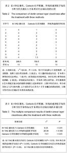
2.1 各组大体观察及探针检查结果 3组去腐后用探针检查窝洞,质地均坚硬,Er:YAG激光组牙本质表面凸凹不平,似山峰状;Carisolv化学机械组牙本质表面光泽较暗,底面较平;传统高速涡轮牙钻组去腐后牙本质表面平整,光亮,并有明显的切割痕迹。 2.2 分析3种方法去腐后扫描电镜观察玷污层清洁度数据 3种方法去除牙本质玷污层清洁度比较差异有显著性意义(P < 0.05),见表2,组间多重比较显示Er:YAG激光组玷污层比Carisolv化学机械组少(P < 0.05),同时也比传统高速涡轮牙钻组少(P < 0.005), Carisolv化学机械组玷污层比传统高速涡轮牙钻组少 (P < 0.05), Er:YAG激光去除玷污层效果最好,见表3。"
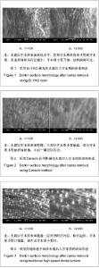
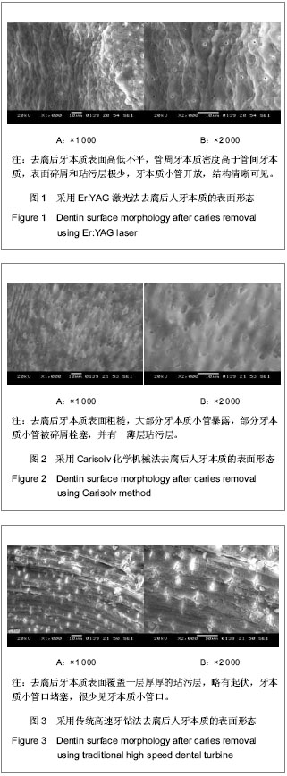
2.3 扫描电镜观察各组牙本质玷污层 见图1-3。 Er:YAG激光组去腐后牙本质表面高低不平,管周牙本质密度高于管间牙本质,表面碎屑和玷污层极少,牙本质小管开放,结构清晰可见,见图1。 Carisolv化学机械组去腐后牙本质表面粗糙,大部分牙本质小管暴露,部分牙本质小管被碎屑栓塞,并有一薄层玷污层,见图2。 传统高速牙钻组去腐后牙本质表面覆盖一层厚厚的玷污层,略有起伏,牙本质小管口堵塞,很少见牙本质小管口,见图3。"
"

| [1] Banerjee A,Watson TF,Kidd EA.Dentine caries excavation:a review of current clinical techniques.Br Dent J.2000;188(9): 476-482. [2] Yazici AR,Ozgunaltay G,Dayangac B.A scanning electron microscopic study of different caries removal techniques on human dentin.Oper Dent.2002;27:360-366. [3] 谢毓秀,王满恩,毛秀萍.国产气涡轮牙钻制备窝洞后人牙髓的即刻反应[J].北京医科大学学报:医学版,1988,20(3):215-217. [4] 许文峰.浅谈微创无痛治疗在龋齿中的研究及应用[J].中国民族民间医药,2009,18(7):44. [5] 吴友农,黄力子,史俊南.根管超声清理效果的扫描电镜评价[J].第四军医大学学报,1989,10(3):154-156. [6] Pai VS,Nadig RR,Jagadeesh T,et al.Chemical analysis of dentin surfaces after Carisolv treatment.J Conserv Dent. 2009; 12(3):118-122. [7] Tsanova STs,Tomov GT.Morphological changes in hard dental tissues prepared by Er:YAG laser (LiteTouch, Syneron), Carisolv and rotary instruments. A scanning electron microscopy evaluation. Folia Med (Plovdiv).2010; 52(3): 46-55. [8] Meller C,Welk A,Zeligowski T,et al.Comparison ofdentin caries excavation with polymer and conventional tungsten carbide burs.Quintessence Int.2007;38(7):565-569. [9] 郑向明,刘筱琳,胡德渝,等.Er:YAG激光在预防性树脂充填中的应用—扫描电镜研究[J].中国激光医学杂志,2003,12(4): 224-228. [10] 吴为良,林实,姜醒,等.Er:YAG激光照射后牙本质粘结界面的剪切强度分析[J].福建医药杂志,2010,32(3):10-12. [11] 欧晓艳,朱洪水.3 种方法去龋后牙本质玷污层扫描电镜观察[J].中国实用口腔科杂志,2008,1(9):538-540. [12] Peric T,Markovic D.In vitro effectiveness of a chemo-mechanical method for caries remova.Eur J Paediatr Dent.2007;8(2):61-67. [13] Subramaniam P,Babu KL,Neeraja G.Comparison of the antimicrobial efficacy of chemomechanical caries removal ( Carisolv) with that of conventional drilling in reducing cariogenic flora.J Clin Pediatr Dent.2008;32(3): 215-219. [14] Bulut G,Zekioglu O,Eronat C,et al.Effect of Carisolv on the human dental pulp: a histological study.J Dent.2004;32(4): 309-314. [15] Aranha AC,De-Paula-Eduardo C,Gutknecht N,et al.Analysis of the interfacial micromorphology of adhesive systems in cavities prepared with Er, Cr: YSGG, Er: YAG laser and bur.Microsc Res Tech.2007;70(8):745-751. [16] 张 笋,陈 涛,葛立宏.扫描电镜下观察铒钇铝石榴石激光备洞后的牙本质形态[J].北京大学学报:医学版,2011,43(5):766-769. [17] Curti M,Rocca JP,Bertrand MF,et al. Morphostructural aspects of Er: YAG prepared class V cavities.J Clin Laser Med Surg. 2004;22(2):119-123. [18] 赵晓一,王世明,张成飞.Er:YAG激光切除牙根断面的扫描电镜观察[J].华西口腔医学杂志,2010,28(5):526-528. [19] 林峰,高萍,郑广宁.激光在牙体牙髓疾病治疗中的研究进展[J].现代口腔医学杂志, 2008,22(4):427-429. [20] Reyhanian A,Parker S,Moshonov J.The use of the erbium yttrium aluminium garnet(2940 nm)in a laser-assisted apicectomy procedure.Br Dent J.2008;205(6):319-323. [21] 贾素慧,胡德渝.Er:YAG激光治疗牙体牙髓病的研究进展[J].国外医学:口腔医学分册,2005,32(5):383-385. [22] Crespi R,Romanos GE,Cassinelli C,et al.Effects of Er:YAG laser and ultrasonic treatment on fibroblast attachment to root.surfaces: an in vitro study.J Periodontol.2006;77(7): 1217-1222. [23] Schwarz F,Bieling K,Venghaus S,et al.Influence of fluorescencecontrolled Er:YAG laser radiation, the vector system and hand instruments on periodontally diseased root surfaces in vivo.J Clin Periodontol.2006;33(3):200-208. [24] Stephanovich M.Scanning electron microscopic study of the effects of Er:YAG lasers and conventional methods of preparation on dentin.SDK and NUS. 2005;4(1):153-156 (Bulgarian) [25] Baraba A,Miletic I,Krmek SJ,et al.Ablative potential of the erbium-doped yttrium aluminium garnet laser and conventional handpieces:a comparative study.Photomed Laser Surg.2009;11:465-504. [26] Krmek SJ,Miletic I,Simeon P,et al.The Temperature Changes in the Pulp Chamber During Cavity Preparation with the Er: YAG Laser Using a Very Short Pulse.Photomed Laser Surg. 2009;27(2):351-355. [27] Jepsen S,Acil Y,Peschel T,et al.Biochemical andmorphological analysis of dentin following selective caries removal with a fluorescence - controlled Er: YAG laser. Lasers Surg Med.2008;40(5):350-357. [28] Kornblit R,Trapani D,Bossu M,et al.The use ofErbium:YAG laser for caries removal in paediatric patients following Minimally Invasive Dentistry concepts.Eur J Paediatr Dent. 2008;9(2):81-87. [29] Louw NP,Pameijer CH,Ackermann WD,et al.Pulp histology after Er:YAG laser cavity preparation in subhuman primates-a pilot study. SADJ.2002;57(8):313-317. [30] 潘蕾,梅陵宣,陈乔尔. Er∶YAG激光制洞对牙髓的生物学效应[J].安徽医科大学学报,2006,41(4):469-471. [31] 郑向明,刘筱琳,胡德渝,等.Er:YAG激光在预防性树脂充填中的应用-扫描电镜研究[J].中国激光医学杂志,2003,12(4):224-228. [32] Eberhard J,Bode K,Hedderich J,et al.Cavity size difference after caries removal by a fluorescence-controlled Er:YAG laser and by conventional bur treatment. Clin Oral Investig. 2008;12(4):311-318. [33] Ceballos L,Toledano M,Osorio R,et al.Bonding to Er–YAG–laser–treated dentin.J Dent Res.2002;81(2): 119-122. [34] Magni E,Ferrari M,Hickel R,et al.Effectofozone gas application on the mechanical properties of dental adhesives bonded to dentin.Dent Mater.2008;24(10):1428-1434. [35] 李子木,仇丽鸿.光固化复合树脂密合度的研究进展[J].中国实用口腔科杂志,2008,1(1):43-46. [36] 贾兴亚,刘婷婷,李心瑶,等.两种牙体预备方法对玻璃离子水门汀与牙本质间粘结力的影响[J].中国医科大学学报,2011,40(3): 234-236. [37] Ramos AC,Esteves Oliveira M,Arana Chavez VE,et al.Adhesives bonded to erbium:yttrium-alumimum-garnet laserirradiated dentin:transmission electronmicroscopy, scanning electron microscppy and tensile bondstrengthanalyses.Lasers Med Sci.2010;25:181-189. [38] 舒文,杨向红.Carisolv去腐方法治疗龋齿的临床对比研究[J].口腔医学研究,2005,21(6):670-671. [39] Katarski G,Kissov K.Contemporary views of Carisolv treatment of dental caries.Clinical cases.SDK and NUS. 2005;4(1):164-172. [40] 梁保刚,张桂荣,赵奇.Carisolv 伢典化学机械去腐法治疗深龋临床疗效研究[J].中国实用口腔科杂志,2009,2(10):621-622. [41] 肖 杰,储冰峰.乳牙Carisolv去龋的扫描电镜观察[J].牙体牙髓牙周病学杂志,2008,18(2):84-86. [42] Banerjee A,Kidd EAM,Walson TF.Scanning electron microscopic observations of human dentine after mechanical caries excavation.J Dent.2008;28:179-186. [43] Cederlund A,Lindskog S,Blomlof J.Effect of a chemomechanical caries removal system(Carisolv) on dentin topography of non-carious dentin.Acta Odontol Scand.1999; 57:185-189. [44] Yazici AR,Ozgunaltay G,Dayangac B.A scanning electron microscopic study of different caries removal techniques on human dentin.Oper Dent.2002;27:360-366. [45] Kinoshita J,Kimura Y,Matsumoto K.Comparative study of carious dentin removal by Er:Cr:YSGG laser and Carisolv.J Clin Laser Med Surg.2003;21:307-315. [46] 许晓燕,于新波,孙慧斌,等.化学-机械去腐法治疗龋齿随访1年的临床研究[C].2007年第七次全国牙体牙髓病学学术会议论文集, 2007:35-37. [47] 夏克俭,李姮,王文梅. Carisolv去龋及酸蚀后牙本质表面形态扫描电镜观察[J].口腔医学,2010,30(1):46-48. [48] 胡林英,宋光泰,钱虹.Carisolv化学机械去龋后牙本质表面的扫描电镜观察[J].武汉大学学报:医学版,2005,26(4):512-514. [49] 陈舟,江勇. Carisolv化学机械法在龋病治疗中的对比研究[J].口腔医学,2006, 26(6):457-459. [50] 陈小贤,葛立宏. Carisolv化学机械去腐技术对微拉伸粘拉力的影响[J].现代口腔医学杂志,2006,20(6):612-614. [51] 于静涛,贾兴亚,乔丽艳,等.铒,铬:钇钪镓石榴石激光切割牙体硬组织效果的扫描电子显微镜观察[J].华西口腔医学杂志,2003, 21(5):356-358. [52] 韦少锋.微创去腐技术治疗龋齿的临床观察[J].中国临床医学, 2010,17(3):434-435. [53] 张兴文,王丽珍.微创无痛治疗在龋齿治疗中的临床观察[J].中国民康医学,2009,21(21):2700-2701. [54] 陈柯,张慧. Er,Cr:YSGG激光治疗口腔疾病的进展[J].中国激光医学杂志,2007,16(1):64-66. [55] Giza S. Comparative studies of carious defects filling using the classical method and dental drill, and using the Carisolv chemomechanical method and the YAG:Er CTL-1601 laser.Ann Acad Med Stetin.2007;53(3):88-99. [56] Tachibana A,Marques MM,Soler JM,et al.Erbium, chromium:yttrium scandium gallium garnet laser for caries removal: influence on bonding of a self-etching adhesive system.Lasers Med Sci. 2008;23(4):435-441. [57] 杨小民.铒激光在口腔医学中的应用[J].实用医学临床杂志,2007, 4(2):33-35. [58] 韩雯斐,苏俭生.激光消融牙体硬组织的研究进展[J].中华老年口腔医学杂志,2008,6(4):239-242. |
| [1] | Zhao Binbin, Zhong Weijian, Ma Guowu, Li Yongqi, Wang Ning. Comparison of the osteogenic effect of three different bone graft materials [J]. Chinese Journal of Tissue Engineering Research, 2021, 25(10): 1507-1510. |
| [2] | Wang Ning, Zhong Weijian. Application and function of autologous blood concentrate in tissue regeneration [J]. Chinese Journal of Tissue Engineering Research, 2021, 25(1): 146-151. |
| [3] | Zheng Shize, Li Ke, Chen Yue, Yan Xiaoyuan, Zhan Desong, Fu Jiale. Effect of different smear layers on the bonding performance and durability of four adhesive systems [J]. Chinese Journal of Tissue Engineering Research, 2020, 24(4): 517-523. |
| [4] |
Li Junqing, Wu Jiayuan.
Effect of mechanical stimulation on dental pulp stem cells [J]. Chinese Journal of Tissue Engineering Research, 2020, 24(25): 4054-4059. |
| [5] | Chen Chaoxiang, Zhou Shuping, Zhang Wei, Xiang Liang, Hou Wei, Wu Junxing. Effects and mechanisms of sodium ibandronate in ovariectomy-induced osteoporosis rats [J]. Chinese Journal of Tissue Engineering Research, 2020, 24(17): 2625-2629. |
| [6] | Li Zhangyi, Liu Fengting, Huang Jianyong, Su Xiaoying, Wang Zhixing, Zheng Quan, Li Yanxia, Liu Xiaozhi. Extension and dentin differentiation potential of dental pulp stem cells from human deciduous teeth on the stiff matrix surface [J]. Chinese Journal of Tissue Engineering Research, 2020, 24(13): 2005-2010. |
| [7] | Feng Ruxue, Cai Rengang, Xie Dandan. Lipopolysaccharide can influence and regulate proliferation and migration of dental pulp stem cells [J]. Chinese Journal of Tissue Engineering Research, 2019, 23(29): 4688-4693. |
| [8] | Zuo Sili. Risk assessment of hemiarthroplasty and internal fixation of proximal femoral nail antirotation for treating hip fractures in older adults [J]. Chinese Journal of Tissue Engineering Research, 2019, 23(28): 4440-4445. |
| [9] | Zhao Binbin, Qiu Zewen, Ma Chenghui, Zhong Weijian, Ma Guowu. Effect of different techniques on the osteogenic property of undecalcified heterogeneous dentin particles [J]. Chinese Journal of Tissue Engineering Research, 2019, 23(26): 4154-4159. |
| [10] | Shao Miaomiao, Liu Zhongxi, Xu Nuo, Liu Qinghua, Wang Dong, He Jianya, Li Xiaojie. Induced pluripotent stem cells in dental tissue regeneration: effect and application [J]. Chinese Journal of Tissue Engineering Research, 2019, 23(1): 151-157. |
| [11] | Cui Ting-ting, Qiu Ze-wen, Shao Yang, Zhong Wei-jian. Osteogenic effects of heterogeneous dentin particles in conjunction with bone marrow concentrate in the maxillary sinus lift [J]. Chinese Journal of Tissue Engineering Research, 2018, 22(30): 4806-4811. |
| [12] | Liang Yun, Shi Jian-jie, Chen Ke, Luo Zhen-shan, Deng Wen-xin. Irradiation with erbium, chromium:yttrium scandium gallium garnet laser alters shear bond strength to primary tooth dentin [J]. Chinese Journal of Tissue Engineering Research, 2018, 22(26): 4208-4214. |
| [13] |
Shen Hao, Liu Jun, Jing Wei-wei, Suo Yong-kuan, Chang Shi-jie, Sha Xian-zheng.
Weak laser effects on the biocompatibility of chitosan and Nafion as implantable glucose sensor outer materials
[J]. Chinese Journal of Tissue Engineering Research, 2018, 22(2): 267-273.
|
| [14] | Lin Bao-shan, Li Ting. An in vivo and in vitro study on the effect of tooth liquid polish sealant on the bond strength of orthodontic brackets [J]. Chinese Journal of Tissue Engineering Research, 2018, 22(18): 2872-2876. |
| [15] |
Cai Yue, Huang Ying, Zhang Hui, Guo Ling .
Stress distribution of the three-dimensional post-crown model with labial and lingual oblique defects of the maxillary central incisor: a finite element analysis
[J]. Chinese Journal of Tissue Engineering Research, 2017, 21(30): 4823-4829.
|
| Viewed | ||||||
|
Full text |
|
|||||
|
Abstract |
|
|||||