Chinese Journal of Tissue Engineering Research ›› 2026, Vol. 30 ›› Issue (33): 8577-8582.doi: 10.12307/2026.315
Relationship of hip bone mineral density and geometry with age and body mass index in women
Gao Haiyan, Zheng Hongyin, Xiao Yi, Huang Jiyuan
- Department of Nuclear Medicine, Sichuan Academy of Medical Sciences · Sichuan Provincial People's Hospital (Affiliated Hospital of University of Electronic Science and Technology of China), Chengdu 610072, Sichuan Province, China
-
Received:2025-04-03Revised:2025-06-14Online:2026-11-28Published:2026-06-08 -
Contact:Huang Jiyuan, MS, Chief physician, Department of Nuclear Medicine, Sichuan Academy of Medical Sciences · Sichuan Provincial People's Hospital (Affiliated Hospital of University of Electronic Science and Technology of China), Chengdu 610072, Sichuan Province, China -
About author:Gao Haiyan, MS, Attending physician, Department of Nuclear Medicine, Sichuan Academy of Medical Sciences · Sichuan Provincial People’s Hospital (Affiliated Hospital of University of Electronic Science and Technology of China), Chengdu 610072, Sichuan Province, China -
Supported by:Clinical Research and Translational Fund of Sichuan Academy of Medical Sciences · Sichuan Provincial People's Hospital, No. 2017LY01 (to HJY); Sichuan Natural Science Foundation, No. 2024NSFSC0667 (to GHY)

CLC Number:
Cite this article
Gao Haiyan, Zheng Hongyin, Xiao Yi, Huang Jiyuan. Relationship of hip bone mineral density and geometry with age and body mass index in women[J]. Chinese Journal of Tissue Engineering Research, 2026, 30(33): 8577-8582.
share this article
Add to citation manager EndNote|Reference Manager|ProCite|BibTeX|RefWorks
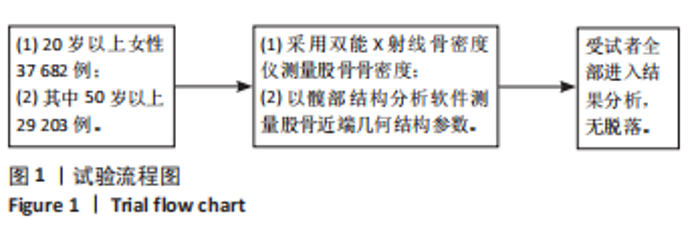
2.1 参与者数量分析 最终纳入符合纳入标准的20岁以上女性37 682例,其中50岁以上29 203例。以10岁为一年龄组,其中80岁及以上为一组,分别计算各组股骨骨密度及几何结构参数。 2.2 试验流程图 见图1。"
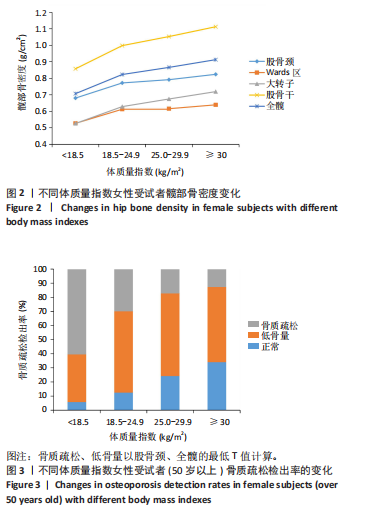
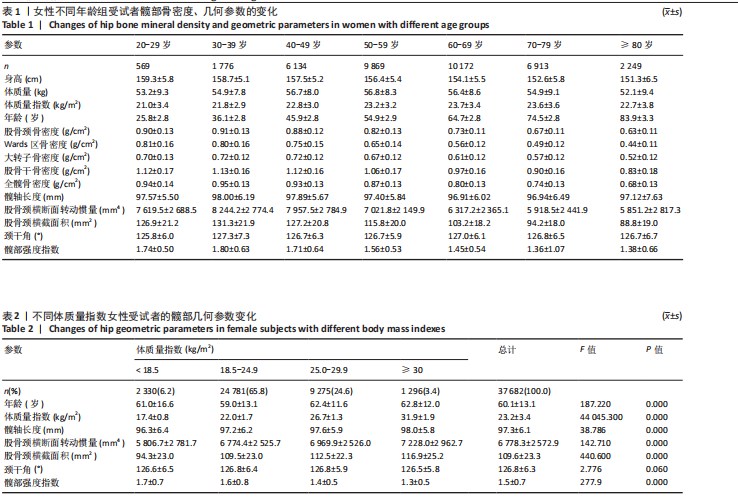
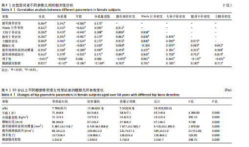
2.3 不同年龄女性受试者髋部骨密度和几何参数的变化 女性股骨颈、大转子、全髋骨密度在30-39岁达到峰值,后随增龄逐渐降低。髋部几何结构参数髋轴长度、股骨颈横断面转动惯量、股骨颈横截面积、髋部强度指数在30-39岁达到峰值,随增龄逐渐降低,见表1。 2.4 不同体质量指数女性受试者髋部骨密度变化 随着体质量指数增加,受试者股骨颈、Wards区、大转子、股骨干、全髋骨密度均逐渐增加,髋部骨质疏松检出率逐渐下降(P < 0.001)。股骨干骨密度最高,其次为全髋、股骨颈,Wards区骨密度最低。体质量指数< 18.5 kg/m2的受试者,骨质疏松症检出率达60.49%;体质量指数> 30.0 kg/m2的受试者,骨质疏松症检出率仅为12.74%。见图2,3。 2.5 不同体质量指数女性受试者的髋部几何参数变化 随着体质量指数增加,髋部髋轴长度、股骨颈横断面转动惯量、股骨颈横截面积逐渐升高(P < 0.001),髋部强度指数逐渐降低(P < 0.001);颈干角随体质量指数变化不明显。见表2。"
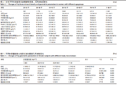
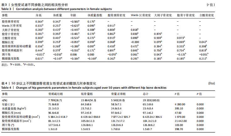
2.6 不同参数之间的相关分析 髋部骨密度与身高、体质量、体质量指数呈正相关(r=0.095-0.425),与年龄呈负相关(r=-0.445至-0.623)。髋轴长度、股骨颈横断面转动惯量、股骨颈横截面积与身高、体质量、体质量指数呈正相关(r=0.059-0.444),与髋部骨密度呈正相关,与年龄呈负相关(r=-0.055至-0.555)。髋部强度指数与年龄、体质量指数呈负相关,与股骨骨密度呈正相关性。见表3。 2.7 50岁以上不同骨密度女性受试者的髋部几何参数变化 骨质疏松组、低骨量组、骨量正常组受试者体质量指数、髋部髋轴长度、股骨颈横断面转动惯量、股骨颈横截面积、颈干角、髋部强度指数相比差异均有显著性意义(P < 0.001);骨量正常组体质量指数、股骨颈横断面转动惯量、股骨颈横截面积、髋部强度指数、髋轴长度高于骨质疏松、低骨量组,年龄、颈干角低于骨质疏松、低骨量组。见表4。"
"
| [1] SI L, WINZENBERG TM, JIANG Q, et al. Projection of osteoporosis‐related fractures and costs in China:2010‐2050.OsteoporosInt. 2015;26(7):1929‐1937. [2] XIA WB, HE SL, XU L, et al. Rapidly increasing rates of hip fracture in Beijing, China. J Bone Miner Res. 2012;27:125-129. [3] 李毅中, 蔡思清, 颜丽笙, 等.不同类型髋部骨折骨密度和髋部结构变化[J].中华骨质疏松和骨矿盐疾病杂志,2015,8(3):243-245. [4] 靳延利,苗强,刘涛,等.女性髋部骨质疏松性骨折患者股骨近端几何结构对患者骨折危险的影响[J].创伤外科杂志,2018,20(3):194-197. [5] 何海洋,杨嘉玲,雷迅.绝经后女性骨质疏松症患病率及影响因素的Meta分析[J].中国全科医学,2024,27(11):1370-1379. [6] 程晓光, 董剩勇, 王亮, 等. 应用双能X线骨密度仪调查中国人群骨密度水平和骨质疏松症患病率[J].中华健康管理学杂志,2019,13(1):51-58. [7] COMPSTON JE, FLAHIVE J, HOSMER DW, et al. Relationship of weight, height, and body mass index with fracture risk at different sites in postmenopausal women: the Global Longitudinal Study of Osteoporosis in Women (GLOW). J Bone Miner Res. 2014;29(2):487-493. [8] LI L, SHEN Y, TAN LH, et al. Association of osteoporotic fractures of femoral neck and femoral neck geometric parameters in native Chinese women. BMC Musculoskelet Disord. 2024;25(1):349. [9] FAJAR JK, TAUFAN T, SYARIF M, et al. Hip geometry and femoral neck fractures: a meta-analysis. J Orthop Translat. 2018;13:1-6. [10] The International Society for Clinical Densitometry (ISCD) 2019 ISCD Official Positions Adult. https://www.iscd.org/officialpositions/2019-iscd-official-positions-adult/. Accessed 22 Jan 2020. [11] 中华医学会骨质疏松和骨矿盐疾病分会. 原发性骨质疏松症诊疗指南(2022)[J]. 中华内分泌代谢杂志,2023,39(5):377-406. [12] DONG Y, ZHANG Y, SONG K, et al. What was the Epidemiology and Global Burden of Disease of Hip Fractures From 1990 to 2019? Results From and Additional Analysis of the Global Burden of Disease Study 2019. Clin Orthop Relat Res. 2023;481(6):1209-1220. [13] MARSHALL D, JOHNELL O, WEDEL H. Meta-analysis of how well measures of bonemineral density predict occurrence of osteoporotic fractures. BMJ. 1996;312:1254-1259. [14] 黄际远,宋文忠,郑洪银,等.成都地区健康人群骨密度的变化及不同T-score截断值对骨质疏松症患病率的影响[J].中国骨质疏松杂志,2016,22(11): 1425-1429,1434. [15] 李琳,伍贤平,张红,等.中国女性与日本女性股骨颈几何参数的比较[J].中南大学学报(医学版),2022,47(3):319-327. [16] 中国老年学和老年医学学会骨质疏松分会妇产科专家委员会与围绝经期骨质疏松防控培训部.围绝经期和绝经后妇女骨质疏松防治专家共识[J].中国临床医生杂志,2020,48(8):903-908. [17] PALERMO A, TUCCINARDI D, DEFEUDIS G, et al. BMI and BMD: The potential interplay between obesity and bone fragility. Int J Environ Res Public Health. 2016;13(6):544. [18] BARRERA G, BUNOUT D, GATTÁS V, et al. A high body mass index protects against femoral neck osteoporosis in healthy elderly subjects. Nutrition. 2004;20(9): 769-771. [19] HLA MM, DAVIS JW, ROSS PD, et al. A multicenter study of the influence of fat and lean mass on bone mineral content: evidence for differences in their relative influence at major fracture sites. Early Postmenopausal Intervention Cohort (EPIC) Study Group. Am J Clin Nutr. 1996;64(3):354-360. [20] MA M, FENG Z, LIU X, et al. The Saturation Effect of Body Mass Index on Bone Mineral Density for People Over 50 Years Old: A Cross-Sectional Study of the US Population. Front Nutr. 2021;8:763677 [21] 路迪迪,张平,刘盼,等. 社区超重绝经后女性代谢综合征与骨转换指标、髋部骨几何参数的研究[J]. 中华内分泌代谢杂志,2023,39(6):479-485. [22] BECK TJ, BROY SB. Measurement of Hip Geometry-Technical Background. J Clin Densitom. 2015;18(3):331-337. [23] 叶云金,李健阳,葛继荣,等.髋部几何力学结合骨折风险预测简易工具预测中老年妇女的骨折风险[J].中国组织工程研究,2020,24(26):4101-4105. [24] 传丰宁,周波.基于双能X线吸收仪的骨折风险评估:骨密度以外的因素[J].中华骨质疏松和骨矿盐疾病杂志,2023,16(4):381-387. [25] ZHANG F, TAN LJ, LEI SF, et al. The differences of femoral neck geometric parameters: effects of age, gender and race. OsteoporosInt. 2010;21(7):1205-1214. [26] GONG J, TANG M, GUO B, et al. Sex- and age-related differences in femoral neck cross-sectional structural changes in mainland Chinese men and women measured using dual-energy X-ray absorptiometry. Bone. 2016;83:58-64. [27] HUNG VW, ZHU TY, CHEUNG WH, et al. Age-related differences in volumetric bone mineral density, microarchitecture, and bone strength of distal radius and tibia in Chinese women: a high-resolution pQCT reference database study. Osteoporos Int. 2015;26(6):1691-1703. [28] 任晓静,蔡思清,吕国荣,等.髋部结构分析在预测髋部脆性骨折的意义[J].中国医学物理学杂志,2017,34(5):513-520. [29] SHEN J, LESLIE WD, NIELSON CM, et al. Associations of Body Mass Index With Incident Fractures and Hip Structural Parameters in a Large Canadian Cohort. J ClinEndocrinolMetab. 2016;101(2):476-484. [30] LESIIE WD, PAHIAVAN PS, TSANG JF, et al. Prediction of hip and other osteoporotic fractures from hip geometry in a large cIinical cohort. Osteoporos Int. 2009;20: 1767-1774. [31] GAO G, ZHANG ZL, ZHANG H, et al. Hip axis length changes in 10,554 males and females and the association with femoral neck fracture. J Clin Densitom. 2008;11(3):360-366. [32] 范吉星,李宁,龚晓峰,等.老年髋部骨折患者股骨近端形态学参数的评估[J].中华医学杂志,2017,97(31):2443-2446. [33] GÓMEZ ALONSO C, DÍAZ CURIEL M, HAWKINS CARRANZA F, et al. Multicenter Project for Research in Osteoporosis. Femoral Bone Mineral Density, Neck-Shaft Angle and Mean Femoral Neck Width as Predictors of Hip Fracture in Men and Women. Osteoporos Int. 2000;11(8):714-720. [34] FAULKNER KG, WACKER WK, BARDEN HS, et al. Femur strength index predicts hip fracture independent ofbone density and hip axis length. Osteoporos Int. 2006;17(4):593-599. [35] ZHANG H, HU YQ, ZHANG ZL. Age trends for hip geometry in Chinese men and women and the association with femoral neck fracture. OsteoporosInt. 2011;22(9):2513-2522. [36] BLAY R, FLORES LE, KUPZYK K, et al. Twelve-month resistance and impact exercise program or risedronate provides a relative benefit to hip bone structure in postmenopausal women: results from a randomized controlled trial. Osteoporos Int. 2024;35(5):877-891. [37] JAARAH N, LAM CFJ, LODHIA N, et al. Differential effects of teriparatide, denosumab and zoledronate on hip structural and mechanical parameters in osteoporosis; a real-life study. J Endocrinol Invest. 2024;47(7):1667-1677. [38] 赵镇,盛笑,成金罗,等.唑来膦酸对骨质疏松患者髋部骨几何结构参数的影响[J].中华骨质疏松和骨矿盐疾病杂志,2022,15(2):175-181. |
| [1] | Chen Huiting, Zeng Weiquan, Zhou Jianhong, Wang Jie, Zhuang Congying, Chen Peiyou, Liang Zeqian, Deng Weiming. Tail anchoring technique of vertebroplasty in treatment of osteoporotic vertebral compression fractures with intravertebral cleft: a finite element analysis [J]. Chinese Journal of Tissue Engineering Research, 2026, 30(9): 2145-2152. |
| [2] | Zeng Xuan, Weng Rui, Ye Shicheng, Tang Jiadong, Mo Ling, Li Wenchao. Two lumbar rotary manipulation techniques in treating lumbar disc herniation: a finite element analysis of biomechanical differences [J]. Chinese Journal of Tissue Engineering Research, 2026, 30(9): 2153-2161. |
| [3] | Cheng Qisheng, Julaiti·Maitirouzi, Xiao Yang, Zhang Chenwei, Paerhati·Rexiti. Finite element analysis of novel variable-diameter screws in modified cortical bone trajectory of lumbar vertebrae [J]. Chinese Journal of Tissue Engineering Research, 2026, 30(9): 2162-2171. |
| [4] | Liu Wenlong, Dong Lei, Xiao Zhengzheng, Nie Yu. Finite element analysis of tibial prosthesis loosening after fixed-bearing unicompartmental knee arthroplasty for osteoporosis [J]. Chinese Journal of Tissue Engineering Research, 2026, 30(9): 2191-2198. |
| [5] | Chen Long, Wang Xiaozhen, Xi Jintao, Lu Qilin. Biomechanical performance of short-segment screw fixation combined with expandable polyetheretherketone vertebral body replacement in osteoporotic vertebrae [J]. Chinese Journal of Tissue Engineering Research, 2026, 30(9): 2226-2235. |
| [6] | Hu Xiongke, Liu Shaohua, Tan Qian, Liu Kun, Zhu Guanghui. Shikonin intervention with bone marrow mesenchymal stem cells improves microstructure of femur in aged mice [J]. Chinese Journal of Tissue Engineering Research, 2026, 30(7): 1609-1615. |
| [7] | Wu Zhilin, , He Qin, Wang Pingxi, Shi Xian, Yuan Song, Zhang Jun, Wang Hao . DYRK2: a novel therapeutic target for rheumatoid arthritis combined with osteoporosis based on East Asian and European populations [J]. Chinese Journal of Tissue Engineering Research, 2026, 30(6): 1569-1579. |
| [8] | Zhang Haiwen, Zhang Xian, Xu Taichuan, Li Chao. Bibliometric and visual analysis of the research status and trends of senescence in osteoporosis [J]. Chinese Journal of Tissue Engineering Research, 2026, 30(6): 1580-1591. |
| [9] | Wen Guangwei, Zhen Yinghao, Zheng Taikeng, Zhou Shuyi, Mo Guoye, Zhou Tengpeng, Li Haishan, Lai Yiyi. Effects and mechanisms of isoginkgetin on osteoclastogenesis [J]. Chinese Journal of Tissue Engineering Research, 2026, 30(6): 1348-1358. |
| [10] | Huang Jie, Zeng Hao, Wang Wenchi, Lyu Zhucheng, Cui Wei. Visualization analysis of literature on the effect of lipid metabolism on osteoporosis [J]. Chinese Journal of Tissue Engineering Research, 2026, 30(6): 1558-1568. |
| [11] | Yang Zhijie, Zhao Rui, Yang Haolin, Li Xiaoyun, Li Yangbo, Huang Jiachun, Lin Yanping, Wan Lei, HuangHongxing. Postmenopausal osteoporosis: predictive values of muscle mass, grip strength, and appendicular skeletal muscle index [J]. Chinese Journal of Tissue Engineering Research, 2026, 30(5): 1073-1080. |
| [12] | Zhou Jian, Zhang Tao, Zhou Weili, Zhao Xingcheng, Wang Jun, Shen Jie, Qian Li, Lu Ming. Effects of resistance training on quadriceps mass and knee joint function in patients with osteoporosis and sarcopenia [J]. Chinese Journal of Tissue Engineering Research, 2026, 30(5): 1081-1088. |
| [13] | Cao Wenqi, Feng Xiuzhi, Zhao Yi, Wang Zhimin, Chen Yiran, Yang Xiao, Ren Yanling. Effect of macrophage polarization on osteogenesis-angiogenesis coupling in type 2 diabetic osteoporosis [J]. Chinese Journal of Tissue Engineering Research, 2026, 30(4): 917-925. |
| [14] | Zeng Hao, Sun Pengcheng, Chai Yuan, Huang Yourong, Zhang Chi, Zhang Xiaoyun. Association between thyroid function and osteoporosis: genome-wide data analysis of European populations [J]. Chinese Journal of Tissue Engineering Research, 2026, 30(4): 1019-1027. |
| [15] | He Qiwang, , , Chen Bo, Liang Fuchao, Kang Zewei, Zhou Yuan, Ji Anxu, Tang Xialin, . Relationship between Alzheimer’s disease and sarcopenia and body mass index: analysis of GWAS datasets for European populations [J]. Chinese Journal of Tissue Engineering Research, 2026, 30(4): 1036-1046. |
| Viewed | ||||||
|
Full text |
|
|||||
|
Abstract |
|
|||||