Chinese Journal of Tissue Engineering Research ›› 2026, Vol. 30 ›› Issue (27): 7015-7022.doi: 10.12307/2026.860
Previous Articles Next Articles
Biomechanical finite element analysis of different ulnar shortening osteotomy techniques in treatment of ulnar impaction syndrome
Zhang Shuai, Han Shichong, Zeng Wenchao
- Department of Hand and Foot Surgery, Jining First People's Hospital, Jining 272000, Shandong Province, China
-
Received:2025-10-15Accepted:2026-01-22Online:2026-09-28Published:2026-04-17 -
Contact:Zeng Wenchao, MS, Chief physician, Master’s supervisor, Department of Hand and Foot Surgery, Jining First People's Hospital, Jining 272000, Shandong Province, China -
About author:Zhang Shuai, MS, Attending physician, Department of Hand and Foot Surgery, Jining First People's Hospital, Jining 272000, Shandong Province, China -
Supported by:Jining Key Research and Development Program of Shandong Province, No. 2023YXNS056 (to ZS)
CLC Number:
Cite this article
Zhang Shuai, Han Shichong, Zeng Wenchao. Biomechanical finite element analysis of different ulnar shortening osteotomy techniques in treatment of ulnar impaction syndrome[J]. Chinese Journal of Tissue Engineering Research, 2026, 30(27): 7015-7022.
share this article
Add to citation manager EndNote|Reference Manager|ProCite|BibTeX|RefWorks
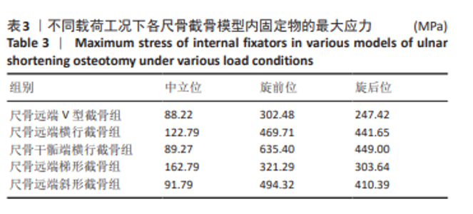
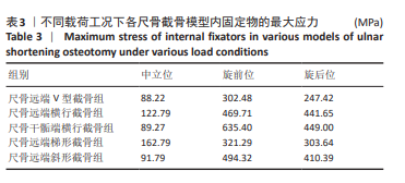
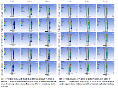
2.1 内固定物的应力及分布 分析5种不同截骨固定方式在3种不同运动即不同载荷下的内固定物应力分布。在中立位单纯肌肉收缩状态下,梯形截骨固定钢板的应力峰值最大,为162.79 MPa,应力集中于钢板中部;V型截骨固定钢板的应力峰值最小,为88.22 MPa,应力也集中于钢板中部。在模拟旋前状态下,干骺端横行截骨固定钢板的应力峰值最大,为635.40 MPa,应力集中于钢板中部;V型截骨固定钢板的应力峰值最小,为302.48 MPa,应力集中于钢板中部。在模拟旋后状态下,干骺端横行截骨固定钢板的应力峰值最大,为449.00 MPa,应力集中于钢板中部;V型截骨固定钢板的应力峰值最小,为247.42 MPa,应力集中于钢板中部。见表3及图4。5种不同截骨内固定方式在3种运动载荷下,V型截骨内固定应力始终最小。说明V形截骨凭借其本身截骨面V形特性,能在一定程度上稳定截骨端并分担内固定物相应的应力。"
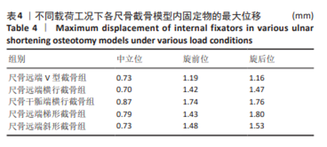
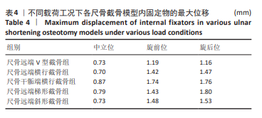
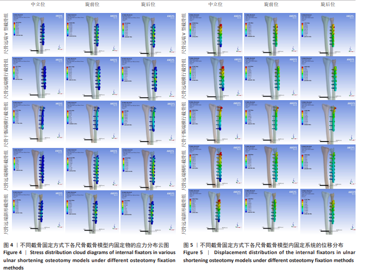
2.2 内固定物的位移及分布 3种不同运动状态下内固定物位移情况差别不明显。在中立位状态下,干骺端横行截骨固定钢板的位移峰值最大,为0.87 mm;远端横行截骨固定钢板的位移峰值最小,为0.70 mm。在模拟旋前状态下,干骺端横行截骨固定钢板的位移峰值最大,为1.74 mm;远端V型截骨固定钢板的位移峰值最小,为1.19 mm。在模拟旋后状态下,远端梯形截骨固定钢板的位移峰值最大,为1.80 mm;远端V型截骨固定钢板的位移峰值最小,为1.16 mm。见表4及图5。以上结果表明,几种截骨固定方式在不同状态下,内固定均较稳定,V形截骨只存在细微优势。"
"
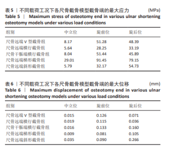
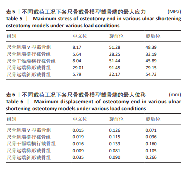
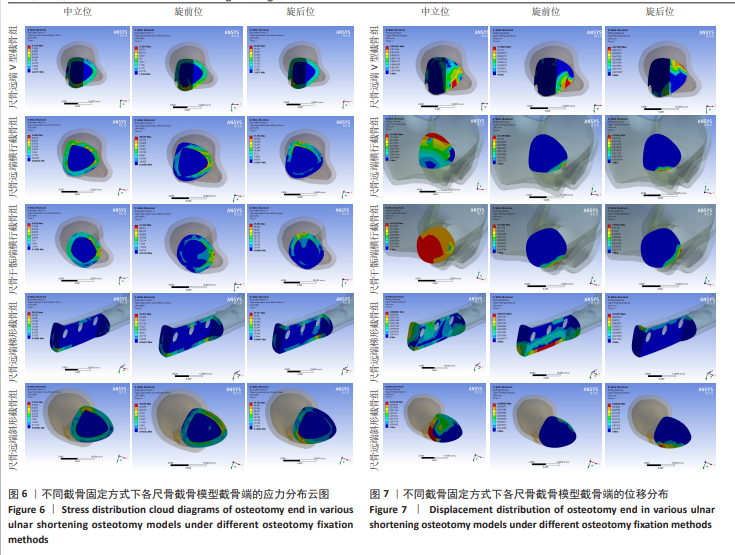
2.3 截骨端的应力及分布 在截骨断端应力方面,在中立位单纯肌肉收缩状态下,远端梯形截骨断端的应力峰值最大,为29.01 MPa;远端横行截骨组截骨断端的应力峰值最小,为5.64 MPa。在模拟旋前状态下,远端梯形截骨组截骨断端的应力峰值最大,为91.45 MPa;远端横行截骨组截骨断端的应力峰值最小,为28.25 MPa。在模拟旋后状态下,远端梯形截骨组截骨断端的应力峰值最大,为79.15 MPa;远端横行截骨组截骨断端的应力峰值最小,为33.19 MPa。见表5及图6。由此可见在骨折断端应力分布中,3种模拟状态下都是远端梯形截骨组截骨断端的应力峰值最大,而远端横行截骨组截骨断端的应力峰值最小。梯形截骨因其截骨面的复杂形状,造成截骨端相应的接触面复杂,因此其应力大;而斜行截骨因其截骨面平滑且与纵轴存在成角的滑面,导致截骨端应力分散,因而应力相对较小。 2.4 截骨端的位移及分布 骨折断端位移数值均较小,保留小数点后3位进行评估比较。在中立位状态下,远端斜形截骨组截骨断端的位移峰值最大,为0.035 mm;远端梯形截骨组截骨断端的位移峰值最小,为0.009 mm。在模拟旋前状态下,尺骨干骺端横行截骨组截骨断端的位移峰值最大,为0.133 mm;尺骨远端梯形截骨组截骨断端的位移峰值最小,为0.081 mm。在模拟旋后状态下,尺骨远端斜形截骨组截骨断端的位移峰值最大,为0.266 mm;尺骨远端横行截骨组截骨断端的位移峰值最小,为0.036 mm。见表6及图7。截骨断端在3种状态下位移均非常小,均在0.3 mm以下。5种截骨方式因其内固定的稳定固定,最终结果均无明显移位,且不同截骨方式间无显著差别。"

"
| [1] MILCH H. Cuff resection of the ulna for malunited Colles’ fracture. JBJS. 1941; 23(2):311-313. [2] RYOO HJ, KIM YB, KWAK D, et al. Ulnar positive variance associated with TFCC foveal tear. Skeletal Radiol. 2023;52(8):1485-1491. [3] 何信坤,阮健,杨科跃,等. 尺骨撞击综合征诊断和治疗方式的选择[J]. 中国骨与关节杂志,2025,14(8):751-757. [4] 郑炜,熊革,李忠哲,等. 尺骨撞击综合征腕骨囊性变的转归及临床分析[J]. 中华手外科杂志,2014,30(4):243-245. [5] SCHMIDLE G, ARORA R, GABL M. Ulnar shortening with the ulna osteotomy locking plate. Oper Orthop Traumatol. 2012;24(3):284-292. [6] 陈时益,高伟阳,汪洋,等.尺骨短缩术治疗尺骨撞击综合征术前术后影像变化及临床意义[J].中华手外科杂志,2014,30(6):451-454. [7] YIN HW, QIU YQ, SHEN YD, et al. Arthroscopic distal metaphyseal ulnar shortening osteotomy for ulnar impaction syndrome: a different technique. J Hand Surg Am. 2013;38(11):2257-2262. [8] 丛晓斌, 李涛, 季伟, 等.尺骨短缩截骨治疗特发性尺骨撞击综合征的疗效分析[J]. 中华手外科杂志,2013,29(1):7-9. [9] 翟晃,赵刚,余炯, 等.尺骨干骺端截骨治疗尺腕撞击综合征[J].中华手外科杂志,2021,37(6):424-428. [10] LUO TD, DE GREGORRIO M, ZUSKOV A, et al. Distal metaphyseal osteotomy allows for greater ulnar shortening compared to diaphyseal osteotomy for ulnar impaction syndrome:a biomechanical study. J Wrist Surg. 2020;9(2):100-104. [11] IMAI H, TAKAHARA M, KONDO M. Ulnar shortening osteotomy for ulnar abutment syndrome: the results of metaphyseal and diaphyseal osteotomies. J Hand Surg Asian Pac Vol. 2020;25(4):474-480. [12] 李学渊, 王扬剑, 孙涛, 等.尺骨斜形截骨短缩技术结合关节镜治疗尺骨撞击综合征[J].中华手外科杂志,2014,30(3):177-179. [13] 张兆毅, 李子华, 黄若强, 等. 尺骨远端斜行缩短截骨术治疗尺骨撞击综合征[J]. 中华手外科杂志,2017,33(3):224-225. [14] 郭明磊, 赵少平, 王振旺.尺骨梯形截骨短缩术治疗尺骨撞击综合征 20 例临床分析[J].泸州医学院学报,2015,38(2):153-156. [15] 于宁, 王彦生, 叶放, 等. V 形尺骨短缩截骨术治疗尺骨撞击综合征[J].中华手外科杂志,2019,35(5):385-386. [16] HUISKES R, CHAO EY. A survey of finite element analysis in orthopedic biomechanics: the first decade. J Biomech. 1983;16(6):385-409. [17] KWON BC, SEO BK, IM HJ, et al. Clinical and radiographic factors associated with distal radioulnar joint “instability in distal radius fractures. Clin Orthop Relat Res. 2012;470(11):3171-3179. [18] LINDUA T, ASPENBERG P. The radioulnar joint in distal radial fractures. Acta Orthop Scand. 2002;73(5):579-588. [19] 陈伟,杨玉亭,王艳,等. 有限元分析弹性髓内钉治疗尺桡骨干双骨折不同进针点的力学差异[J]. 中国组织工程研究,2026,30(15):3772-3779. [20] 王小阵, 席金涛, 陈龙, 等. Wiltse入路经椎间孔外椎体间融合术治疗单节段Ⅰ、Ⅱ度退变性腰椎滑脱症[J]. 生物骨科材料与临床研究,2025,22(3):14-19. [21] MENA A, WOLLSTEIN R, YANG J. Development of a finite element model of the human wrist joint with radial and ulnar axial force distribution and radiocarpal contact validation. J Biomech Eng. 2025;147(3):031006. [22] 张波,魏蓝,赵振国,等.尺骨头干骺端截骨与尺骨干截骨内固定治疗尺腕撞击综合征的疗效分析[J].中华手外科杂志,2024,40(1):48-52. [23] 赵驰,林平,徐柯烽,等. 两种不同截骨方式治疗尺骨撞击综合征[J]. 中华手外科杂志,2023,39(2):170-173. [24] SHARMA A. A study of titanium elastic nailing in forearm fractures in elderly patients. J Orthop Spine Trauma. 2024;10(2):82-86. [25] FACAN JB, MAINARD N, FACYR PA, et al. Results of isolated ulnar shaft shortening osteotomy in the treatment of idiopathic ulnocarpal impaction syndrome. Hand Surg Rehabil. 2022;41(5):589-594. [26] 郭欣,樊瑜波,李宗明. 掌骨受轴向压力作用下的腕部生物力学分析[J]. 航天医学与医学工程,2008,21(1):45-49. [27] ANDERSON DD,DANIEL TD. A contact-coupled finite element analysis of the radio carpal joint. Semin Arthroplasty. 1995;6(1):30-36. [28] TENCER AF, VIEGASSF, CANTRELL J, et al. Pressure distribution in the wrist joint. J Orthop Res. 1988;6(4):509-517. [29] NISHIWAKI M, NAKAMURA T, NAGURA T, et al. Ulnar-shortening effect on distal radioulnar joint pressure: a biomechanical study. J Hand Surg Am. 2008; 33(2)198-205. [30] SCHUIND F, AN KN, BERGLUND L, et al. The distal radioulnar ligaments: A Biomechanical study. J Hand Surg Am. 1991;16(6):1106-1114. [31] SHORT WH, WERNER FW, FORTINO MD, et al. The stabilizing mechanism of the distal radioulnar joint during pronation and supination. J Hand Surg Am. 1995; 20(6):930-936. [32] 钦斌,黄永火,欧阳羽,等.轴向应力作用下的舟骨有限元分析[J]. 第三军医大学学报,2010,32(11):1213-1215. [33] 周晓宁. 腕关节三维有限元模型的建立及桡骨远端骨折发生机制的生物力学分析[D]. 北京:北京中医药大学,2014. [34] PIROKO JM, YAO J. Minimally invasive approaches to ulnar-sided wrist disorders. Hand Clin. 2014;30(1):77-89. [35] 马炜,田文. 尺骨撞击综合征诊断与治疗[J]. 中国骨与关节杂志,2014,3(3): 213-215. [36] 王凯,郑奕,周黎辉. 尺骨撞击综合征研究进展[J]. 沈阳医学院学报,2024, 26(2):200-203. [37] PALMER AK, WERENG FW. Biome-chanics of the distal radioulnar joint. Clin Orthop. 1984;187(7):8. [38] PALMER AK. Triangular fibrocartilage complex lesions: a classification. J Hand Surg Am. 1989;14(4):594-606. [39] SLUTSKY DJ. Arthroscopic management of ulnocarpal impaction syndrome and ulnar styloid impaction syndrome. Hand Clin. 2017;33(4):639-650. [40] 孙广峰, 金文虎, 聂开瑜,等.尺骨短缩术治疗尺骨撞击综合征的体会[J]. 中华手外科杂志,2016,32(2):149-150. [41] VAJGEL A, CAMARGO IB, WILLMERSDORF RB, et al. Comparative finite element analysis of the biomechanical stability of 2.0 fixation plates in atrophic mandibular fractures. J Oral Maxillofac Surg. 2013;71(2):335-342. [42] LIANG C, JIANG F, KAWAGUCHI D, et al. A Biomechanical Simulation of Forearm Flexion Using the Finite Element Approach. Bioengineering. 2023;11(1):23. [43] 徐天宇,陈默迪,谢明茹,等.内、外踝韧带缺损对邻近核心肌腱生物力学影响的有限元分析[J].中国组织工程研究,2025,29(33):7223-7230. [44] 张子峥,罗旺,刘长路,等.膝内侧间室骨关节炎单髁置换中有限元分析的应用价值[J]. 中国组织工程研究,2026,30(9):2313-2322. [45] WANG H, YAO G, HE K, et al. ACL reconstruction combined with anterolateral structures reconstruction for treating ACL rupture and knee injuries: a finite element analysis. Front Bioeng Biotechnol. 2024;12:1437684-1437684. [46] LING Q, HE E, ZHANG H, et al. A novel narrow surface cage for full endoscopic oblique lateral lumbar interbody fusion: A finite element study. J Orthop Sci . 2019;24(6):991-998. [47] MENEGAZ GL, GOMIDE LC, ARAUJO CA. Biomechanical evaluation of acromioclavicular joint reconstructions using a 3-dimensional model based on the finite element method. Clin Biomech (Bristol, Avon). 2019;70:170-176. [48] SHENG W, JI A, FANG R, et al. Finite Element- and Design of Experiment-Derived Optimization of Screw Configurations and a Locking Plate for Internal Fixation System. Comput Math Methods Med. 2019;2019:5636528. [49] SOGANCI UNSAL G, HASANOGLU ERBASAR GN, AYKENT F, et al. Evaluation of Stress Distribution on Mandibular Implant-supported Overdentures with Different Bone Heights and Attachment Types: A 3D Finite Element Analysis. J Oral Implantol. 2019;45(5):363-370. [50] OZCAN M, ACAR E, BASCI O, et al. Minimial clinically important difference values in distal metaphyseal ulnar shortening for ulnar impaction syndrome and assessment of the relationship between level of the osteotomy and bone union time. Acta Orthop Traumatol Turc. 2024;58(1):27-33. [51] 贺凯,邢文华,李峰,等.有限元法在脊柱胸腰段骨折生物力学分析中的应用及发展方向[J]. 中国组织工程研究,2025,29(15):3244-3252. |
| [1] | Zhang Zizheng, Luo Wang, Liu Changlu. Application value of finite element analysis on unicompartmental knee arthroplasty for medial knee compartmental osteoarthritis [J]. Chinese Journal of Tissue Engineering Research, 2026, 30(9): 2313-2322. |
| [2] | Liu Wenlong, Dong Lei, Xiao Zhengzheng, Nie Yu. Finite element analysis of tibial prosthesis loosening after fixed-bearing unicompartmental knee arthroplasty for osteoporosis [J]. Chinese Journal of Tissue Engineering Research, 2026, 30(9): 2191-2198. |
| [3] | Zheng Wangyang, Fei Ji, Yang Di, Zhao Lang, Wang Lingli, Liu Peng, Li Haiyang. Finite element analysis of the force changes of the supraspinatus tendon and glenohumeral joint during the abduction and flexion of the humerus [J]. Chinese Journal of Tissue Engineering Research, 2026, 30(9): 2199-2207. |
| [4] | Cai Qirui, Dai Xiaowei, Zheng Xiaobin, Jian Sili, Lu Shaoping, Liu Texi, Liu Guoke, Lin Yuanfang. Mechanical effects of Long’s traction orthopedic method on cervical functional units: quantitative analysis of biomechanical model of head and neck [J]. Chinese Journal of Tissue Engineering Research, 2026, 30(9): 2208-2216. |
| [5] | Rao Jingcheng, Li Yuwan, Zheng Hongbing, Xu Zhi, Zhu Aixiang, Shi Ce, Wang Bing, Yang Chun, Kong Xiangru, Zhu Dawei. Biomechanical differences between the new proximal femoral stable intramedullary nail and traditional intramedullary nail#br# [J]. Chinese Journal of Tissue Engineering Research, 2026, 30(9): 2217-2225. |
| [6] | Chen Long, Wang Xiaozhen, Xi Jintao, Lu Qilin. Biomechanical performance of short-segment screw fixation combined with expandable polyetheretherketone vertebral body replacement in osteoporotic vertebrae [J]. Chinese Journal of Tissue Engineering Research, 2026, 30(9): 2226-2235. |
| [7] | Cheng Qisheng, Julaiti·Maitirouzi, Xiao Yang, Zhang Chenwei, Paerhati·Rexiti. Finite element analysis of novel variable-diameter screws in modified cortical bone trajectory of lumbar vertebrae [J]. Chinese Journal of Tissue Engineering Research, 2026, 30(9): 2162-2171. |
| [8] | Wu Hongxu, Liu Xuanyu, Wang Taoyu, Wang Shiyao, Cheng Jingyi, Zhang Mingwen, Zhang Yinxia, Liu Zhihua, Wang Xiaojie. Finite element simulation of scoliosis with muscle unit introduction: verification of correction effect under bidirectional load [J]. Chinese Journal of Tissue Engineering Research, 2026, 30(9): 2172-2181. |
| [9] | Liu Jiafu, Ren Ruxia, Liao Zhouwei, Zhou Xiali, Wu Yihong, Zhang Shaoqun. Three-dimensional finite element analysis of cervical spine biomechanical characteristics in a rat model of cervical vertigo [J]. Chinese Journal of Tissue Engineering Research, 2026, 30(9): 2182-2190. |
| [10] | Zhang Nan, Meng Qinghua, Bao Chunyu. Characteristics and clinical application of ankle joint finite element models [J]. Chinese Journal of Tissue Engineering Research, 2026, 30(9): 2343-2349. |
| [11] | Chen Huiting, Zeng Weiquan, Zhou Jianhong, Wang Jie, Zhuang Congying, Chen Peiyou, Liang Zeqian, Deng Weiming. Tail anchoring technique of vertebroplasty in treatment of osteoporotic vertebral compression fractures with intravertebral cleft: a finite element analysis [J]. Chinese Journal of Tissue Engineering Research, 2026, 30(9): 2145-2152. |
| [12] | Zeng Xuan, Weng Rui, Ye Shicheng, Tang Jiadong, Mo Ling, Li Wenchao. Two lumbar rotary manipulation techniques in treating lumbar disc herniation: a finite element analysis of biomechanical differences [J]. Chinese Journal of Tissue Engineering Research, 2026, 30(9): 2153-2161. |
| [13] | Yang Xuetao, Zhu Menghan, Zhang Chenxi, Sun Yimin, Ye Ling. Applications and limitations of antioxidant nanomaterials in oral cavity [J]. Chinese Journal of Tissue Engineering Research, 2026, 30(8): 2044-2053. |
| [14] | Zheng Xuying, Hu Hongcheng, Xu Libing, Han Jianmin, Di Ping. Stress magnitude and distribution in two-piece cement-retained zirconia implants under different loading conditions and with varying internal connection shapes [J]. Chinese Journal of Tissue Engineering Research, 2026, 30(8): 1979-1987. |
| [15] |
Dong Chunyang, Zhou Tianen, Mo Mengxue, Lyu Wenquan, Gao Ming, Zhu Ruikai, Gao Zhiwei.
Action mechanism of metformin combined with Eomecon chionantha Hance dressing in treatment of deep second-degree burn wounds#br#
#br#
[J]. Chinese Journal of Tissue Engineering Research, 2026, 30(8): 2001-2013.
|
| Viewed | ||||||
|
Full text |
|
|||||
|
Abstract |
|
|||||