Chinese Journal of Tissue Engineering Research ›› 2026, Vol. 30 ›› Issue (34): 9088-9094.doi: 10.12307/2026.865
Previous Articles Next Articles
Future medical research on brain organoids: interdisciplinary training, bioengineering technologies, and optimized model maturity
He Renda1, Ma Wei2, Sun Yongsi1, Mo Xueni2
- 1The First Clinical Medical College of Guangxi University of Chinese Medicine, Nanning 530200, Guangxi Zhuang Autonomous Region, China; 2Development Planning Office of Guangxi University of Chinese Medicine, Nanning 530200, Guangxi Zhuang Autonomous Region, China
-
Received:2025-10-29Revised:2026-01-22Online:2026-12-08Published:2026-04-15 -
Contact:Mo Xueni, PhD, Professor, Master’s supervisor, Development Planning Office of Guangxi University of Chinese Medicine, Nanning 530200, Guangxi Zhuang Autonomous Region, China -
About author:He Renda, MS candidate, The First Clinical Medical College of Guangxi University of Chinese Medicine, Nanning 530200, Guangxi Zhuang Autonomous Region, China -
Supported by:National Natural Science Foundation of China (General Program), No. 81874453 (to MXN); Guangxi Natural Science Foundation, No. 2020GXSFAA297270 (to MXN); Guangxi University of Chinese Medicine Youth Fund, No. 2023QN006 (to MW); Guangxi University of Chinese Medicine Key Project, No. 2024ZD006 (to MXN)
CLC Number:
Cite this article
He Renda, Ma Wei, Sun Yongsi, Mo Xueni. Future medical research on brain organoids: interdisciplinary training, bioengineering technologies, and optimized model maturity[J]. Chinese Journal of Tissue Engineering Research, 2026, 30(34): 9088-9094.
share this article
Add to citation manager EndNote|Reference Manager|ProCite|BibTeX|RefWorks
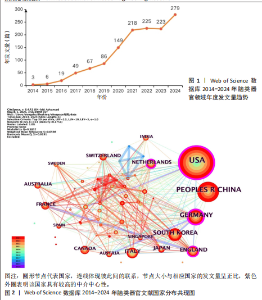
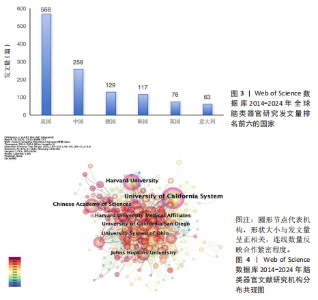
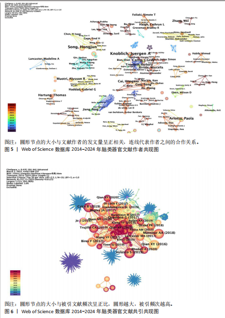
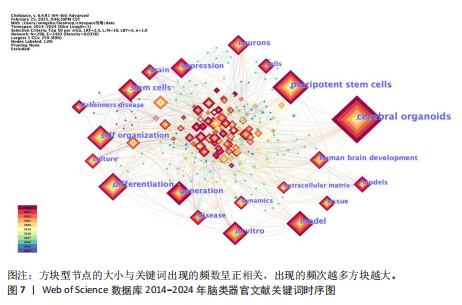
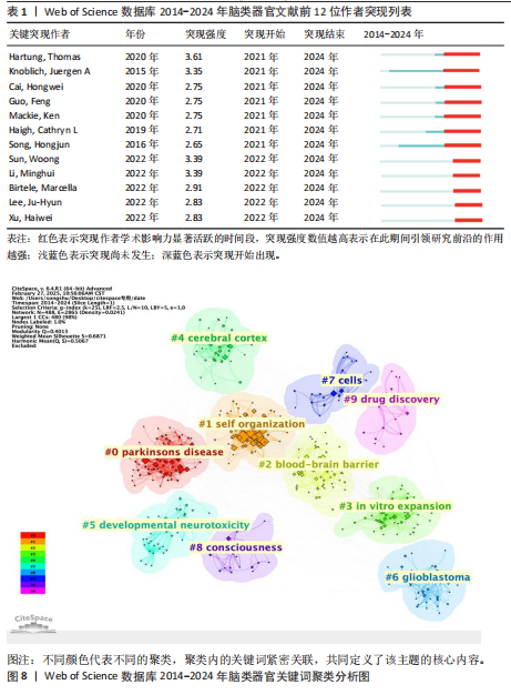
2.1 脑类器官年发文量分析 如图 1所示:从2014-2024年,脑类器官领域的研究文献共计发表了1 324篇,整体呈现上升趋势。2014-2021年,文献数量从3篇迅速增长至218篇,显示出该领域的快速发展;2022-2023年,文献发表量趋于平稳,而2024年则再次出现显著增长,这表明脑类器官医学持续受到广泛关注,具有巨大的发展潜力。 2.2 脑类器官文献国家分布 根据脑类器官文献的国家共现分析结果(图2,3),2014-2024年间,共有64个国家在该领域发表了相关文献。其中,发文量排名前六的国家分别为美国(568篇)、中国(258篇)、德国(129篇)、韩国(117篇)、英国(76篇)和意大利(63篇)。进一步分析中介中心性,排名前六的国家依次为美国(0.4)、德国(0.24)、英国(0.17)、荷兰(0.11)、伊朗(0.08)和韩国(0.08)。中介中心性作为衡量节点影响力的重要指标,其值高于0.1表明该国家在合作网络中与其他节点联系紧密[8],具有较高的学术影响力。由此可见,美国和德国在脑类器官医学研究领域处于核心地位,研究成果对全球该领域的发展具有重要引领作用。尽管中国发文量较多,但中介中心性相对较低,这表明中国在该领域的核心枢纽作用仍有提升空间。相比之下,荷兰和伊朗等国的发文总量虽未跻身前列,却表现出较高的中介中心性,显示出这些国家在合作网络中扮演了不可或缺的桥梁作用。 2.3 脑类器官文献机构分布 发文量是衡量科研机构实力的重要指标之一,能够直观反映机构在特定领域的研究活跃度和影响力。如图4所示,全球共有372个机构参与了脑类器官领域的研究,其中发文量排名前十的机构依次为:加州大学系统(94篇)、哈佛大学(57篇)、中国科学院(44篇)、俄亥俄大学系统(39篇)、加州大学圣地亚哥分校(38篇)、哈佛大学医学附属机构(35篇)、约翰斯·霍普金斯大学(35篇)、马克斯·普朗克学会(31篇)、哈佛医学院(29篇)以及美国国立卫生研究院(NIH)(28篇)。从机构间的合作网络来看,绝大部分机构之间的连线较为紧密,表明该领域内机构间的合作交流频繁,形成了较为广泛的科研协作网络。 2.4 脑类器官文献作者分布与作者突现分析 基于作者共现分析(图5)的结果,N值为398,E值为503,表明脑类器官研究的广泛性与复杂性,同时网络密度为0.006 4,说明研究者之间的合作关系较为分散,未形成高度集中的核心群体。图中可以直观发现高发文量作者之间的合作频率较低,而低发文量作者与高发文量作者之间则表现出较为密切的合作关系。这一现象表明,研究团队内部形成了稳定的合作网络,但不同研究团队之间尚未建立广泛的合作关系。具体而言,发文量排名前三的作者分别为Knoblich, Juergen A(尤尔根·A·克诺布利希,13篇)、Song, Hongjun(宋洪军,12篇)和Arlotta, Paola(保拉·阿洛塔,9篇),这些作者在领域内具有较高的学术影响力。根据普赖斯定律(Price’s Law)计算得出核心作者最低发文量为2.701篇,取整后为3篇,得到脑类器官研究领域的核心作者人数为77人,占总作者人数的19.34%[9]。进一步进行作者突现分析(表1),研究识别出前12位突现作者,突现强度分布较为均衡。这一结果表明,脑类器官研究领域具有较高的主题多样性,研究热点分布均匀,且研究者的活跃程度相对一致。此外,研究热点的集中爆发期主要出现在2021-2024年,这一时间段的学术成果显著增加,表明脑类器官研究领域在近年来进入了快速发展的新阶段,这一发展趋势可能得益于技术进步、"

"

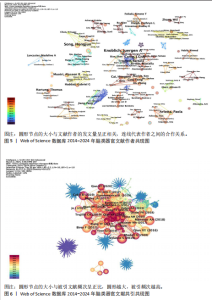
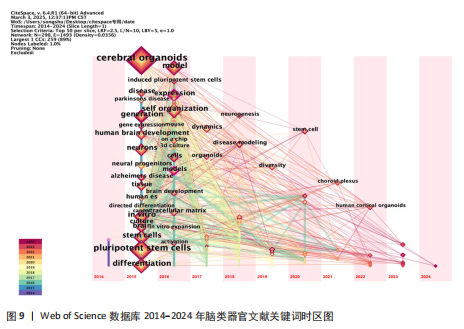
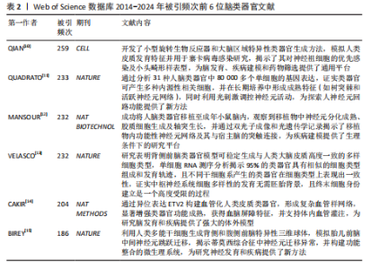
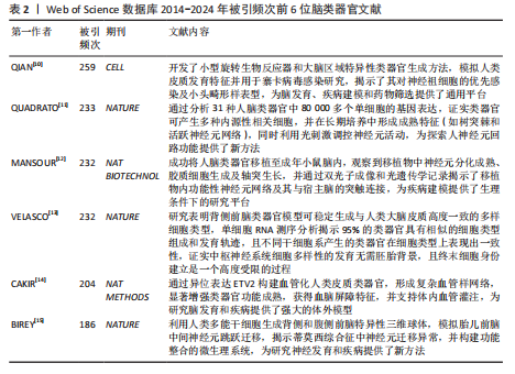
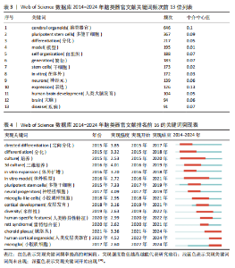
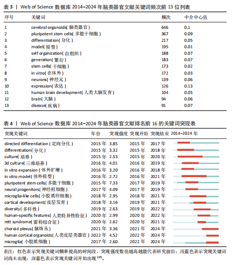
研究方法的创新以及跨学科合作的深化。 2.5 脑类器官文献共引分析 将脑类器官文献进行文献共引分析得到网络图谱,见图6,共引频次最高的6篇文章分别是Brain-Region-Specific Organoids Using Mini-bioreactors for Modeling ZIKV Exposure(使用微型生物反应器模拟 ZIKV 暴露的大脑区域特异性类器官,259次),Cell diversity and network dynamics in photosensitive human brain organoids(光敏人脑类器官中的细胞多样性和网络动力学,233次),An in vivo model of functional and vascularized human brain organoids(一种功能性和血管化人脑类器官的体内模型,232次),Individual brain organoids reproducibly form cell diversity of the human cerebral cortex(单个大脑类器官可重复地形成人类大脑皮质的细胞多样性,232次),Engineering of human brain organoids with a functional vascular-like system(具有功能性类血管系统的人脑类器官工程,204次),Assembly of functionally integrated human forebrain spheroids(功能整合的人类前脑球体的组装,186次),见表2[10-15],这些文献的高共引频次表明在脑类器官研究领域具有广泛认可度和重要的参考价值。 2.6 脑类器官关键词共现及聚类分析 将关键词、近义词合并后对数据进行共现分析,见图7,将出现频次排序前13位的关键词进行统计,见表3,通过聚类分析,得到Q值为0.505 5,S值为0.797 8的关键词聚类图,见图8,模块值Q和轮廓值S是衡量聚类结构合理性的2个重要指标,Q > 0.3说明聚类结构显著,S > 0.5表明聚类结果同质性较高[16],由此可见图8的聚类结构合理,代表性显著,根据关键词聚类图将近10年脑类器官领域的研究划分为以下10个主要研究主题:Parkinson’s disease(帕金森病),self-organization (自组织),blood-brain barrier (血脑屏障),in vitro expansion (体外扩增),cerebral cortex (大脑皮质),developmental neurotoxicity (发育性神经毒性),glioblastoma (胶质母细胞瘤),cells (细胞),consciousness (意识),drug discovery (药物发现)。 2.7 脑类器官文献关键词突现分析 对脑类器官文献关键词进行突现分析,见表4[17],表中pluripotent stem cells (多能干细胞)的突现强度最强,达到7.33,该方向在2017-2019年的热度较高,in vitro model(体外模型)、culture (培养)突现持续时间最长,表明in vitro model、culture发展潜力大,是该领域的长期研究热点,近年热点研究方向则为choroid plexus (脉络丛)、human cortical organoids (人类皮质类器官)以及microglia(小胶质细胞)。 2.8 脑类器官研究领域发展趋势 通过关键词时区图能快速把握领域的研究动态和趋势,如图9所示,节点N=290,连线E=1 493,密度为0.035 6,表明关键词之间存在显著关联但并非完全联通,2014-2017年,研究聚焦于基础技术,如induced pluripotent stem cells(多能干细胞诱导)、differentiation(分化)以及culture (培养),建立脑类器官生成的基础方法并使用动物模型验证技术可行性[10,18],self-organization "

"

(自组织)节点的出现,强调类器官模拟脑发育动态的能力显现[12,19];2018-2021年研究方向转变为stem cell(干细胞)、disease modeling(疾病模型的构建)等多样性研究,如利用患者诱导多能干细胞构建脑类器官,模拟阿尔茨海默病等神经炎症以及β-淀粉样蛋白沉积[20];从2022年开始,研究热点逐渐偏向human cortical organoids(人大脑皮质类器官)等更为复杂的脑网络功能发育模拟系统,同时通过跨学科融合,推动脑类器官从“结构模型”向“功能模型”的升级[21-23]。"

"
"
"
| [1] STILES J, JERNIGAN TL. The basics of brain development. Neuropsychol Rev. 2010;20(4):327-348. [2] CHEN H, JIN X, LI T, et al. Brain organoids: Establishment and application. Front Cell Dev Biol. 2022;10:1029873. [3] KIM H, JANG EJ, SANKPAL NV, et al. Recent Development of Brain Organoids for Biomedical Application. Macromol Biosci. 2023;23(3): e2200346. [4] LANCASTER MA, RENNER M, MARTIN CA, et al. Cerebral organoids model human brain development and microcephaly. Nature. 2013; 501(7467):373-379. [5] KOKOL P, BLAŽUN VOŠNER H, ZAVRŠNIK J. Application of bibliometrics in medicine: a historical bibliometrics analysis. Health Info Libr J. 2021;38(2):125-138. [6] 王勇,李宏宇,刘雨航,等.股骨头坏死手术治疗知识图谱:2005-2024数据的文献计量学分析[J].中国组织工程研究,2025,29(33):7250-7260. [7] 徐灿丽,何文星,汪磊,等.肝脏类器官研究的文献计量学分析[J].中国组织工程研究,2024, 28(7):1099-1104. [8] BAI X, SONG Z, ZHOU Y, et al. Bibliometrics and Visual Analysis of the Research Status and Trends of Postpartum Depression From 2000 to 2020. Front Psychol. 2021;12:665181. [9] YAO S, TANG Y, YI C, et al. Research Hotspots and Trend Exploration on the Clinical Translational Outcome of Simulation-Based Medical Education: A 10-Year Scientific Bibliometric Analysis From 2011 to 2021. Front Med (Lausanne). 2022;8: 801277. [10] QIAN X, NGUYEN HN, SONG MM, et al. Brain-Region-Specific Organoids Using Mini-bioreactors for Modeling ZIKV Exposure. Cell. 2016;165(5):1238-1254. [11] QUADRATO G, NGUYEN T, MACOSKO EZ, et al. Cell diversity and network dynamics in photosensitive human brain organoids. Nature. 2017;545(7652):48-53. [12] MANSOUR AA, GONÇALVES JT, BLOYD CW, et al. An in vivo model of functional and vascularized human brain organoids. Nat Biotechnol. 2018; 36(5):432-441. [13] VELASCO S, KEDAIGLE AJ, SIMMONS SK, et al. Individual brain organoids reproducibly form cell diversity of the human cerebral cortex. Nature. 2019;570(7762):523-527. [14] CAKIR B, XIANG Y, TANAKA Y, et al. Engineering of human brain organoids with a functional vascular-like system. Nat Methods. 2019;16(11):1169-1175. [15] BIREY F, ANDERSEN J, MAKINSON CD, et al. Assembly of functionally integrated human forebrain spheroids. Nature. 2017;545(7652): 54-59. [16] 梁红,李浩宇.基于CiteSpace的国内术语翻译研究可视化分析(2001—2020)[J].中国科技术语,2022,24(1):87-96. [17] 赵思思,刘勇.基于CiteSpace的人工智能在脑卒中领域应用进展的可视化分析[J].重庆医学, 2024,53(24):3706-3713+3719. [18] LANCASTER MA, KNOBLICH JA. Generation of cerebral organoids from human pluripotent stem cells. Nat Protoc. 2014;9(10):2329-2340. [19] PAŞCA AM, SLOAN SA, CLARKE LE, et al. Functional cortical neurons and astrocytes from human pluripotent stem cells in 3D culture. Nat Methods. 2015;12(7):671-678. [20] PARK J, WETZEL I, MARRIOTT I, et al. A 3D human triculture system modeling neurodegeneration and neuroinflammation in Alzheimer’s disease. Nat Neurosci. 2018;21(7):941-951. [21] PARK Y, HERNANDEZ S, HERNANDEZ CO, et al. Modulation of neuronal activity in cortical organoids with bioelectronic delivery of ions and neurotransmitters. Cell Rep Methods. 2024; 4(1):100686. [22] REVAH O, GORE F, KELLEY KW, et al. Maturation and circuit integration of transplanted human cortical organoids. Nature. 2022;610(7931):319-326. [23] ZHANG XP, WANG XY, WANG SN, et al. The generation and properties of human cortical organoids as a disease model for malformations of cortical development. Neural Regen Res. 2023; 18(10):2119-2126. [24] ZHAO HH, HADDAD G. Brain organoid protocols and limitations. Front Cell Neurosci. 2024;18: 1351734. [25] 祖勉,王瑛,刘伟,等.美国“脑计划”实施特点分析及启示[J].中国科学院院刊,2023, 38(2):302-314. [26] 李新钢,张鑫,陈安静.当代脑计划研究进展[J].山东大学学报(医学版),2020,58(8):5-9+21. [27] ACHARYA P, CHOI NY, SHRESTHA S, et al. Brain organoids: A revolutionary tool for modeling neurological disorders and development of therapeutics. Biotechnol Bioeng. 2024;121(2): 489-506. [28] ZUSHIN PH, MUKHERJEE S, WU JC. FDA Modernization Act 2.0: transitioning beyond animal models with human cells, organoids, and AI/ML-based approaches. J Clin Invest. 2023; 133(21):e175824. [29] AILI Y, MAIMAITIMING N, WANG Z, et al. Brain organoids: A new tool for modelling of neurodevelopmental disorders. J Cell Mol Med. 2024;28(17):e18560. [30] HONGXI W, RUTING W, YIYANG L, et al. Human Brain Organoids: Development and Applications. J Microbiol Biotechnol. 2025;35:e2411040. [31] XUE J, CHU Y, HUANG Y, et al. A tumorigenicity evaluation platform for cell therapies based on brain organoids. Transl Neurodegener. 2024; 13(1):53. [32] YU S, JIANG L, SONG M, et al. Regulation of BBB function and pathological evolution of PD by microenvironment “spatiotemporal gradient”: unique advantages of microfluidic chips. Front Aging Neurosci. 2025;17:1599509. [33] SCHICKEL E, BENDER T, KAYSAN L, et al. Human cerebral organoids model tumor initiation and infiltration in an autologous astrocyte-supported setting. iScience. 2025;28(9):113334. [34] QIAN X, JACOB F, SONG MM, et al. Generation of human brain region-specific organoids using a miniaturized spinning bioreactor. Nat Protoc. 2018;13(3):565-580. [35] SHAJI M, TAMADA A, FUJIMOTO K, et al. Deciphering potential vascularization factors of on-chip co-cultured hiPSC-derived cerebral organoids. Lab Chip. 2024;24(4):680-696. [36] KONG D, PARK KH, KIM DH, et al. Cortical-blood vessel assembloids exhibit Alzheimer’s disease phenotypes by activating glia after SARS-CoV-2 infection. Cell Death Discov. 2023;9(1):32. [37] LENIN S, PONTHIER E, SCHEER KG, et al. A Drug Screening Pipeline Using 2D and 3D Patient-Derived In Vitro Models for Pre-Clinical Analysis of Therapy Response in Glioblastoma. Int J Mol Sci. 2021;22(9):4322. [38] LI Z, XU J, LANG Y, et al. JMX0207, a Niclosamide Derivative with Improved Pharmacokinetics, Suppresses Zika Virus Infection Both In Vitro and In Vivo. ACS Infect Dis. 2020;6(10):2616-2628. [39] ZIFFRA RS, KIM CN, ROSS JM, et al. Single-cell epigenomics reveals mechanisms of human cortical development. Nature. 2021; 598(7879):205-213. [40] FENG Y, ZHENG H, TANG J, et al. Protocol for generating in vitro glioma models using human-induced pluripotent- or embryonic-stem-cell-derived cerebral organoids. STAR Protoc. 2023; 4(3):102346. [41] UZQUIANO A, KEDAIGLE AJ, PIGONI M, et al. Proper acquisition of cell class identity in organoids allows definition of fate specification programs of the human cerebral cortex. Cell. 2022;185(20):3770-3788.e27. [42] BOWLES KR, SILVA MC, WHITNEY K, et al. ELAVL4, splicing, and glutamatergic dysfunction precede neuron loss in MAPT mutation cerebral organoids. Cell. 2021;184(17):4547-4563.e17. [43] DAO L, YOU Z, LU L, et al. Modeling blood-brain barrier formation and cerebral cavernous malformations in human PSC-derived organoids. Cell Stem Cell. 2024;31(6):818-833.e11. [44] ANTÓN-BOLAÑOS N, FARAVELLI I, FAITS T, et al. Brain Chimeroids reveal individual susceptibility to neurotoxic triggers. Nature. 2024;631(8019):142-149. [45] FUMADÓ NAVARRO J, CRILLY S, CHAN WK, et al. Cerebral Organoids with Integrated Endothelial Networks Emulate the Neurovascular Unit and Mitigate Core Necrosis. Adv Sci (Weinh). 2025; 12(43):e07256. [46] NARAZAKI G, MIURA Y, PAVLOV SD, et al. Scalable production of human cortical organoids using a biocompatible polymer. Nat Biomed Eng. 2025; 9(12):2115-2123. [47] KIM H, KANG S, CHO B, et al. Parkinson’s Disease Modeling Using Directly Converted 3D Induced Dopaminergic Neuron Organoids and Assembloids. Adv Sci (Weinh). 2025;12(14):e2412548. [48] LI C, FLECK JS, MARTINS-COSTA C, et al. Single-cell brain organoid screening identifies developmental defects in autism. Nature. 2023;621(7978):373-380. [49] KOCH LS, CHOY BUENTELLO D, BROERSEN K. Robust Tissue Fabrication for Long-Term Culture of iPSC-Derived Brain Organoids for Aging Research. J Vis Exp. 2023;(195). doi: 10.3791/64586. [50] AKKOUH IA, UELAND T, SZABO A, et al. Longitudinal Transcriptomic Analysis of Human Cortical Spheroids Identifies Axonal Dysregulation in the Prenatal Brain as a Mediator of Genetic Risk for Schizophrenia. Biol Psychiatry. 2024;95(7): 687-698. [51] QIAN X, SU Y, ADAM CD, et al. Sliced Human Cortical Organoids for Modeling Distinct Cortical Layer Formation. Cell Stem Cell. 2020;26(5):766-781.e9. [52] BOUTOM SM, SILVA TP, PALECEK SP, et al. Central nervous system vascularization in human embryos and neural organoids. Cell Rep. 2024; 43(12):115068. [53] LI M, GAO L, ZHAO L, et al. Toward the next generation of vascularized human neural organoids. Med Res Rev. 2023;43(1):31-54. [54] PAGLIARO A, ARTEGIANI B, HENDRIKS D. Emerging approaches to enhance human brain organoid physiology. Trends Cell Biol. 2025;35(6):483-499. [55] GEIDIES A, MEDAR ML, BEYER HM. Engineering organoids as cerebral disease models. Curr Opin Biotechnol. 2025;92:103253. [56] HAO YX, LI CR, LU ZJ, et al. Remodeling and repair of the damaged brain: the potential and challenges of organoids for ischaemic stroke. J Transl Med. 2025;23(1):767. [57] JONES HE, ROBERTSON GL, BODNYA C, et al. Leptomeningeal Neural Organoid Fusions as Models to Study Meninges-Brain Signaling. Stem Cells Dev. 2025;34(7-8):152-163. |
| [1] | Xu Canli, He Wenxing, Wang Yuping, Ba Yinying, Chi Li, Wang Wenjuan, Wang Jiajia. Research context and trend of TBK1 in autoimmunity, signaling pathways, gene expression, tumor prevention and treatment [J]. Chinese Journal of Tissue Engineering Research, 2026, 30(在线): 1-11. |
| [2] | Zhu Xiaolong, Zhang Wei, Yang Yang. Visualization analysis of research hotspots and cutting-edge information in the field of intervertebral disc regeneration and repair [J]. Chinese Journal of Tissue Engineering Research, 2026, 30(9): 2391-2402. |
| [3] | Wen Fayan, Li Yan, Qiang Tianming, Yang Chen, Shen Linming, Li Yadong, Liu Yongming. Unilateral biportal endoscopic technology for treatment of lumbar degenerative diseases: global research status and changing trends [J]. Chinese Journal of Tissue Engineering Research, 2026, 30(9): 2380-2390. |
| [4] | Lai Yu, Chen Yueping, Zhang Xiaoyun. Research hotspots and frontier trends of bioactive materials in treating bone infections [J]. Chinese Journal of Tissue Engineering Research, 2026, 30(8): 2132-2144. |
| [5] | Huang Jie, Zeng Hao, Wang Wenchi, Lyu Zhucheng, Cui Wei. Visualization analysis of literature on the effect of lipid metabolism on osteoporosis [J]. Chinese Journal of Tissue Engineering Research, 2026, 30(6): 1558-1568. |
| [6] | Bu Fanchen, Hua Zhen, , Li Xiaolong, Lyu Jinye, Man Hao, Wang Jianwei, . Triangular fibrocartilage complex injuries: a visualization analysis of treatment hotspots and frontiers [J]. Chinese Journal of Tissue Engineering Research, 2026, 30(34): 9095-9102. |
| [7] | Zhang Anqi, Hua Haotian, Cai Tianyuan, Wang Zicheng, Meng Zhuo, Zhan Xiaoqian, Chen Guoqian . Pain after total knee arthroplasty: current status and trend analysis [J]. Chinese Journal of Tissue Engineering Research, 2026, 30(3): 795-804. |
| [8] | Wang Degang, Mei Junhua, Wang Junli, Zheng Li, Chen Guohua. Bibliometric and visualization analysis of the mechanism of osteogenic factors and neurotransmitters in the bone-brain axis [J]. Chinese Journal of Tissue Engineering Research, 2026, 30(29): 7724-7731. |
| [9] | Wang Lei, Hu Baoyang, Fang Fang. Bibliometric analysis of research hotspots on mitochondria and spinal cord injury treatment [J]. Chinese Journal of Tissue Engineering Research, 2026, 30(29): 7764-7772. |
| [10] | Wang Xueting, Yang Wei, Wang Pengqin. Post-stroke rehabilitation robotics: current research status and hot topics in and outside China [J]. Chinese Journal of Tissue Engineering Research, 2026, 30(28): 7404-7409. |
| [11] | Zhang Jingyi, Zhi Liang, Yang Zeyu, Li Yaning, Hu Jia, Wang Jia, Wang Yulong, Long Jianjun. Extracorporeal shock wave therapy: current research status, hotspots, and trends [J]. Chinese Journal of Tissue Engineering Research, 2026, 30(28): 7410-7417. |
| [12] | Yue Yuhang, Xie Liangyu, Shi Liupeng, Yin Zuozhen, Cao Shengnan, Shi Bin, Sun Guodong. Bibliometric analysis of application of artificial intelligence in orthopedic imaging diagnosis [J]. Chinese Journal of Tissue Engineering Research, 2026, 30(28): 7418-7427. |
| [13] | Guo Jun, Lu Zheng, Yu Jinling, Hao Yuanyuan, Liu Kaishun, Liu Xuexia, Huang Yourong. Oxidative stress and osteoporosis: a bibliometric analysis of literature from SCI core database [J]. Chinese Journal of Tissue Engineering Research, 2026, 30(28): 7428-7436. |
| [14] | Xu Canli, He Wenxing, Wang Yuping, Ba Yinying, Chi Li, Wang Wenjuan, Wang Jiajia. Research context and trend of TANK binding kinase 1 in autoimmunity and tumor prevention and treatment [J]. Chinese Journal of Tissue Engineering Research, 2026, 30(28): 7456-7464. |
| [15] | Liu Yan, Zuo Qingchun, Li Weiying, Wu Xubo. Research hotspots and trends of optogenetics in behavioral neuroscience [J]. Chinese Journal of Tissue Engineering Research, 2026, 30(28): 7396-7403. |
| Viewed | ||||||
|
Full text |
|
|||||
|
Abstract |
|
|||||